
Cornea Dr Soujanya K MBBS MS DNB FPRS
Cornea Dr. Soujanya K, MBBS, MS, DNB, FPRS Assistant Professor YMCH
Specific learning objectives: • • Basic gross anatomy of cornea Layers of cornea and its importance Why is cornea transparent? Cornea pathology: – Keratitis – Abrasion vs ulcer – Corneal opacity
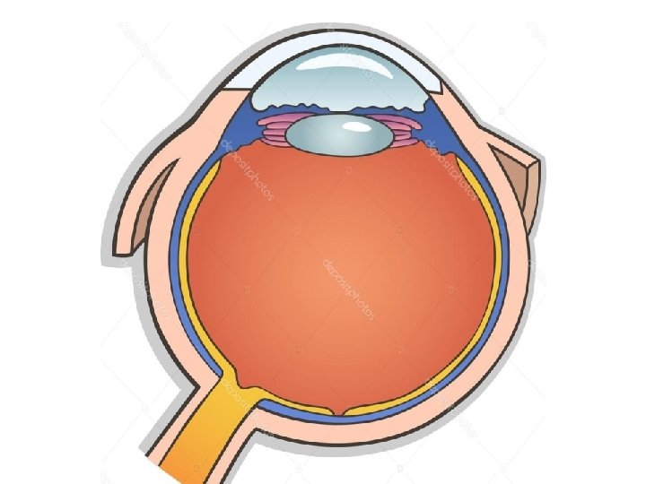
What is cornea?
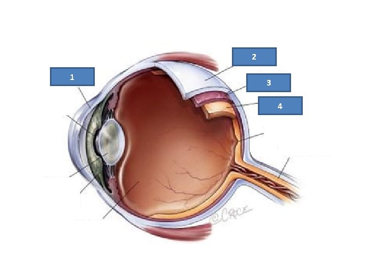
2 1 3 4
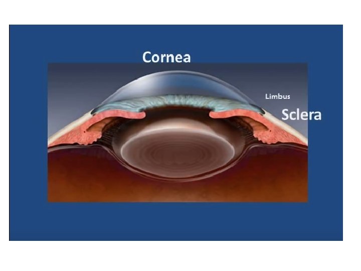

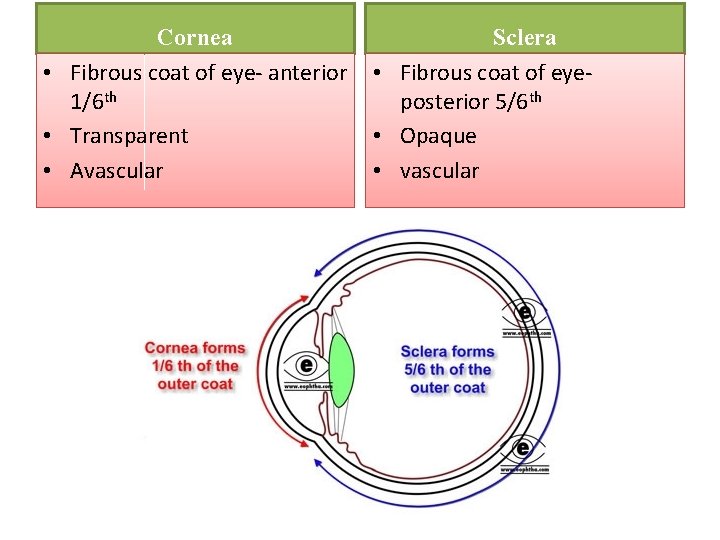
Cornea • Fibrous coat of eye- anterior 1/6 th • Transparent • Avascular Sclera • Fibrous coat of eyeposterior 5/6 th • Opaque • vascular
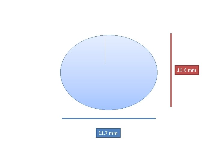
10. 6 mm 11. 7 mm

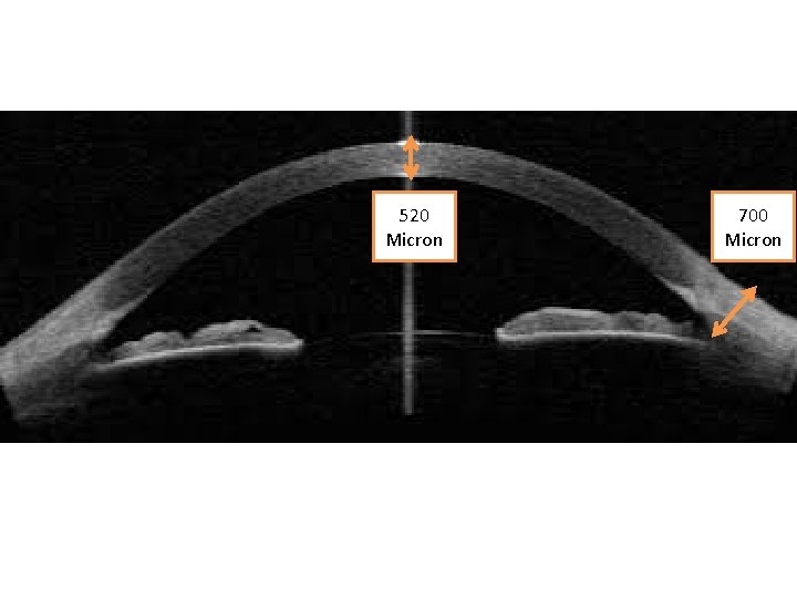
520 Micron 700 Micron
What are the main functions of cornea? • Integrity of the eye – fibrous coat • Transmission of light into the eye – transparent. • Most important refractive media of the eye – acts like a converging lens
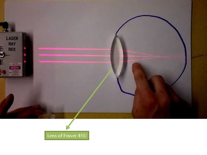
Lens of Power 45 D
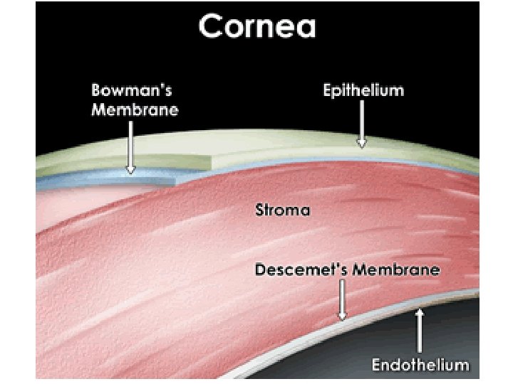
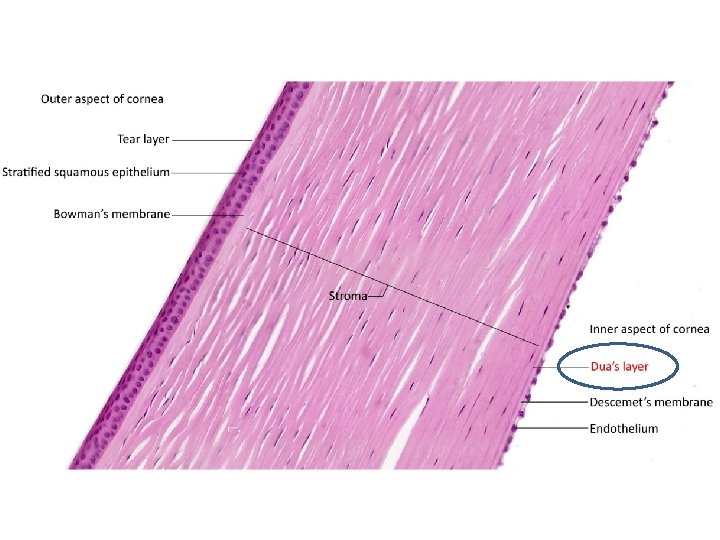
Layers of cornea
• A : Anterior Epithelium • B : Bowman’s Layer • • • : Corneal stroma D : Descmets membrane : Endothelium
Epithelium
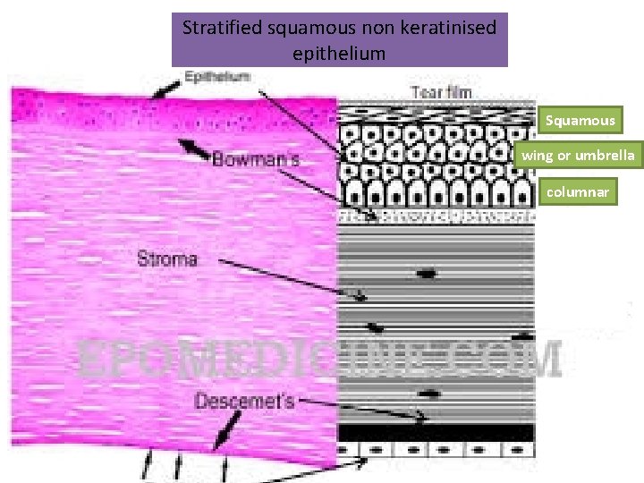
Stratified squamous non keratinised epithelium Squamous wing or umbrella columnar
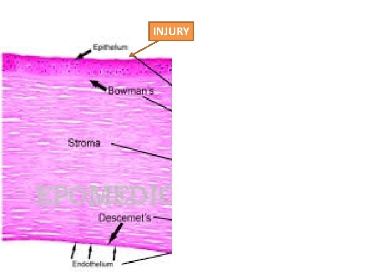
INJURY
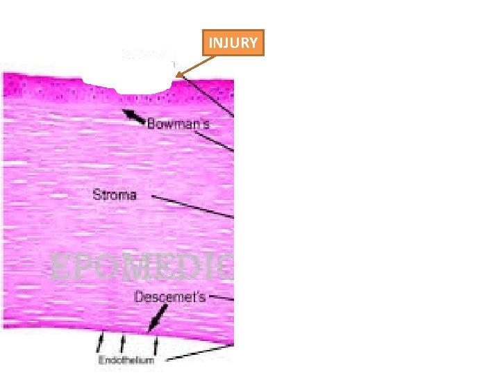
INJURY
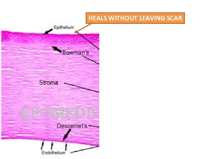
HEALS WITHOUT LEAVING SCAR

Bowman’s Layer
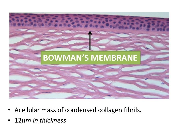
BOWMAN’S MEMBRANE • Acellular mass of condensed collagen fibrils. • 12μm in thickness
BOWMAN’S MEMBRANE – resists infection
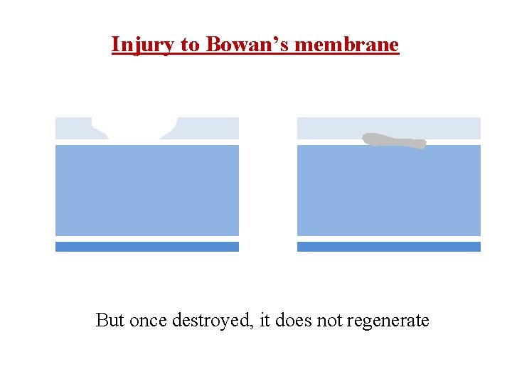
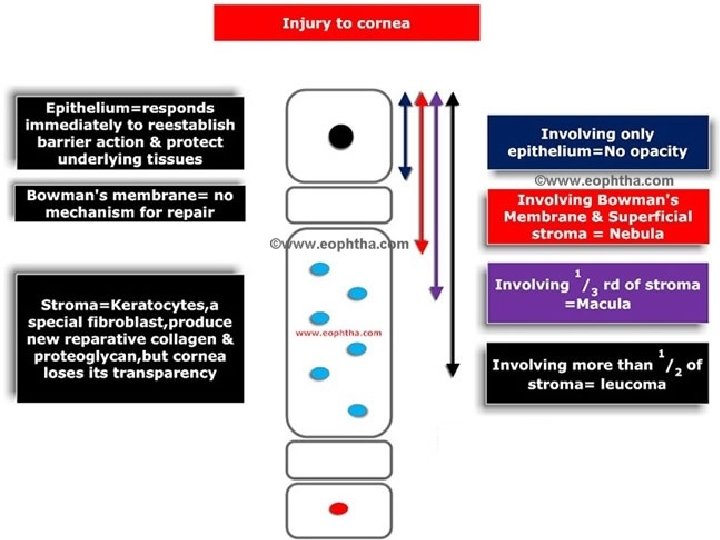
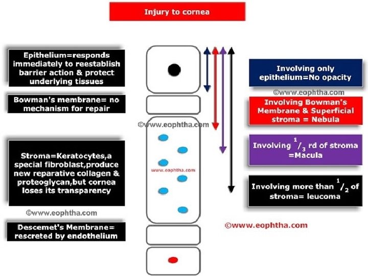
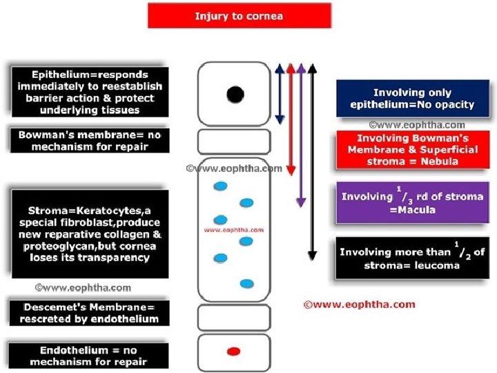
Injury to Bowan’s membrane But once destroyed, it does not regenerate

Stroma
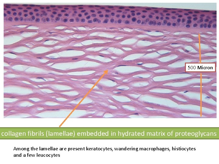
500 Micron collagen fibrils (lamellae) embedded in hydrated matrix of proteoglycans Among the lamellae are present keratocytes, wandering macrophages, histiocytes and a few leucocytes
Descemet’s membrane
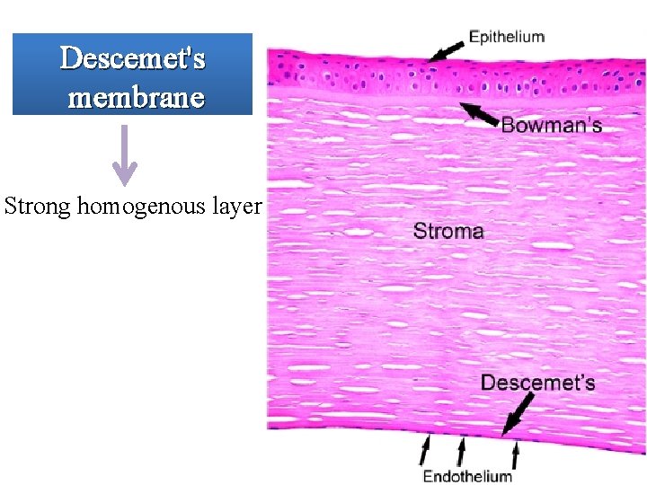
Descemet's membrane Strong homogenous layer
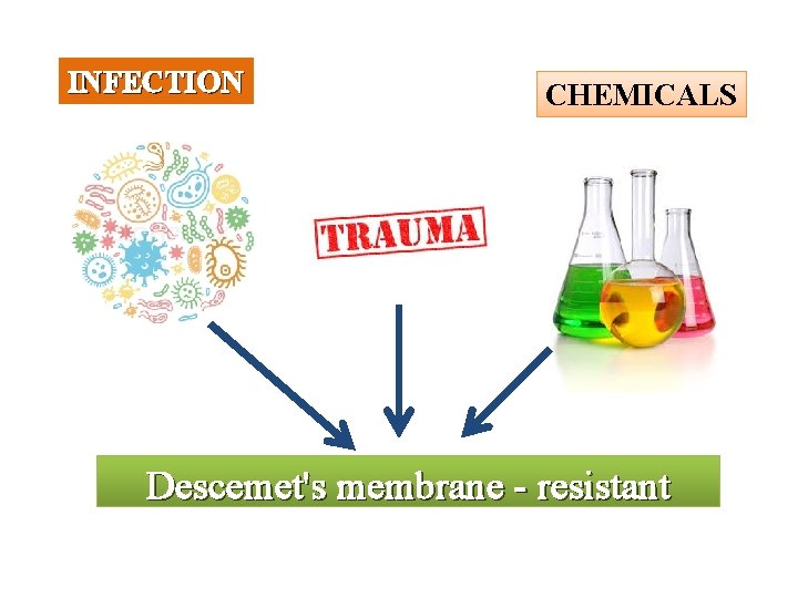
INFECTION CHEMICALS Descemet's membrane - resistant
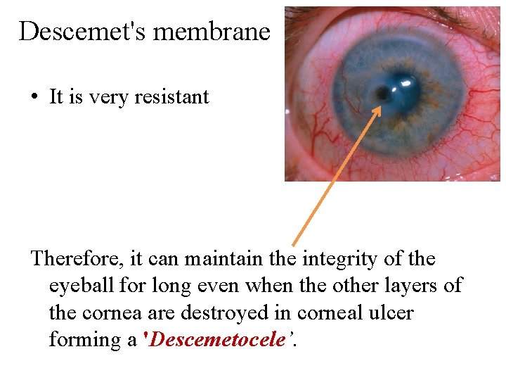
Descemet's membrane • It is very resistant Therefore, it can maintain the integrity of the eyeball for long even when the other layers of the cornea are destroyed in corneal ulcer forming a 'Descemetocele’.
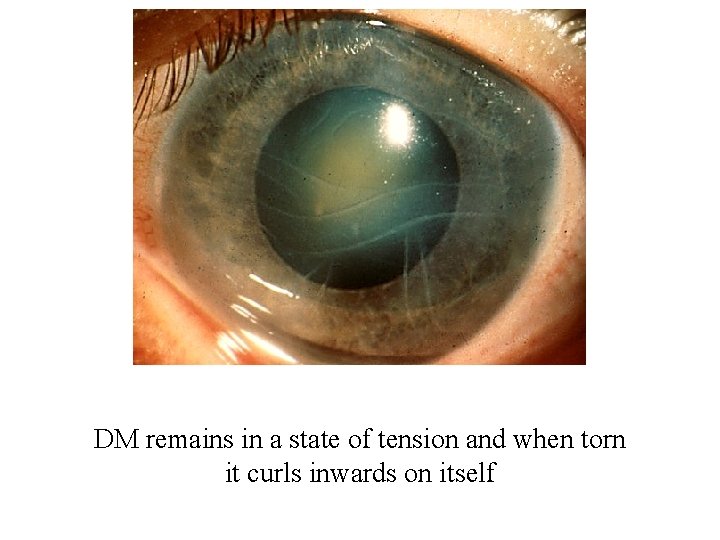
DM remains in a state of tension and when torn it curls inwards on itself
Endothelium
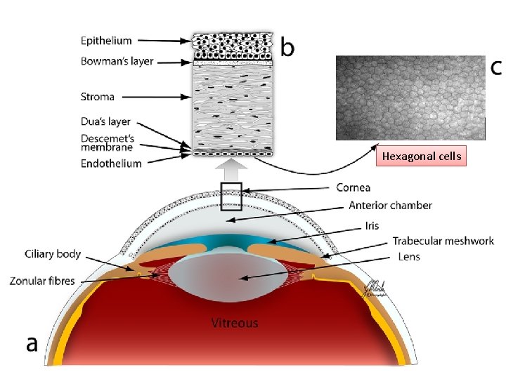
Hexagonal cells
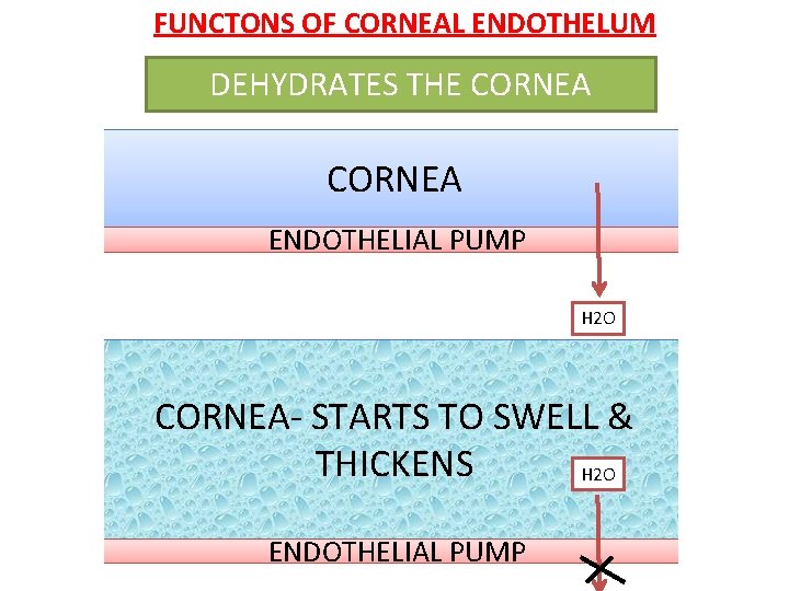
FUNCTONS OF CORNEAL ENDOTHELUM DEHYDRATES THE CORNEA ENDOTHELIAL PUMP H 2 O CORNEA- STARTS TO SWELL & THICKENS H 2 O ENDOTHELIAL PUMP
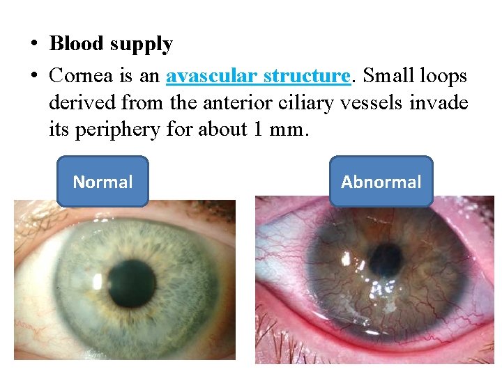
• Blood supply • Cornea is an avascular structure. Small loops derived from the anterior ciliary vessels invade its periphery for about 1 mm. Normal Abnormal
Nerve supply • Cornea is supplied by anterior ciliary nerves which are branches of ophthalmic division of the 5 th cranial nerve. • After going about 2 mm in cornea the nerves lose their myelin sheath and divide dichotomously and form three plexuses — the stromal, subepithelial and intraepithelial.
Why is cornea transparent? • Relatively dehydrated state, • Absence of blood vessels and pigment, • Uniform refractive index of all the layers • Uniform spacing of the collagen fibrils in the stroma.

2 Theories – Maurice Theory – Goldmann Theory

Maurice theory • The collagen fibrils are arranged in a regular lattice, separated by less than the wavelength of light so that any irregularly refracted rays of light are eliminated by destructive interference.

Normal Corneal edema
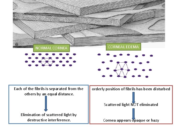
NORMAL CORNEA Each of the fibrils is separated from the others by an equal distance. CORNEAL EDEMA orderly position of fibrils has been disturbed Scattered light NOT eliminated Elimination of scattered light by destructive interference. Cornea appears opaque or hazy
Goldmann Theory • Cornea is transparent because fibrils are small in relation to light and don’t interfere with light transmission.
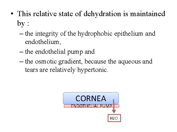
• This relative state of dehydration is maintained by : – the integrity of the hydrophobic epithelium and endothelium, – the endothelial pump and – the osmotic gradient, because the aqueous and tears are relatively hypertonic. CORNEA ENDOTHELIAL PUMP H 2 O
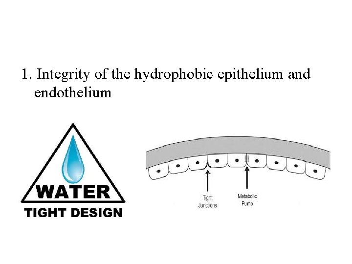
1. Integrity of the hydrophobic epithelium and endothelium
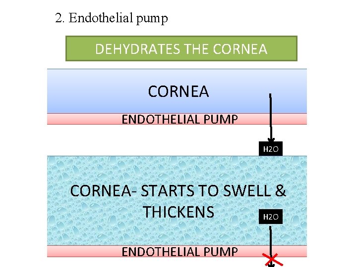
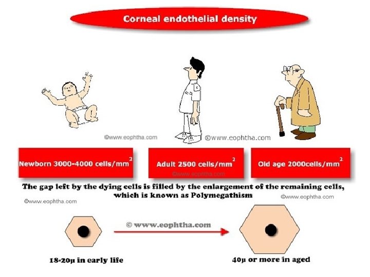
2. Endothelial pump DEHYDRATES THE CORNEA ENDOTHELIAL PUMP H 2 O CORNEA- STARTS TO SWELL & THICKENS H 2 O ENDOTHELIAL PUMP
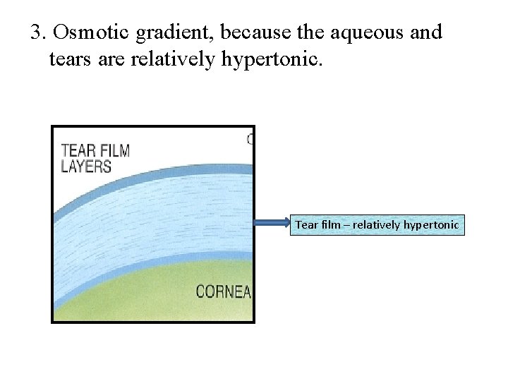
3. Osmotic gradient, because the aqueous and tears are relatively hypertonic. Tear film – relatively hypertonic

Pathology
• The major pathological changes that occur are broadly categorized as – keratitis, and corneal 'ulceration‘ – 'scarring‘ and 'opacification'.
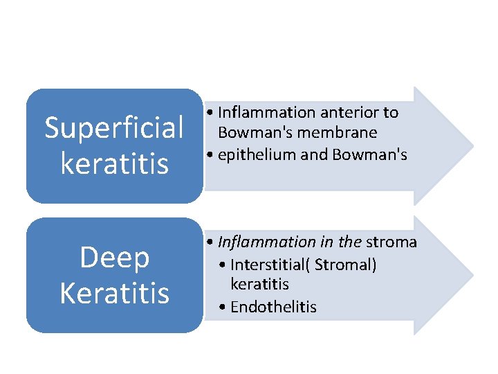
Superficial keratitis • Inflammation anterior to Bowman's membrane • epithelium and Bowman's Deep Keratitis • Inflammation in the stroma • Interstitial( Stromal) keratitis • Endothelitis
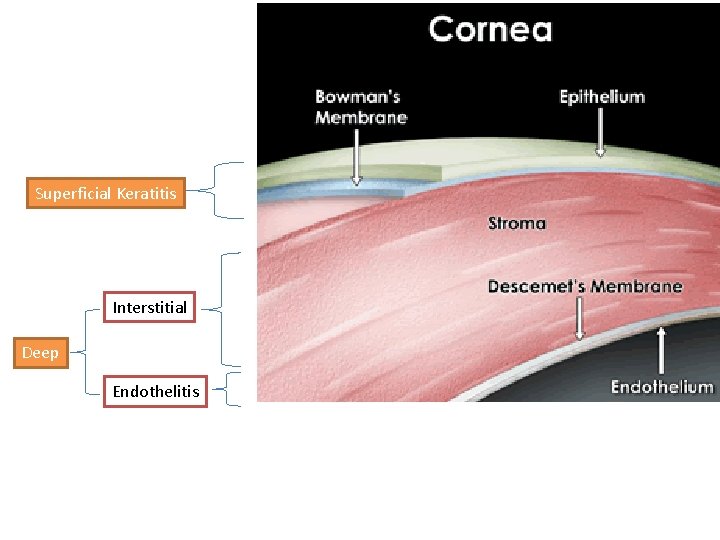
Superficial Keratitis Interstitial Deep Endothelitis
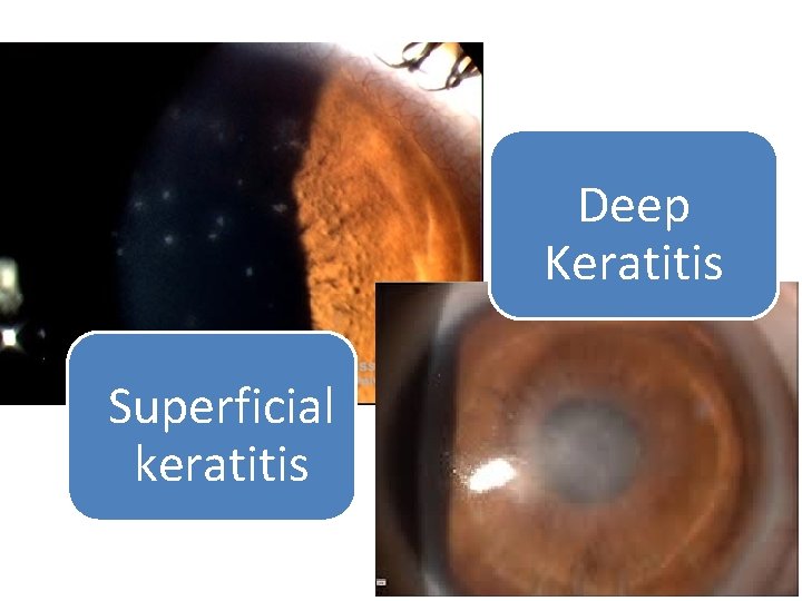
Deep Keratitis Superficial keratitis
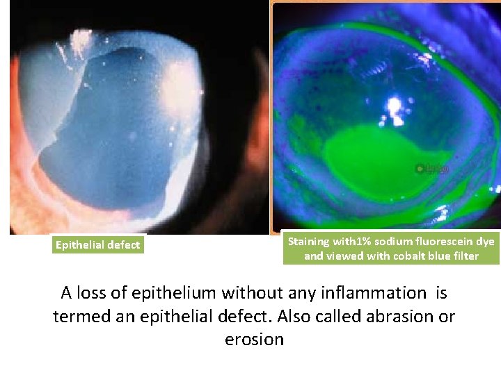
Epithelial defect Staining with 1% sodium fluorescein dye and viewed with cobalt blue filter A loss of epithelium without any inflammation is termed an epithelial defect. Also called abrasion or erosion
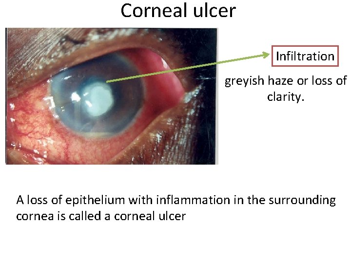
Corneal ulcer Infiltration greyish haze or loss of clarity. A loss of epithelium with inflammation in the surrounding cornea is called a corneal ulcer
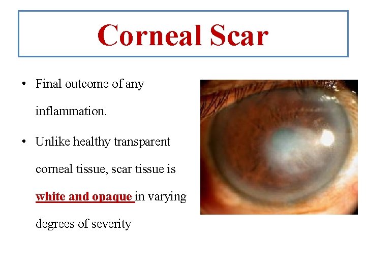
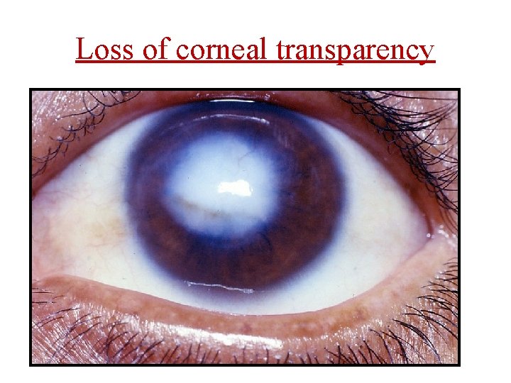
Corneal Scar • Final outcome of any inflammation. • Unlike healthy transparent corneal tissue, scar tissue is white and opaque in varying degrees of severity
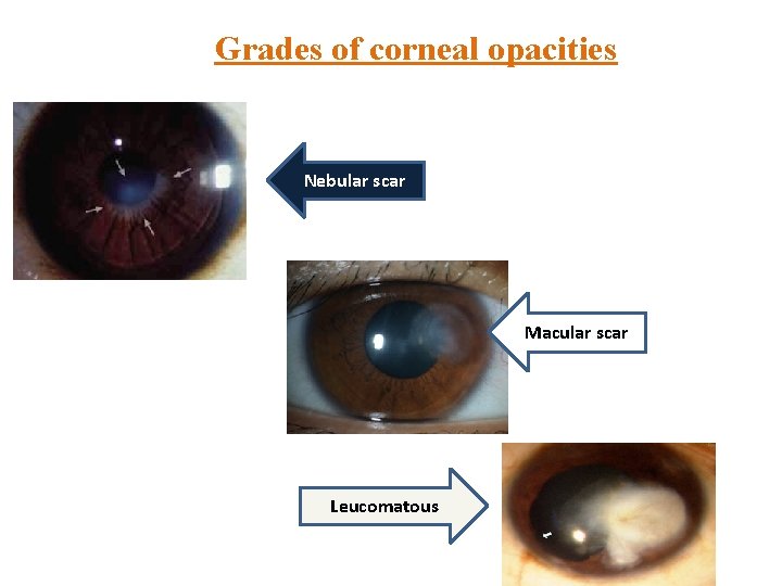
Grades of corneal opacities Nebular scar Macular scar Leucomatous
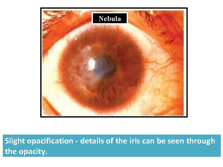
Nebula Slight opacification - details of the iris can be seen through the opacity.
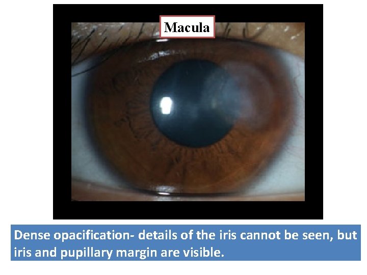
Macula Dense opacification- details of the iris cannot be seen, but iris and pupillary margin are visible.
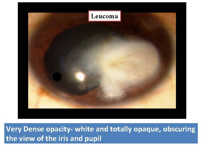
Leucoma Very Dense opacity- white and totally opaque, obscuring the view of the iris and pupil
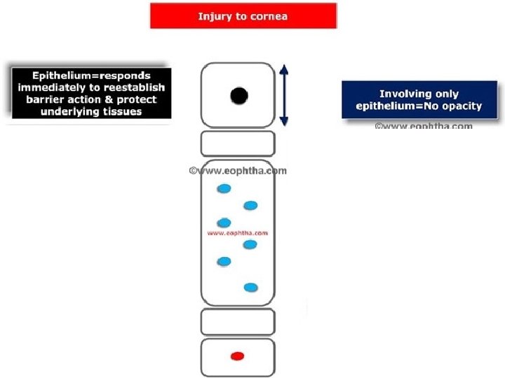
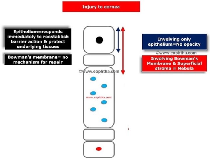
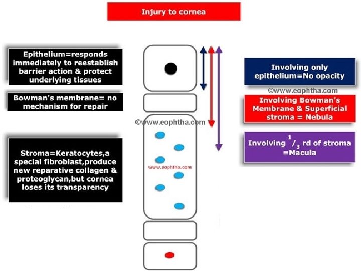
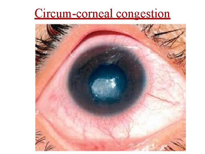
Symptoms of corneal disease • • Decreased vision Lacrimation Redness Photophobia ‘Foreign body’ sensation Pain Visible ‘whiteness’ of the cornea
Signs of corneal disease
Loss of corneal transparency
Circum-corneal congestion
Corneal surface
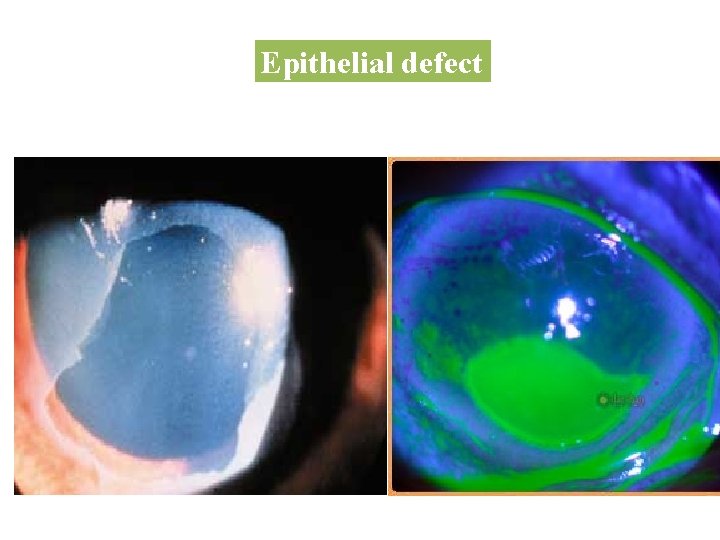
Epithelial defect
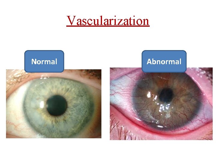
Vascularization Normal Abnormal
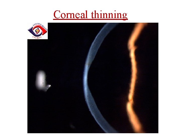
Corneal thinning
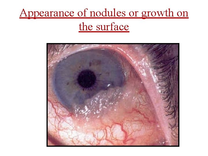
Appearance of nodules or growth on the surface
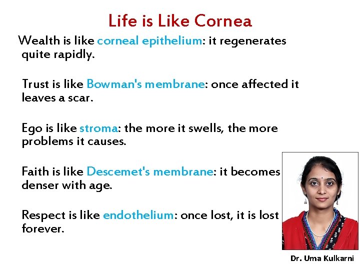
Life is Like Cornea Wealth is like corneal epithelium: it regenerates quite rapidly. Trust is like Bowman's membrane: once affected it leaves a scar. Ego is like stroma: the more it swells, the more problems it causes. Faith is like Descemet's membrane: it becomes denser with age. Respect is like endothelium: once lost, it is lost forever. Dr. Uma Kulkarni
Questions • 1. Name the layers of cornea • 2. Write briefly about the anatomical and physiological factors responsible for corneal transparency. • 3. Name and define the 3 grades of corneal opacity. • 4. Write briefly about corneal response to injury.

Useful internet links • Cornea : 1. anatomy : Adel Abdelshafik https: //www. youtube. com/watch? v=i. Vov. Wu 7 x. Sj. Q • Cornea 1: A Remarkable Structure https: //www. youtube. com/watch? v=Xf 21 nk 2 w 8 g 0
QUIZ • Thickest layer of cornea is _____ • Bowman’s layer regenerates after injury : True or false • Refractive power of the cornea is _____ • The most important refractive media of the eye is _____ • Cornea is supplied by branches of the mandibular division of 5 th cranial nerve: True or false
- Slides: 80