CORE Case 9 Workshop Erin OConnor MD Temple


- Slides: 50
CORE Case 9 Workshop Erin O’Connor MD Temple University
Learning Objectives 1. Review the anatomy of the intracranial compartments. 2. Know the appropriate imaging work up for patients with head trauma 3. Recognize the appearance of intracranial hemorrhage on CT 4. Describe the various types and causes of brain herniation
Learning Objectives 5. Recognize the typical changes in appearance of intracranial blood products with age on CT 6. List criteria that are used in spinal trauma to determine if CT of the cervical spine should be performed 7. Describe the role of plain radiographs, CT and MRI in cervical spine trauma 8. Evaluate cervical spine alignment on imaging
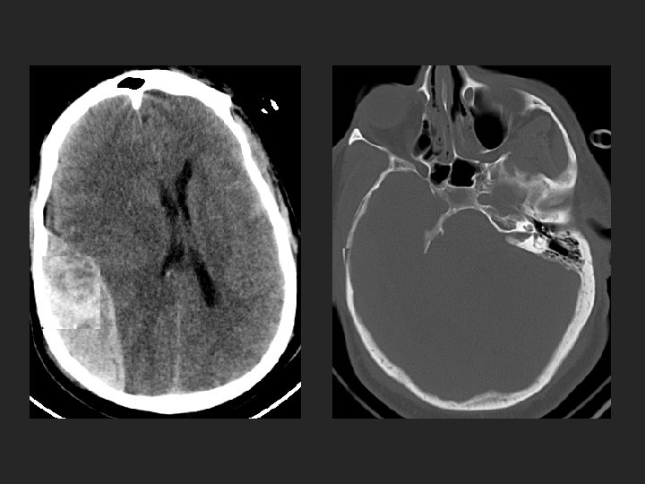
Trauma to the head – 49 year old male, s/p assault with baseball bat to head What imaging modality would you use?
Why not use MRI in the setting acute trauma to head?
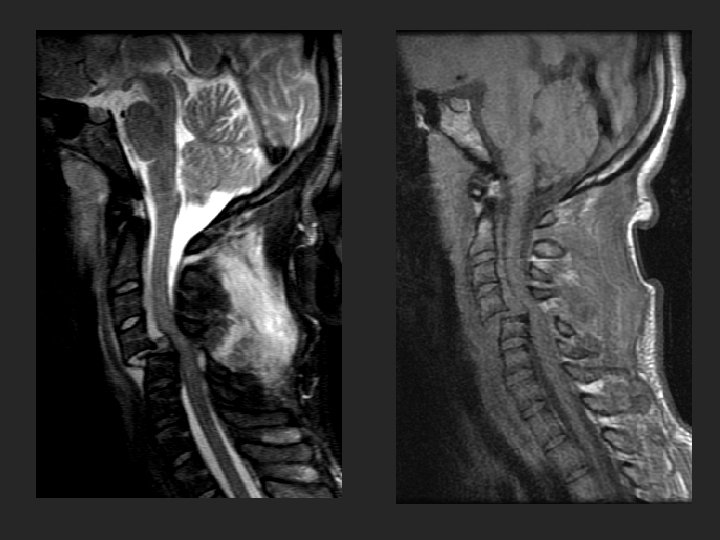
35 year old male with new onset of seizures, now referred to neurologist. Head trauma 3 months ago. What imaging modality should be used?
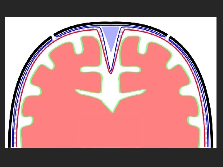
superior sagittal sinus epidural space suture subdural space subarachnoid space periosteum Meninges: pia mater arachnoid mater dura mater
subarachnoid space
subdural space
What is subfalcine herniation?
falx tentorial incisura tentorium cerebelli
What is uncal herniation?
falx tentorial incisura tentorium cerebelli
What are the potential sequela of herniation?
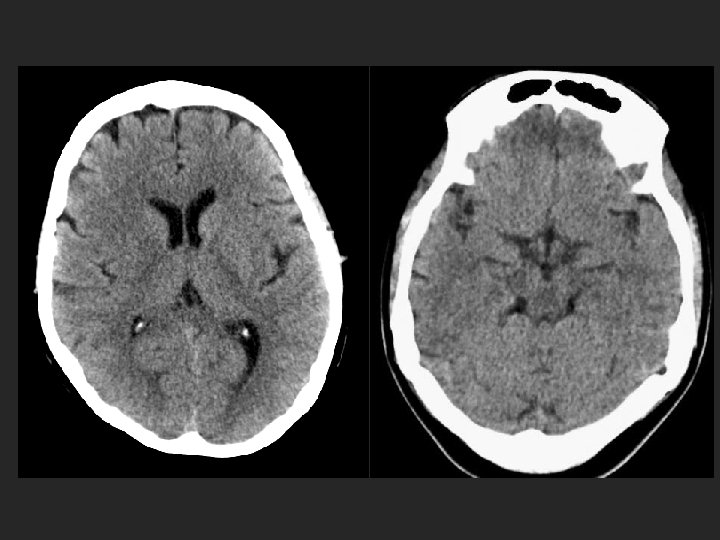
normal What type of herniation is seen on the left image?
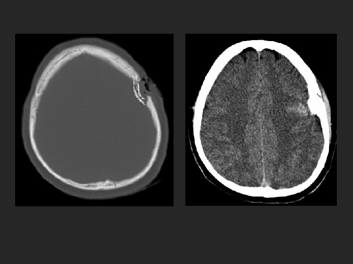
BLOOD Products of Varying AGES on CT
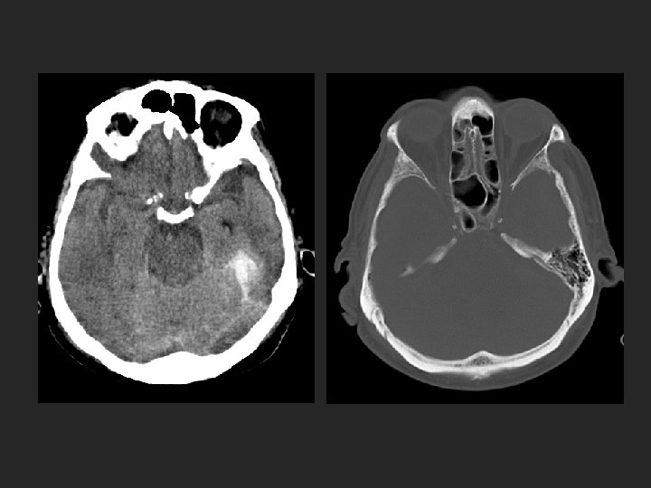
What are brain contusions?
Where do brain contusions typically occur?
What is coup contre-coup injury?
62 yo male, s/p seizure with trauma to head
Evolution of blood products with age
23 yo male, s/p fall down steps
10 mos. old female, rolled off of bed
40 yo male, s/p fall down stairs
59 yo female, driver in MVC
Normal Cervical Spine Alignment
spinolaminar line anterior vertebral line posterior spinous line
35 yo female, s/p bicycle collision with posterior midline cervical tenderness Would you image this patient?
What guidelines exist to help clinicians determine if patients who experience trauma should have C-spine imaging?
Nexus Criteria C-spine imaging in all patients with neck trauma unless of these criteria are met: • • • No posterior midline cervical tenderness No evidence of intoxication Normal level of alertness (GCS=15) No focal neurologic deficit No painful distracting injury
What should be done if this patient complains of upper extremity numbness and tingling?
27 yo male, s/p low speed rear end collision, GCS = 15 Would you image this patient?
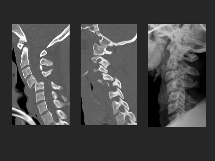
36 yo male s/p fall down flight of steps while intoxicated, +LOC X-table lateral Images courtesy of Je
36 yo male s/p fall down flight of steps while intoxicated, +LOC X-table lateral Images courtesy of Je What features guided the decision to obtain a CT of the cervical spine?
36 yo male s/p fall down flight of steps while intoxicated, +LOC X-table lateral Images courtesy of Je Can you name other mechanisms which are considered high risk by the Canadian C spine rules?
36 yo male s/p fall down flight of steps while intoxicated, +LOC Images courtesy of Je Can you explain why CT angiography was ordered on this patient?
What type of injury do you think this patient experienced?