
CONTROLLED HYDROSTATIC PRESSURE IN UTEROVAGINAL CANAL BY USING
CONTROLLED HYDROSTATIC PRESSURE IN UTERO-VAGINAL CANAL BY USING CONDOM, I. V. SET AND I. V. FLUID SHIVKAR’S PACK

USES ØOBSTETRICAL 1. PPH (POSTPARTUM HEAMORRHAGE) 2. INVERSION OF UTERUS – For reposition & maintenance ØGYNAECOLOGICAL HEMORRHAGE 1. UTERINE- (Menorrhagia D. U. B. ) 2. CERVICAL- (Traumatic, Ca-Cervix)

(SHIVKAR’S PACK) REQUIREMENTS 1. CONDOM 2. I. V. SET 3. I. V. FLUID 4. A PAIR OF SURGICAL SCISSORS 5. CURVED ARTERY FORCEPS 6. ROLLER PACK 7. THREAD
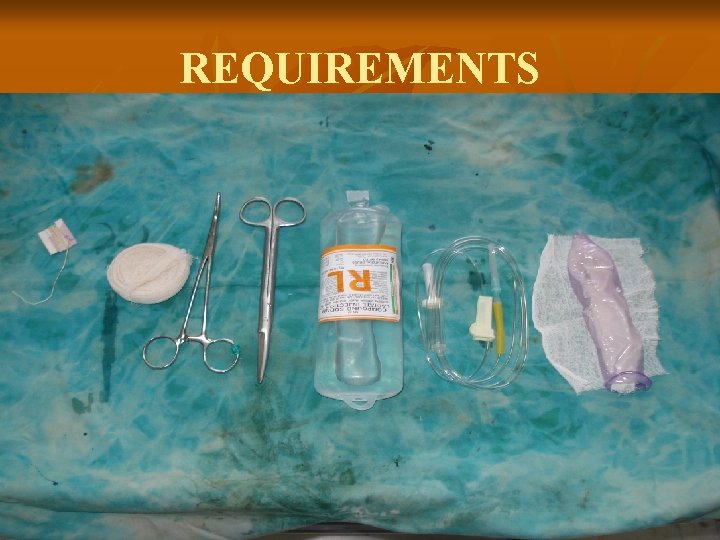
REQUIREMENTS

CONDOM Ø PREFERRABLY WASHED & AUTOCLAVED Ø ACCORDING TO EMERGENCY a) STERILIZED IN CHEMICALS, BOILING WATER OR ALCOHOL AND WASHED TO REMOVE CHEMICALS. b) WITHOUT STERILIZATION IF NEED IS THAT URGENT (AS CONDOMS ARE MANUFACTURED & PACKED IN HYGENIC ENVIRONMENT).
I. V. SET Commonly used I. V. Set is selected as its total length does not exceed 150 c. m. & it has small portion of elastic tube which can be utilized for fixing condom to I. V. Set (Alternative to tying with a thread). 0. 5 cm elastic band withstands 60 cm hydrostatic pressure and 1 cm elastic band can withstand over 100 cm hydrostatic pressure before leakage. I. V. FLUID Normal saline or any I. V. Fluid is used for safety, in case there is a leakage in the system & fluid goes into circulation.

SURGICAL SCISSORS CURVED ARTERY FORCEPS Medium size artery forceps is used to stretch & apply the elastic ring over the condom at various level. (An alternative for tying with a thread)
ROLLER PACK Medium size roller pack is used to pack vagina to prevent displacement of condom pack. THREAD A long & strong thread or a long narrow roller bandage to manipulate height of fluid source.
ASSEMBLY OF APPARATUS (SHIVKAR’S PACK) 1) Detach the elastic portion of the I. V. Set. 2) Make an extra hole on plastic tubing of the I. V. Set near its end. 3) Fix the condom by an elastic band obtained from detached portion in the 1 st step to I. V. Set. 4) Attach I. V. Set to fluid source. Inflate condom to 200 ml. 5) Manipulate to remove trapped air in condom & tubing. Empty the condom with syphon action. 6) Close the flow controller, when condom is in collapsed state.
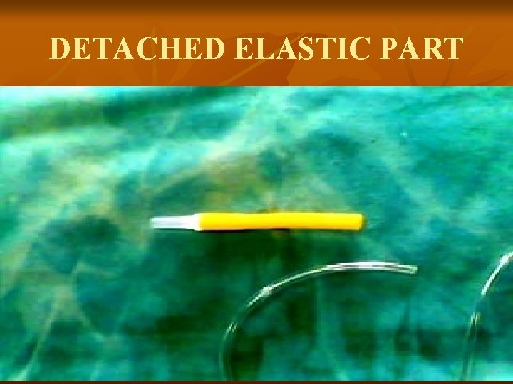
DETACHED ELASTIC PART
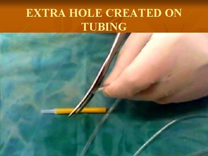
EXTRA HOLE CREATED ON TUBING
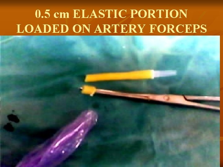
0. 5 cm ELASTIC PORTION LOADED ON ARTERY FORCEPS
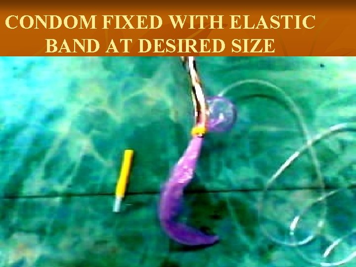
CONDOM FIXED WITH ELASTIC BAND AT DESIRED SIZE
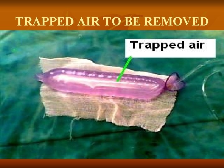
TRAPPED AIR TO BE REMOVED
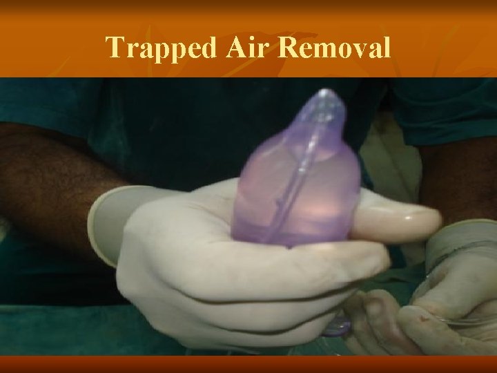
Trapped Air Removal
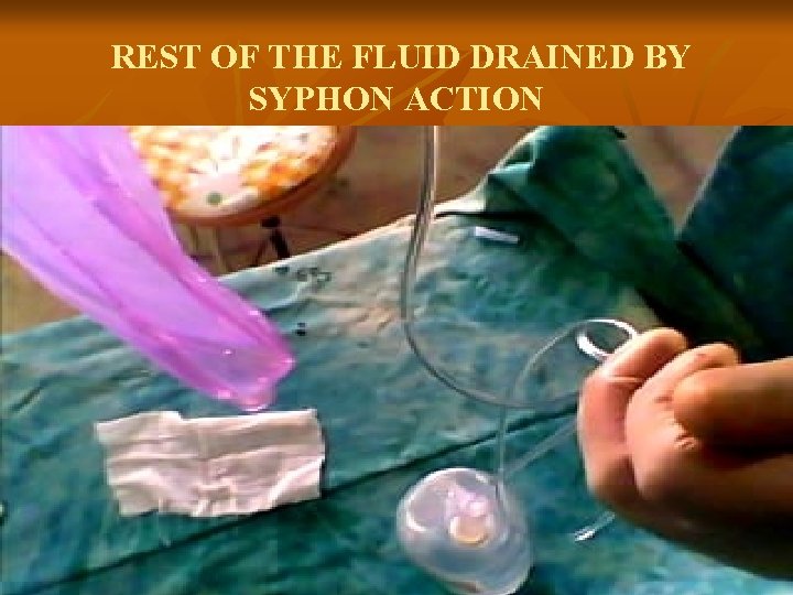
REST OF THE FLUID DRAINED BY SYPHON ACTION
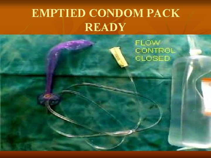
EMPTIED CONDOM PACK READY
OPERATION 1)Visualize & immobilize the cervix. It is not necessary in an uncomfortable situation in immediate post partum period, as cervix is wide open & balloon placed in vagina will find its way into the uterus on inflation. 2)Condom along with tubing inserted into the uterus up to its elastic band. With hands or with sponge holder, hold it lightly else condom may get punctured. (Replace with a new one if it happens).
OPERATION 3)Pack rest of the vagina loosely with roller pack. Take care not to kink the plastic tubing. Make patient’s position comfortable by removing lithotomy position if given. 4)Keep the fluid source at 60 cm above the abdomen with patient in lying down position. 5)Apply airway needle to fluid source to open the system to environmental pressure. Now we are ready to inflate the condom balloon.
APPLICATION OF CONTROLLED HYDROSTATIC PRESSURE IN P. P. H. ØMethod 1: - Predetermined pressure may be applied (not recommended ordinarily). It is only for experts, who are going to be with patient all the time & know their way to restrict uterine size by various methods. ØMethod 2: - Balance the hydrostatic pressure with uterine tone (recommended even for inexperienced).
BALANCING THE HYDROSTATIC PRESSURE WITH UTERINE TONE (Shivkar’s Pack) 1 After releasing the flow controller, fluid will rush through the tubing, inflating & pressurizing the utero-vaginal canal. As the pressure develops in the condom balloon the fluid flow will be reduced & eventually stops if sufficient utero-vaginal tone exists.
BALANCING THE HYDROSTATIC PRESSURE WITH UTERINE TONE 2 If the flow continues indefinitely or eventually increases then we are going in the overstretched utero-vaginal condition. This condition will invite more hemorrhage. Lower the fluid source gradually, till the fluid flow stops completely or preferably the reversal of fluid flow is observed. This will ensure against overstretched utero-vaginal condition.
Mechanism of haemostasis in balancing controlled hydrostatic pressure with uterine tone. n n n When intrauterine pressure is raised myometrium is ‘stretched’ which in turn compresses arterioles passing through it. This results into reduction of blood pressure of uterine surface bleeders. Further fall in B. P. of surface bleeders can be expected if uterus is allowed to contract over the balloon pack. Direct pressure applied on surface bleeders by condom balloon. In due time course vessels near surface undergo thrombosis.
PRACTICAL NOTES 1 After inflating to 300 to 400 ml, fluid source is lowered to 25 cm from the abdomen to see the reversal of flow. If there is no reversal of flow further lowering by 5 cm every 5 min. s is advised to see the reversal of flow. After observing the reversal of flow height of fluid source may be increased if necessary & possible without further appreciable distension (not > 50 ml)

PRACTICAL NOTES 2 It is essential to continue all the measures to increase uterine tone i. e. , uterotonics, blood transfusion, oxygen, calcium, etc. Measure the blood loss by keeping a kidney-tray at perineum after packing. If blood loss is less than 25 ml/hr …ignore. If it is up to 50 ml check the availability of blood for transfusion. If it is more than 50 ml, search for an alternative treatment.

PRACTICAL NOTES 3 Keep the pack in place for next 6 hrs (count the time after coagulation time has become normal).
PRACTICAL NOTES 4 Removal of pack – Not to remove at once. Reduce the fluid source height by 5 cm at every 10 mins interval till the fluid source level goes below patients bed level. Then remove roller pack & condom pack.

PRACTICAL NOTES 5 Failure of the pack has been observed in placenta previa with morbid adhesion and forceful removal at LSCS. Alternative measures should be considered. In placenta previa with morbid adhesions I preferred removal of placenta at latter date. The pack was effective in attempted but abandoned removal of placenta with morbid adhesions.
PRACTICAL NOTES 6 Recently ‘Dr. Ashok Anand’s GMC Stitch’ applied during LSCS, after delivery of baby but before delivery of placenta, in cases of all types of placenta previa, has shown promising results in controlling bleeding. It was presented at 53 rd & 54 th All India Congress of Obst. & Gynec. Dr. Ashok Anand, Prof. Obstetrics & Gynec. , of Grant Medical College, Mumbai, has submitted it for the publication to British Journal of Obstetrics and Gynecology.

THANK YOU Continue if interested. . .

PRESENTED BY DR. K. S. SHIVKAR M. D. , D. G. O Retired Associate Prof. Grant Medical College, & Sir, J. J. Group of Teaching Hospitals, Mumbai, Maharashtra, India. ………. . Continued for interested
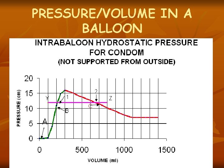
PRESSURE/VOLUME IN A BALLOON
Shivkar’s Pack n n Any elastic balloon will exibit Phase A (dead space), Phase B (in this Pressure rises with volume) and Phase C (in this Pressure in the balloon falls with further expansion). Magnitudes are variable in each balloon. Simillerly utero-vaginal canal may be considered as an elastic, multilayered and live balloon. Magnitudes may vary from time to time and in each case. But Phases A, B & C will persist.
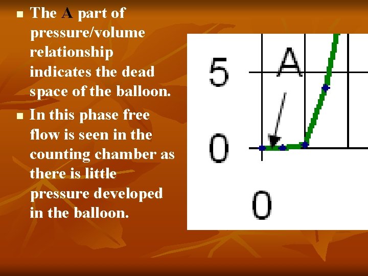
n n The A part of pressure/volume relationship indicates the dead space of the balloon. In this phase free flow is seen in the counting chamber as there is little pressure developed in the balloon.

In Phase B balloon is on stretch and pressure rises with inflation. n Gradual slowing of fluid flow is observed in counting chamber as intra balloon pressure rises. n Fluid flow stops completely if intra balloon pressure equals level of fluid source. n
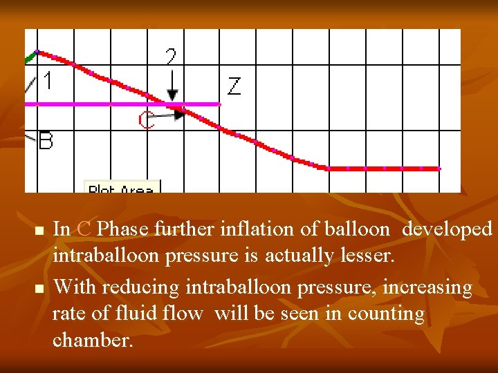
n n In C Phase further inflation of balloon developed intraballoon pressure is actually lesser. With reducing intraballoon pressure, increasing rate of fluid flow will be seen in counting chamber.
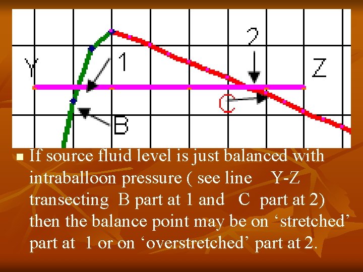
n If source fluid level is just balanced with intraballoon pressure ( see line Y-Z transecting B part at 1 and C part at 2) then the balance point may be on ‘stretched’ part at 1 or on ‘overstretched’ part at 2.
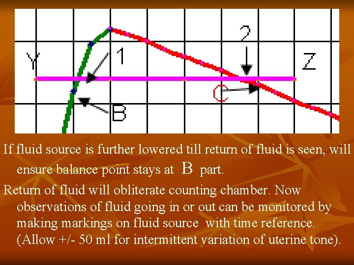
If fluid source is further lowered till return of fluid is seen, will ensure balance point stays at B part. Return of fluid will obliterate counting chamber. Now observations of fluid going in or out can be monitored by making markings on fluid source with time reference. (Allow +/- 50 ml for intermittent variation of uterine tone).
CONTROLLED HYDROSTATIC PRESSURE IN UTERINE INVERSION (SHIVKAR’S PACK) Inverted uterus can be easily pushed manually into the vagina. Fluid source used should be one liter at 60 cm. No sedation or anesthesia is required. Shivkar's Pack simplifies O’Sullivan’s Method of correction of Inversion of Uterus.
OPERATION • After pushing the uterus as high as possible, deflated condom balloon up to its elastic ring is placed into the vagina and lower part of the vagina packed with roller pack. • Make patient’s position comfortable by removing lithotomy position if given. • Keep fluid source at 60 cm from patient’s abdomen. Apply air way to fluid source. • Now we are ready to inflate the balloon pack.
APPLICATION OF CONTROLLED HYDROSTATIC PRESSURE IN UTERINE INVERSION (SHIVKAR’S PACK) 1 n n Open the flow controller. Fluid will flow in the tubing tamponading vagina and start pushing the inversion mass in the abdomen. Attending Doctor should keep one hand on the patient’s abdomen to feel the mass of inversion.
APPLICATION OF CONTROLLED HYDROSTATIC PRESSURE IN UTERINE INVERSION 2 n n n Attending Doctor should keep the whole mass below umbilicus of patient by hands. As the pressure develops ring of inversion can be appreciated by the Doctor. Gradually the ring increases in size and disappears. Gradually uterine contour can be appreciated and corrected uterine shape felt per abdomen.
APPLICATION OF CONTROLLED HYDROSTATIC PRESSURE IN UTERINE INVERSION 3 n n As soon as uterine inversion is corrected fluid flow increases. Now lower the fluid source to 25 cm height from patient’s abdomen allowing return of the fluid into the fluid source. Now start drugs to increase uterine tone. Keep fluid source at 20 -25 cm level from patient’s abdomen to prevent re-inversion.
APPLICATION OF CONTROLLED HYDROSTATIC PRESSURE IN UTERINE INVERSION 4 n n Reposition of inversion of uterus takes about 5 minutes to two hours with little discomfort to the patient and does not require any sedation or anesthesia. Keep patience if vital signs are OK. If height of fluid source increased above 60 cm them reposition is faster but there is appreciable discomfort to the patient.
APPLICATION OF CONTROLLED HYDROSTATIC PRESSURE IN UTERINE INVERSION 5 n n After 4 to 6 hrs. fluid source is gradually lowered by 5 cm/10 minutes interval and fluid allowed to return to source bottle. When the fluid source goes below patient’s bed level, roller pack and condom pack is removed.
CONTROLLED HYDROSTATIC PRESSURE IN GYNAECOLOGY (SHIVKAR’S PACK) n n DUB FIBROIDS MENORRHAGIA DUE TO COAGULOPATHY, THOMBOCYTOPENIA CERVICAL HAEMORRHAGES i. e. TRAUMATIC, POST CAUTERISATION, FOLLOWING CERVICAL AMPUTATION. CA-CERVIX etc. In these cases vagina is packed with Shivkar’s pack.
CONTROLLED HYDROSTATIC PRESSURE IN GYNAECOLOGICAL UTERINE HAMEMORRHGES (Shivkar’s Pack) n n I. V. set tubing with condom can be easily inserted through gynaec. Cervical canal without requiring anaesthesia or sedation or manual dilatation. Elastic band is applied on 1/4 th of the condom as gynaec. uterus is smaller. Let the patient empty the bladder before the operation. Preoperative Atropine 0. 6 mg is given.
OPERATION n n Cervix is visualized and immobilized with vulsellum. Endometrial Biopsy may be taken. Condom with tubing inserted through cervical canal by holding it at the elastic band with a large curved artery forceps till even the elastic band goes in through cervical canal. Roller pack is applied below cervix to prevent the displacement of balloon pack. Remove vulsellum and pack rest of the vagina roller pack. Take care not to kink tubing. Make the patient’s position comfortable.
APPLICATION OF CONTROLLED HYDROSTATIC PRESSURE IN GYNAEC UTERUS 1 n n n Keep the fluid source at 60 cm above the patient’s abdomen. Ensure the airway of fluid source. Open the flow controller. Observe the free fluid flow for short period as gynaec. Uterus is small. Gradually drop by drop even gynaec uterus distends with 60 cm hydrostatic pressure.
APPLICATION OF CONTROLLED HYDROSTATIC PRESSURE IN GYNAEC UTERUS 2 n n If the uterus distends beyond 100 ml. then bring the fluid source down to balance with the uterine tone. The intrauterine pressure is maintained for four hours. Periodically the fluid source is lowered till it goes below patients bed. Remove the roller pack and then the condom pack.
Mechanism of haemostasis in gynaec. Uterus n n n Pressure on the endometrium kinks the bleeders at stratum spongiosum level. The kinked vessels undergo thrombosis in due course. Endometrium superficial to stratum basalis undergoes ischemic necrosis and subsequently shades if still present like normal menses. Then haemostasis is observed. Why subsequent cycles tend to become Normal? Then it is especially useful in perimenopausal group.
PRACTICAL NOTES 1 n n n Haemostasis observed if the intrauterine pressure is maintained for 3 to 4 hrs. After removal of pack a) complete haemostasis, b)continued but less than menstrual bleeding or c)passage of clot or endometrial caste followed by haemostasis observed. It is interesting to know that in the subsequent menstrual cycles the bleeding tends to become normal specially in the cases with distensible uterus. These patients were followed up over period of two years (80% success rate).
PRACTICAL NOTES 2 n n In the Uterus with myometrial hyperplasia and no distention the subsequent cycles continued to remain heavy. If Shivkar’s Pack used premenstrually in patients with H/O heavy menses, then subsequent menstrual blood loss is near Normal. No anaesthetics or sedatives used during the procedure. To satisfy scientific community on long term effects more studies are required.
Chronology n n Around 1980 -81, I saw a case of tight uterine packing done by one of our consultant and realized the high degree of skill required for the purpose. Scanning of literature on uterovaginal packing revealed various machines , methods and their pitfalls. I was ready with the Method by 1981, in which I corrected most of the pitfalls. I used it to pack vagina in cases of vaginal lacerations, Ca-cervix bleeding and post cervical surgery. Nursing staff used to call it Shivkar’s vaginal drip.
n Second week of November 1984: First time Shivkar’s Pack was used successfully in case of PPH following delivery of Abruptio Placenta , failure of traditional pack, due to failure of coagulation of blood. When I started ordering in professional manner to get me a condom, I. V. Set, etc. Residents were surprised and curious about my actions which I completed in Five minutes from the first order.
n n November 1984 Departmental Clinical meeting in OBGY the case was discussed. The Meeting was attended by Prof. & H. O. D. Dr. (Mrs. ) Kamal Deshmukh, all consultants and residents of OBGY of Grant medical college, & Sir, J. J. Group of Hospitals, Mumbai. 14/01/1985 ‘Dr. K. S. Shivkar’s(India) Method of Packing Uterovaginal Canal’ was submitted to Research Society’s Journal for publication of Grant Medical College, & Sir, J. J. Group of Hospitals, which was subsequently published.
n 28/12/1985 29 th All India Congress of Obst. & Gynecology held at Mumbai: Shivkar’s Pack was presented as ‘Pressure Balloon Pack (for hollow viscous bleeding)’ Authored by Dr. K. S. Shivkar & Dr. B. J. Wadia (Hon. Prof. & Unit Head ) of Grant Medical College, Mumbai. The abstract of the same was published in the Souvenir published at that time.
n However, the complete Paper Titled ‘DR. K. S. SHIVKAR’S (INDIA) METHOD FOR PACKING UTEROVAGINAL CANAL’ was submitted on 24/10/85 (Paper/934/86) before 29 th AICOG to The Journal of Obstetrics and Gynecology of India, failed to appeal the publisher and was not accepted for publication.
21/08/1985 Patent Application No. 218/ BOM/85 submitted to Government of India Patent Office Branch at Lower Parel, Mumbai. n It was not pursued for final Patent. n As in real life Shivkar’s pack will be required rarely unless misused. n Manufacturing it as per patent specification will make it extra ordinarily costly to the end user, ( poor women) commonly affected group. n
I feel life saving inventions should not be patentable. As they become costly because of patent. n Hence the idea of patenting Shivkar’s Pack was abandoned. n The Shivkar’s Pack was published by teaching resident Doctors and other Doctors from all over India attending Dr. B. J. Wadia’s annual CME workshop in the dept. of Obst. & Gynecology at Sir, J. J. Hospital from 1986 to 1995. n
n In 1987 Shivkar’s Pack was used first time in St. Georges Hospital, Mumbai, affiliated to Grant Medical College, to correct Inversion of uterus immediately following delivery, by Dr. Palep (prof. & unit head) and Dr. S. K. Patil (Lecturer). Fluid source height used was 100 cm. Volume used was 1000 ml. with no anesthesia to patient. The correction was successful in Five minutes.
n February 22, 2003. Dr. Vinita Salvi (Prof. Obst & Gyn. Seth G. S. Medical college, K. E. M. Hospital , Mumbai) Presented ‘Shivkar’s Pack and Internal Iliac Artery Embolization – Indigenous innovation couples with Modern Technology to save Lives’ in the Conference of the South Asian Federation of Obstetrics & Gynecology 2003.
n. Express Healthcare Management Newspaper, India, issue dated 16 th to 31 May 2003 published 'Condom Useful in arresting post delivery bleeding' on Internet http: //www. expresshealthcaremgmt. com/200 30531/focus. shtml n. Finally Shivkar’s Pack was published as ‘Pressure Balloon Therapy in Uncontrolled Obstetrical Hemorrhage’ Authored Shivkar Krishna S. , Khadilkar Suvarna S, Gandhewar Manisha. J. Obstet. Gynaecol Ind Vol. 53, No. 4, July/August 2003.

SHIVKAR’S PACK IS DEDICATED TO DEVELOPING COUNTRIES BY DR. KRISHNA SHANKAR SHIVKAR
- Slides: 64