Control of eye movement Third Nerve Palsy Eye

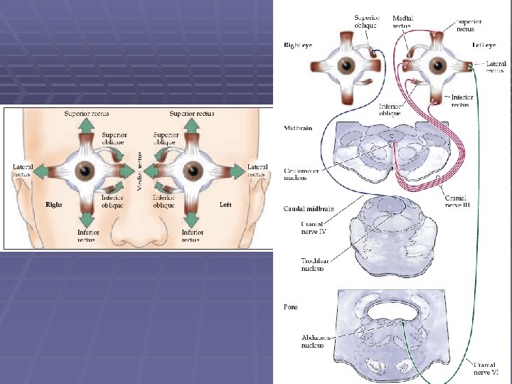
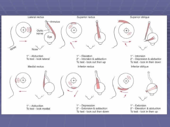


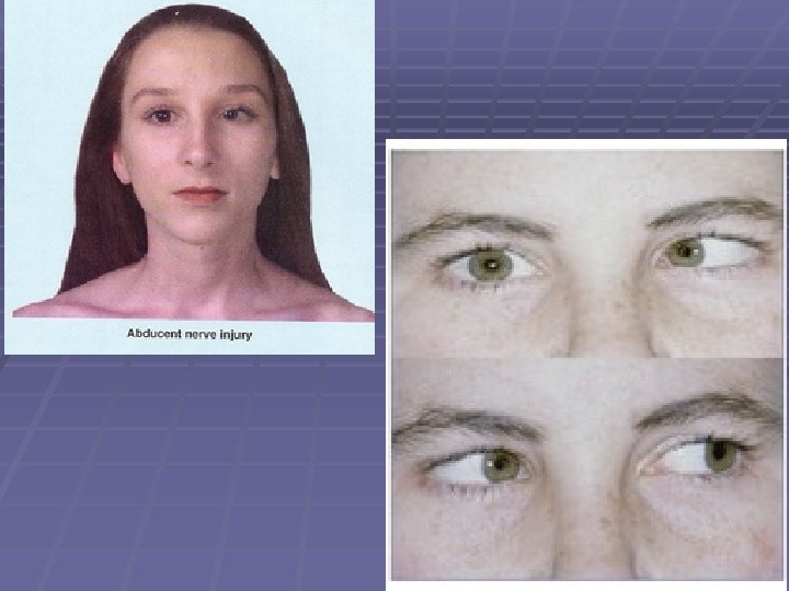
 Figure 28 -11 Vertical eye movements")


 Temporal bone overlying the anterior or the posterior semicircular canal")

- Slides: 53
Control of eye movement
Third Nerve Palsy Eye “down and out”
Trochlear Nerve Palsy Note: Right eye • Instead of intorsion and depression action of superior oblique • See extorsion and elevation Observe how the axes over the right eye shift when patient generates a compensatory head movement Attempted Correction: • Patient tilts head to her left • Tucks chin to foveate on object • Left eye will align accordingly
(Also known as Field of Forel) Figure 28 -11 Vertical eye movements
Atlas 6 -23
VI VII nuc. VI
Fig. 28 -13
Basic pathway for controlling saccadic eye movements
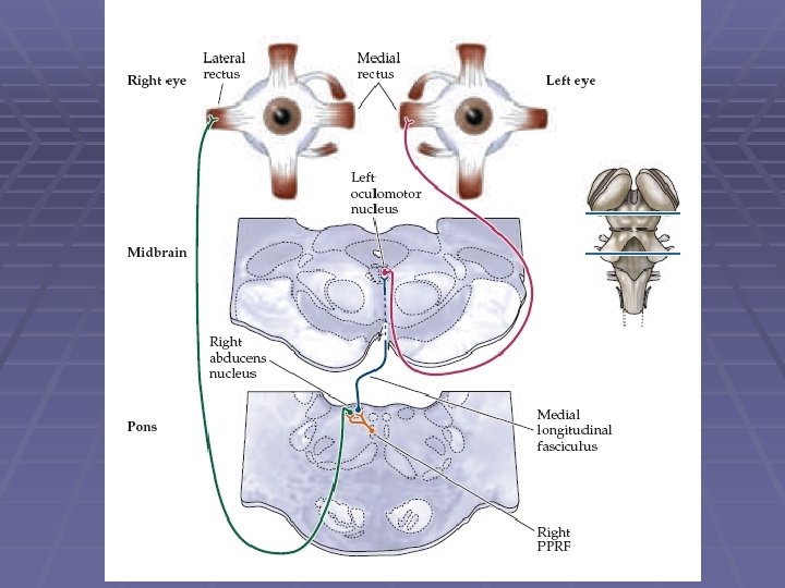
MLF
L R
L R Disconjugate eye Conjugate eye movements
L Conjugate eye R X movements Internuclear Ophthalmoplegia Disconjugate eye movements
L R ONE-AND-A-HALF SYNDROME
The Vestibular System
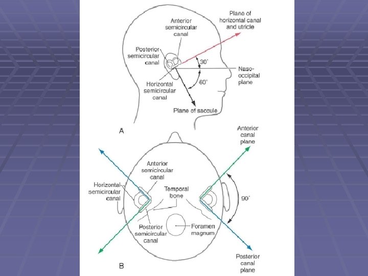
Anatomy of the ear
Anatomy of the ear
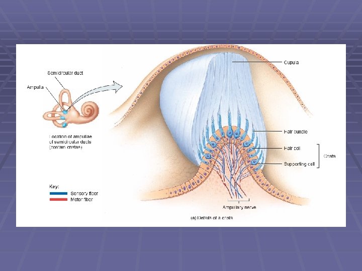
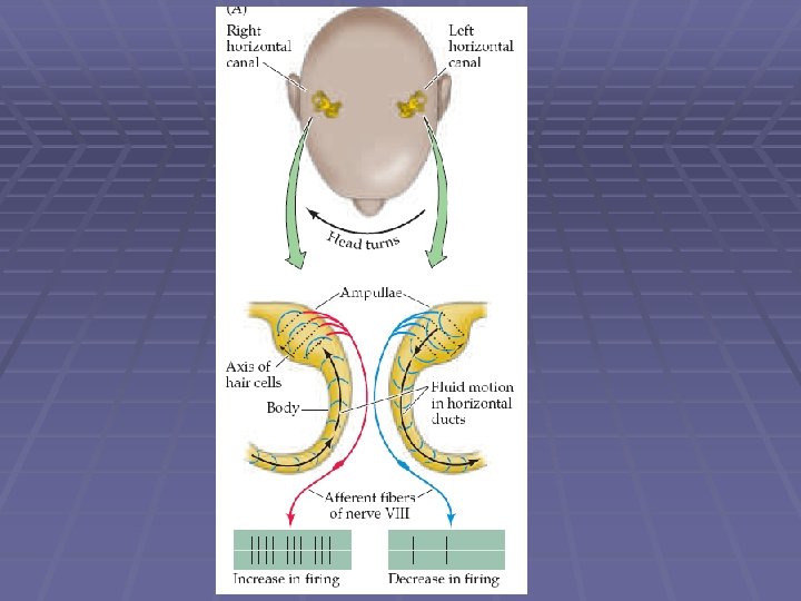
Ampulla of Semicircular canal
Anatomy of the ear
Anatomy of the ear
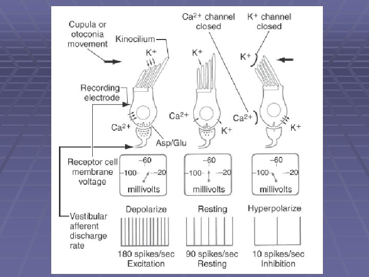
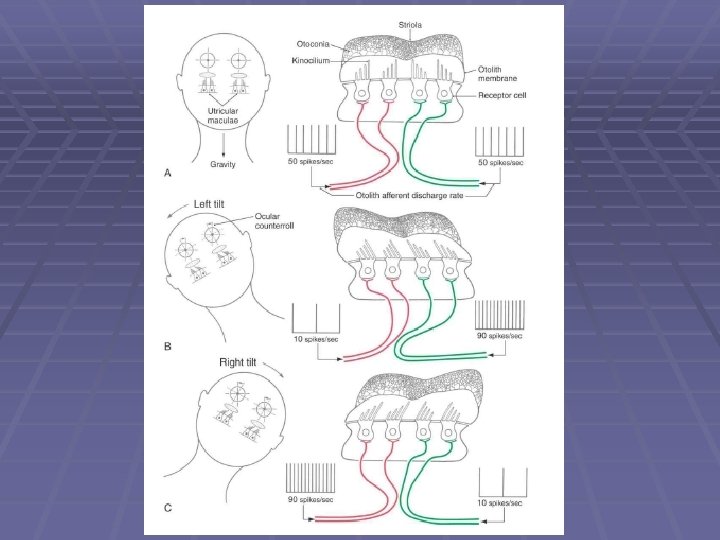
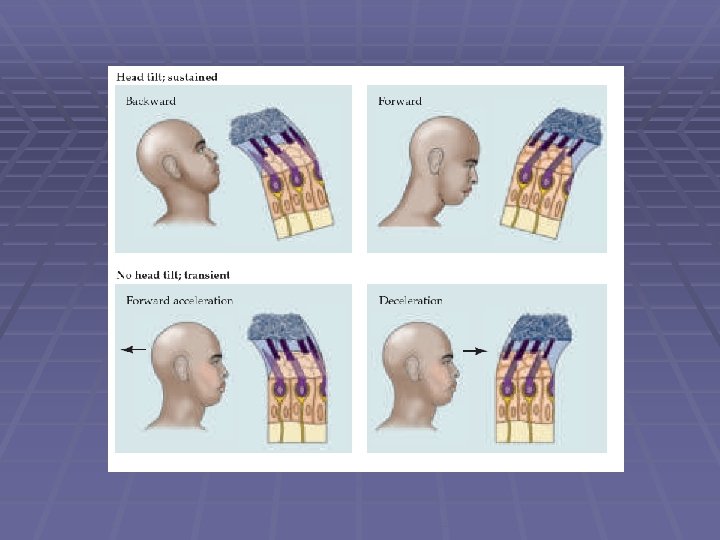
Macula and otolith organ
Macula and otolith organ
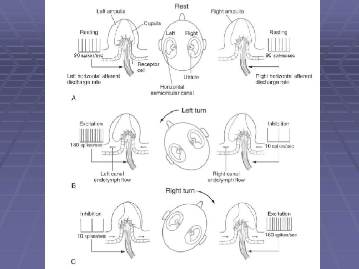
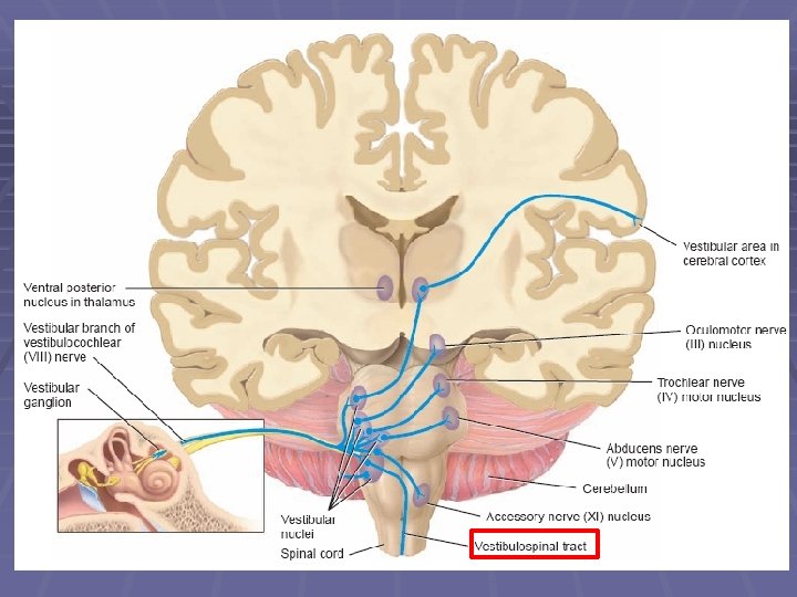
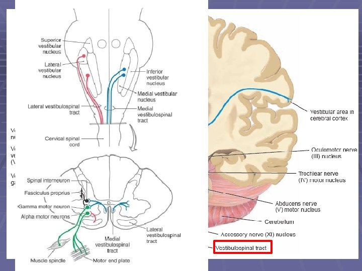
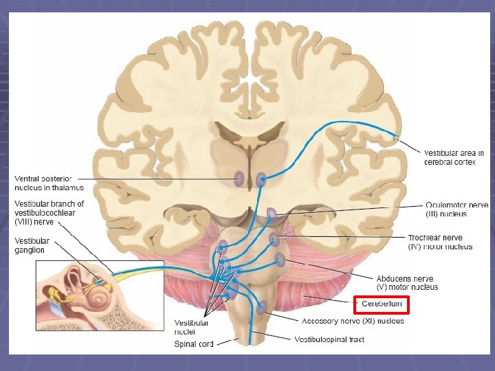
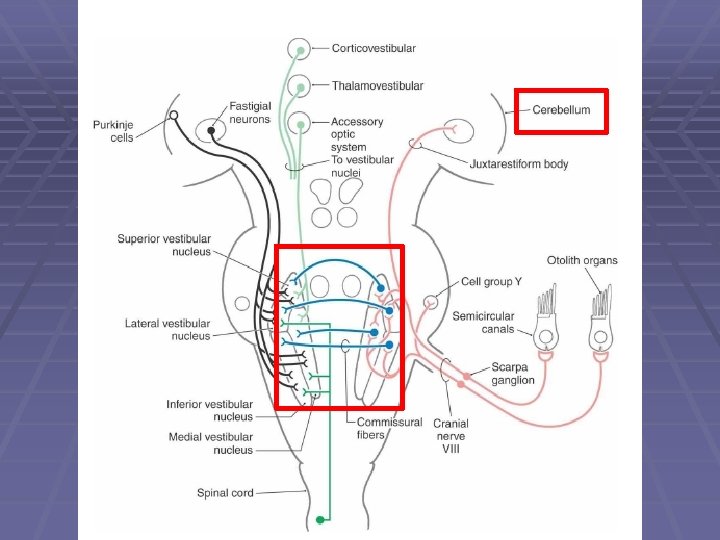
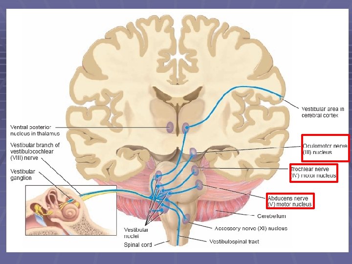
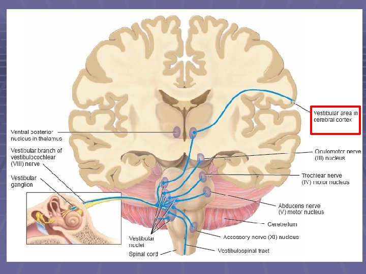
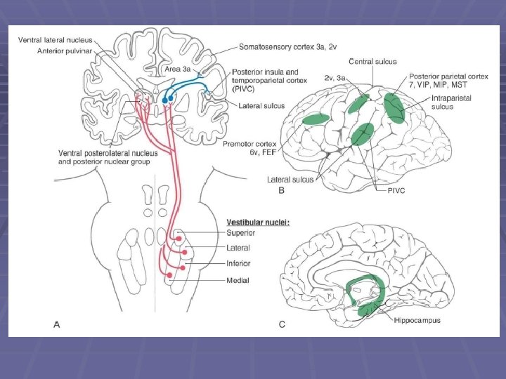
VESTIBULAR PATHWAY
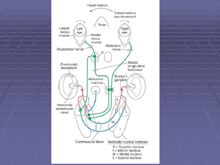
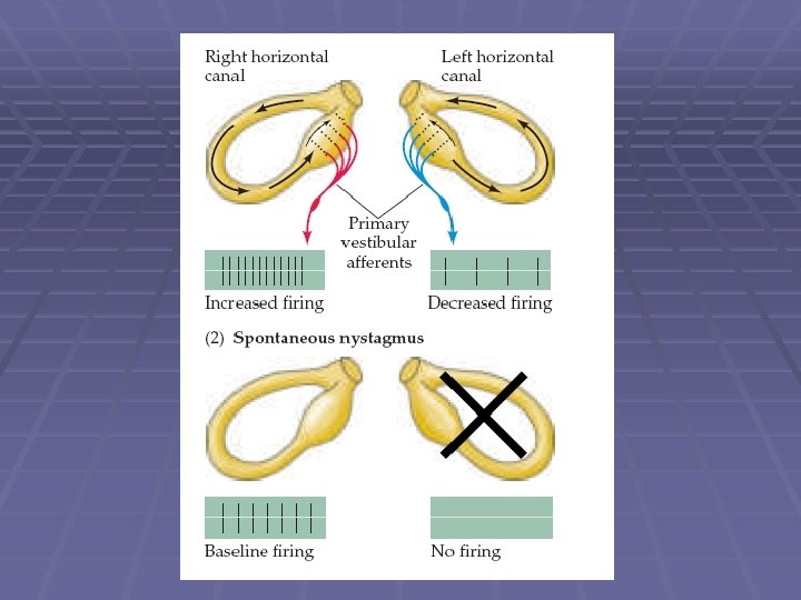
VESTIBULOOCULAR REFLEX § Compensatory for head movements § Rotational Reflex § Linear Reflex
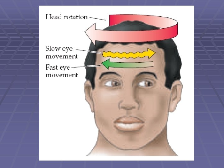
VESTIBULOOCULAR REFLEX § Compensatory for head movements § Rotational Reflex § Linear Reflex § Nystagmus
Caloric test
Ménière Disease results from a disruption of normal endolymph volume Symptoms include: Severe vertigo Positional nystagmus (when head in a particular position) Nausea Affected individuals can also experience-unpredictable attacks of auditory & vestibular symptoms: Vomiting Tinnitus (ringing in ears) Inability to make head movements Inability to stand passively Low frequency hearing loss Treatment: administration of a diuretic (hydrochlorothiazide) & a salt restricted diet Persistent condition: shunt implantation into swollen endolymphatic sac, or delivery of a vestibulotoxic agents (gentamicin) into erilymph.
Benign Paroxysmal Positional Vertigo § common clinical disorder. § condition characterized by brief episodes of vertigo that coincide with particular changes in body position. § pathophysiology poorly understood. § posterior canal abnormalities are implicated. § otoconia crystals in the utricle may separate from the otolith membrane and become lodged in the cupula, causing abnormal cupula deflections. AND/OR partial inflammation of cranial nerve VIII
Dix-Hallpike test The definitive diagnostic test for benign paroxysmal positional vertigo • Patient from sitting to supine position. • Head turned 450 to one side and extended 200 backward. • Observe eyes for nystagmus (30 sec. ). • Bring back to a sitting position. • Small delay, test other side. • A positive test consists of a burst of nystagmus. • Posterior canal BPPV (more common) – eyes jump upward.
Dizziness: non-specific term. generally means spatial disorientation. may or may not involve feelings of movement. may be accompanied by nausea or postural instability. may be caused by factors other than vestibular dysfunction. Vertigo: specific term. perception of body motion. spinning or turning sensation when no real motion is taking place. Tinnitus Some of these causes include high blood pressure, diabetes, listening to loud music, a tumor, thyroid conditions, and medications / antidepressants, sedatives, antibiotics, antiinflammatories, and aspirin.
Semicircular Canal Dehiscence (opening) Temporal bone overlying the anterior or the posterior semicircular canal thins, creating an opening/dehiscence next to the dura. Dehiscence over left superior canal Text Fig. 22 -5 CT scan of the temporal bone projected into the plane of the left superior/anterior canal, in a patient with superior canal dehiscence syndrome. The dehiscense exposes the bony labyrinth to the extradural space. Symptoms: vertigo and oscillopsia in response to loud sounds (Tullio Phenomenon), or in response to maneuvers that change middle ear or intracranial pressure. Nystagmus evoked by these stimuli aligns with the plane of the dehiscent superior canal. Treatment: Surgical closure of the defect by bone replacement. © 2005 Elsevier
Vestibular Neuritis § § § severe vertigo, nausea, vomiting no hearing loss or other CNS abnormalities possible edema of the vestibular nerve/ganglion. thought to be produced by acute viral infection. treated with anitemetics, vestibular suppressants, corticosteroids, & antiviral agents.