
Control of cell growth Cells in a multicellular


- Slides: 59
Control of cell growth Cells in a multicellular organism communicate through chemical signals Hormones act over a long range Local mediators are secreted into the local environment Some cells communicate through direct cell contact
• Cells are stimulated when extra cellular signalling molecules bind to a receptor • Each receptor recognises a specific protein (ligand) • Receptors act as transducers that convert the signal from one physical form to another.
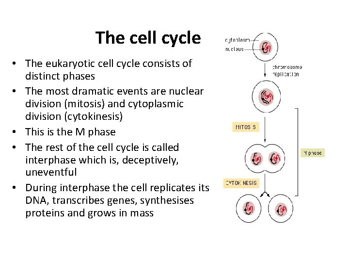
The cell cycle • The eukaryotic cell cycle consists of distinct phases • The most dramatic events are nuclear division (mitosis) and cytoplasmic division (cytokinesis) • This is the M phase • The rest of the cell cycle is called interphase which is, deceptively, uneventful • During interphase the cell replicates its DNA, transcribes genes, synthesises proteins and grows in mass
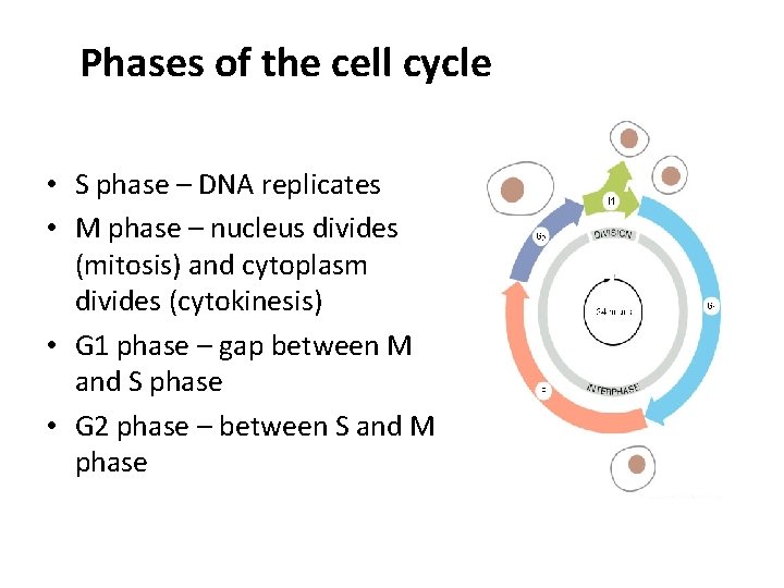
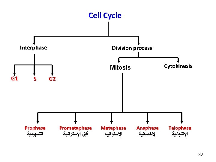
Phases of the cell cycle • S phase – DNA replicates • M phase – nucleus divides (mitosis) and cytoplasm divides (cytokinesis) • G 1 phase – gap between M and S phase • G 2 phase – between S and M phase
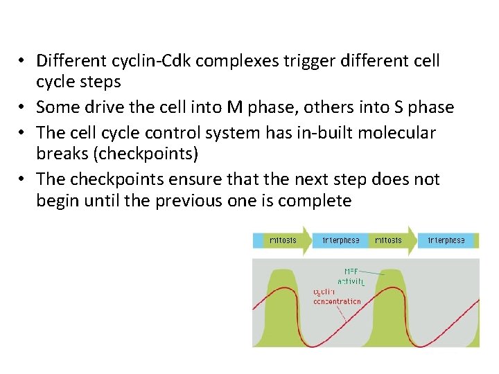
Cell cycle control • Cell cycle machinery is subordinate to a cell cycle control system • The control system consists mainly of protein complexes • These complexes consist of a cyclin subunit and a Cdk subunit • The cyclin has regulatory function, the Cdk catalytic function • Cdk expression is constant, but cyclin concentrations rise and fall at specific times in the cell cycle • The Cdks are cyclically activated by cyclin binding and by phosphorylation status • Once activated, Cdks phosphorylate key proteins in the cell
• Different cyclin-Cdk complexes trigger different cell cycle steps • Some drive the cell into M phase, others into S phase • The cell cycle control system has in-built molecular breaks (checkpoints) • The checkpoints ensure that the next step does not begin until the previous one is complete
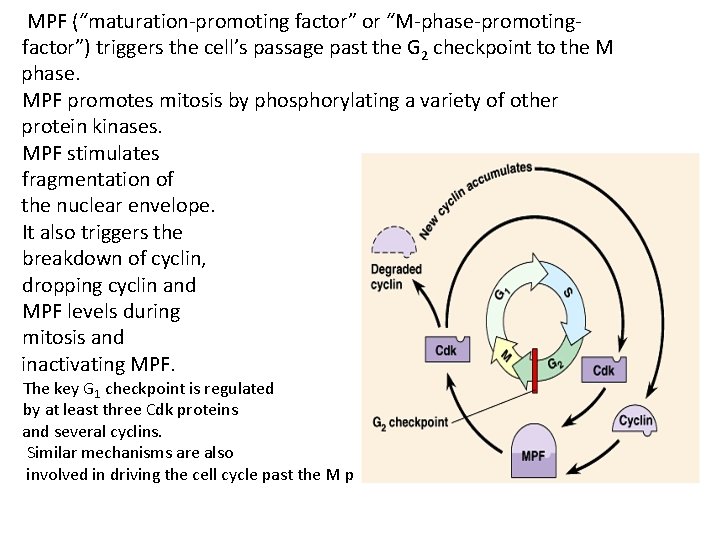
MPF (“maturation-promoting factor” or “M-phase-promotingfactor”) triggers the cell’s passage past the G 2 checkpoint to the M phase. MPF promotes mitosis by phosphorylating a variety of other protein kinases. MPF stimulates fragmentation of the nuclear envelope. It also triggers the breakdown of cyclin, dropping cyclin and MPF levels during mitosis and inactivating MPF. The key G 1 checkpoint is regulated by at least three Cdk proteins and several cyclins. Similar mechanisms are also involved in driving the cell cycle past the M phase checkpoint.
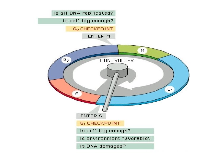
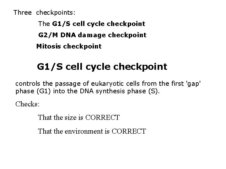
Three checkpoints: The G 1/S cell cycle checkpoint G 2/M DNA damage checkpoint Mitosis checkpoint G 1/S cell cycle checkpoint controls the passage of eukaryotic cells from the first 'gap' phase (G 1) into the DNA synthesis phase (S). Checks: That the size is CORRECT That the environment is CORRECT
G 1/S cell cycle checkpoint How do they do that? Major proteins involved: Cyclins (proteins) - level fluctuate in the cell cycle. & Cyclin dependent KINASES* (Cdks) They add phosphate groups to proteins that control processes in the cell cycle. They only do this when the cyclins are present.
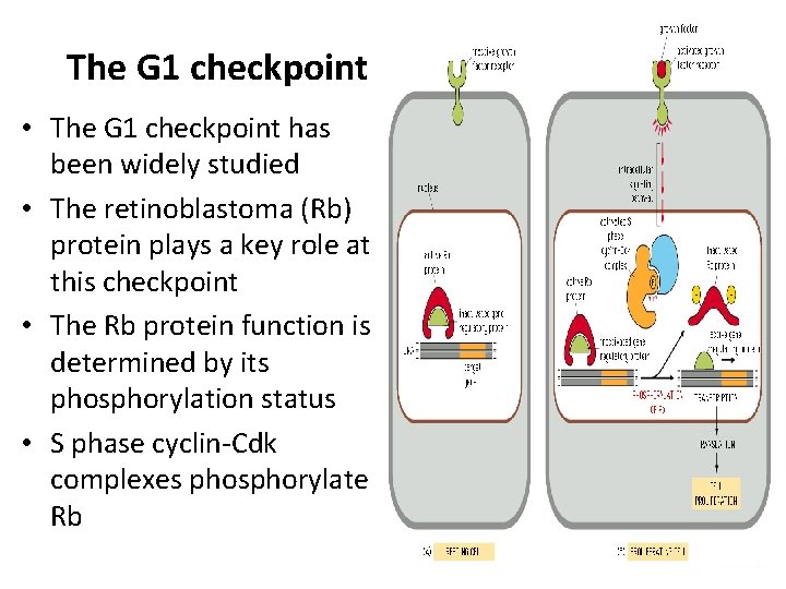
The G 1 checkpoint • The G 1 checkpoint has been widely studied • The retinoblastoma (Rb) protein plays a key role at this checkpoint • The Rb protein function is determined by its phosphorylation status • S phase cyclin-Cdk complexes phosphorylate Rb
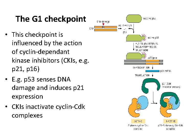
The G 1 checkpoint • This checkpoint is influenced by the action of cyclin-dependant kinase inhibitors (CKIs, e. g. p 21, p 16) • E. g. p 53 senses DNA damage and induces p 21 expression • CKIs inactivate cyclin-Cdk complexes
G 2/M DNA damage checkpoint The G 2/M DNA damage checkpoint prevents the cell from entering mitosis (M phase) if the genome is damaged. It also checks if the cell is big enough (i. e. has the resources to undergo mitosis) Almost exclusively, internally controlled M checkpoint The M checkpoint is where the attachment of the spindle fibres to the centromeres is assessed. Only if this is correct can mitosis proceed. Failure to attach spindle fibres correctly would lead to failure to separate chromosomes
Cellular adaptations of growth and differentiation • Cells must respond to a variety of stimuli that may be hormonal, paracrine or through direct cell contact • These stimuli may arise under physiological or pathological conditions • The way that cells adapt in terms of growth and differentiation depends in part on their ability to divide
Cellular proliferative capacity • Tissues can be classified according to the ability of their cells to divide • Some tissues contain a pool of cells that move rapidly from one cell cycle to the next. These are labile cells • Some cells dismantle their cell cycle control machinery and exit the cell cycle • These cells are said to be in G 0. • Some of these cells can re-enter the cell cycle when stimulated, e. g. by growth factors. These are stable cells • Others are unable to re-enter the cell cycle. These are permanent cells
Growth and differentiation responses 1 - Hyperplasia 2 - Hypertrophy 3 - Atrophy 4 - Metaplasia 1 - Hyperplasia: • Increase in the number of cells in an organ or tissue, which may then have an increased size
2 - Hypertrophy: • An increase in cell size, and resultant increase in organ size 3 - Atrophy: • Shrinkage in cell size by loss of cell substance 4 - Metaplasia: • Reversible change of one adult cell type to another adult cell type
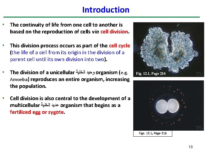
Introduction • The continuity of life from one cell to another is based on the reproduction of cells via cell division. • This division process occurs as part of the cell cycle (the life of a cell from its origin in the division of a parent cell until its own division into two). • The division of a unicellular ﻭﺣﻴﺪ ﺍﻟﺨﻠﻴﺔ organism (e. g. Amoeba) reproduces an entire organism, increasing the population. Fig. 12. 1, Page 216 • Cell division is also central to the development of a multicellular ﻋﺪﻳﺪ ﺍﻟﺨﻠﻴﺔ organism that begins as a fertilized egg or zygote. Figs. 12. 1, Page 216 18
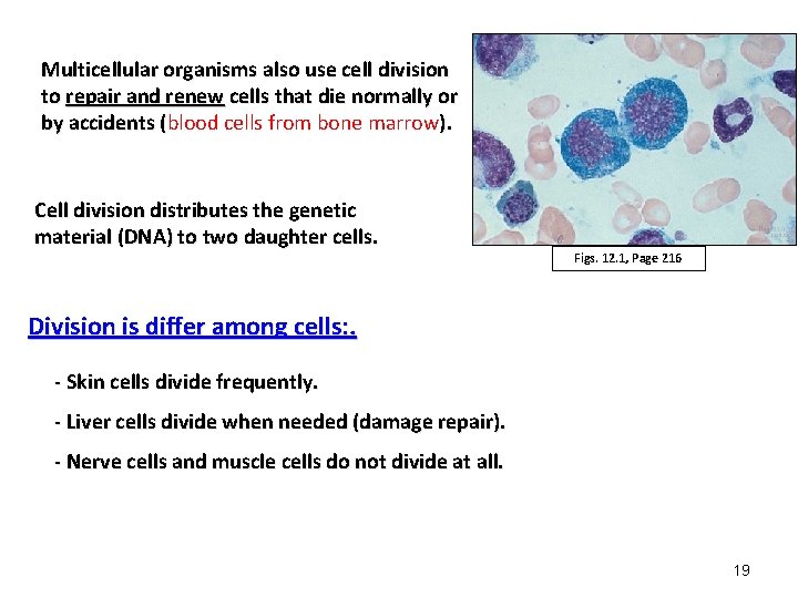
Multicellular organisms also use cell division to repair and renew cells that die normally or by accidents (blood cells from bone marrow). ( Cell division distributes the genetic material (DNA) to two daughter cells. Figs. 12. 1, Page 216 Division is differ among cells: . - Skin cells divide frequently. - Liver cells divide when needed (damage repair). - Nerve cells and muscle cells do not divide at all. 19
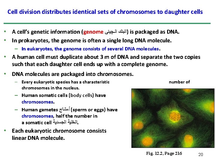
Cell division distributes identical sets of chromosomes to daughter cells • A cell’s genetic information (genome )ﺍﻟﺒﻨﻚ ﺍﻟـﭽﻴﻨﻰ is packaged as DNA. • In prokaryotes, the genome is often a single long DNA molecule. – In eukaryotes, the genome consists of several DNA molecules. • A human cell must duplicate about 3 m of DNA and separate the two copies such that each daughter cell ends up with a complete genome. • DNA molecules are packaged into chromosomes. – Every eukaryotic species has a characteristic chromosomes in the nucleus. – Human somatic cells (body cells) have chromosomes. – Human gametes ( ﺃﻤﺸﺎﺝ sperm or eggs) have chromosomes, half the number in a somatic cell ﺍﻟﺨﻠﻴﺔ ﺍﻟﺠﺴﺪﻳﺔ. number of 46 23 • Each eukaryotic chromosome consists linear DNA molecule. of a long, Fig. 12. 2, Page 216 20
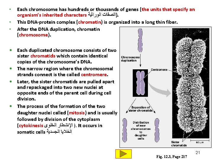
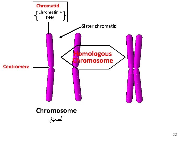
• • • Each chromosome has hundreds or thousands of genes (the units that specify an organism’s inherited characters )ﺍﻟﺼﻔﺎﺕ ﺍﻟﻮﺭﺍﺛﻴﺔ. This DNA-protein complex (chromatin) is organized into a long thin fiber. After the DNA duplication, chromatin condenses form (chromosome). • Each duplicated chromosome consists of two sister chromatids which contain identical copies of the chromosome’s DNA. • The narrow region where the chromosomal strands connect is the called centromere. • Later, the sister chromatids are pulled apart and repackaged into two new nuclei at opposite ends of the parent cell during cell division. • The process of the formation of the two daughter nuclei called (mitosis) and is usually followed by division of the cytoplasm (cytokinesis ) ﺍﻹﻧﺸﻄﺎﺭ ﺍﻟﺨﻠﻮﻯ. It occurs in somatic cells ﺍﻟﺨﻼﻳﺎ ﺍﻟﺠﺴﺪﻳﺔ Fig. 12. 3, Page 217 21
Chromatid Chromatin + DNA Sister chromatid Centromere Homologous Chromosome ﺍﻟﺼﺒﻎ 22
• In the gonads ﺍﻟﻤﻨﺎﺳﻞ , cells undergo a meiosis division, which yields four daughter cells, each with half the chromosomes number of the parent cell. – In humans, meiosis reduces the number of chromosomes from 46 to 23. • Each of us inherited 23 chromosomes from each parent: one set in an egg and one set in a sperm during meiosis. • gametes (ﺍﻷﻤﺸﺎﺝ eggs or sperm) are produced only in gonads ( ﺍﻟﻤﻨﺎﺳﻞ ovaries or testes). • The fertilized egg undergoes trillions of cycles of mitosis and cytokinesis to produce a fully developed multicellular human. • These processes continue every day to replace dead and damaged cell. • Fertilization fuses two gametes together and doubles the number of chromosomes to 46 again. 23
Definitions Genes: The units that specify an organism’s inherited characters. Chromatin: A DNA-protein complex which is organized into a long thin fiber Chromosome: The package that formed from a condensed, coiled and folded chromatin. Chromatids: Two sister arms (chromatids) formed from each duplicated chromosome. They contain identical copies of the chromosome’s DNA Centromere: The narrow region at which the chromosomal strands are connect together. Mitosis: Is the division process which forms two daughter nuclei Cytokinesis: ﺍﻹﻧﺸﻄﺎﺭ ﺍﻟﺨﻠﻮﻯ Is the division stage of the cytoplasm which usually follow the mitosis. Meiosis: A division process that occurs In the gonads ﺍﻟﻤﻨﺎﺳﻞ , and yields four daughter cells, each with half the chromosomes of the parent. 24
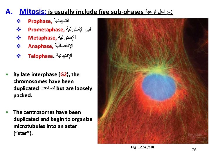
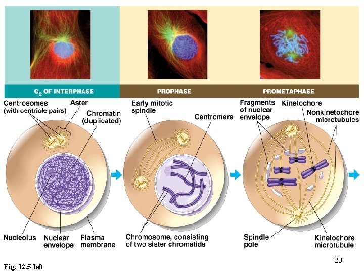
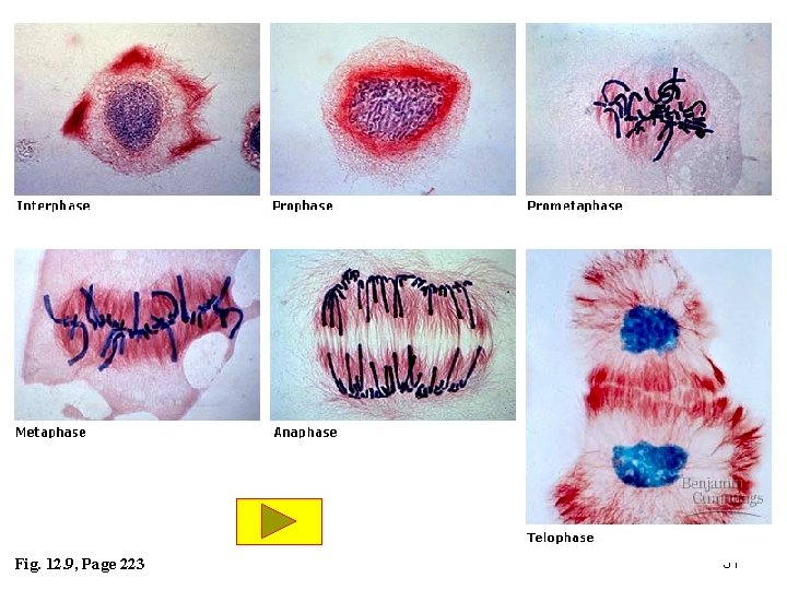
A. Mitosis: is usually include five sub-phases ﻣﺮﺍﺣﻞ ﻓﺮﻋﻴﺔ : v v Prophase, ﺍﻟﺘﻤﻬﻴﺪﻳﺔ Prometaphase, ﻗﺒﻞ ﺍﻹﺳﺘﻮﺍﺋﻴﺔ Metaphase, ﺍﻹﺳﺘﻮﺍﺋﻴﺔ Anaphase, ﺍﻹﻧﻔﺼﺎﻟﻴﺔ v Telophase. ﺍﻹﻧﺘﻬﺎﺋﻴﺔ • By late interphase (G 2), the chromosomes have been duplicated ﺗﻀﺎﻋﻔﺖ but are loosely packed. • The centrosomes have been duplicated and begin to organize microtubules into an aster (“star”). Fig. 12. 5 a, 218 25
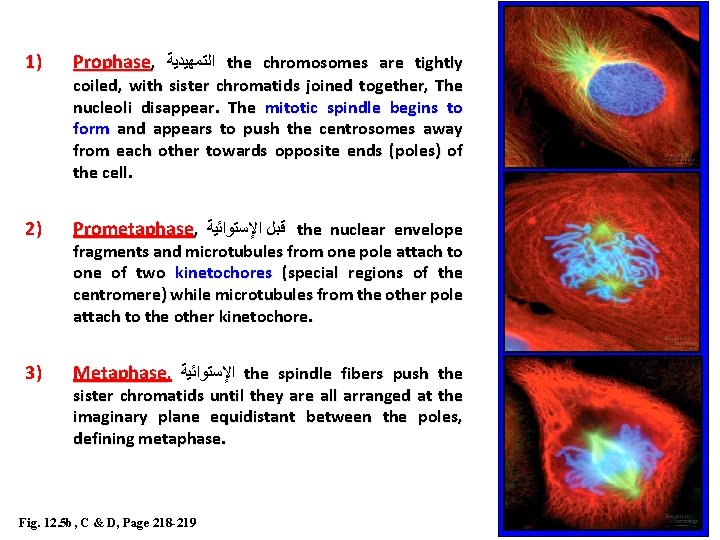
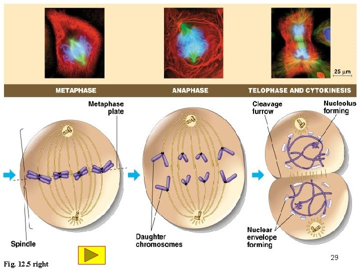
1) Prophase, ﺍﻟﺘﻤﻬﻴﺪﻳﺔ the chromosomes are tightly 2) Prometaphase, ﻗﺒﻞ ﺍﻹﺳﺘﻮﺍﺋﻴﺔ the nuclear envelope 3) Metaphase, ﺍﻹﺳﺘﻮﺍﺋﻴﺔ the spindle fibers push the coiled, with sister chromatids joined together, The nucleoli disappear. The mitotic spindle begins to form and appears to push the centrosomes away from each other towards opposite ends (poles) of the cell. fragments and microtubules from one pole attach to one of two kinetochores (special regions of the centromere) while microtubules from the other pole attach to the other kinetochore. sister chromatids until they are all arranged at the imaginary plane equidistant between the poles, defining metaphase. Fig. 12. 5 b, C & D, Page 218 -219 26
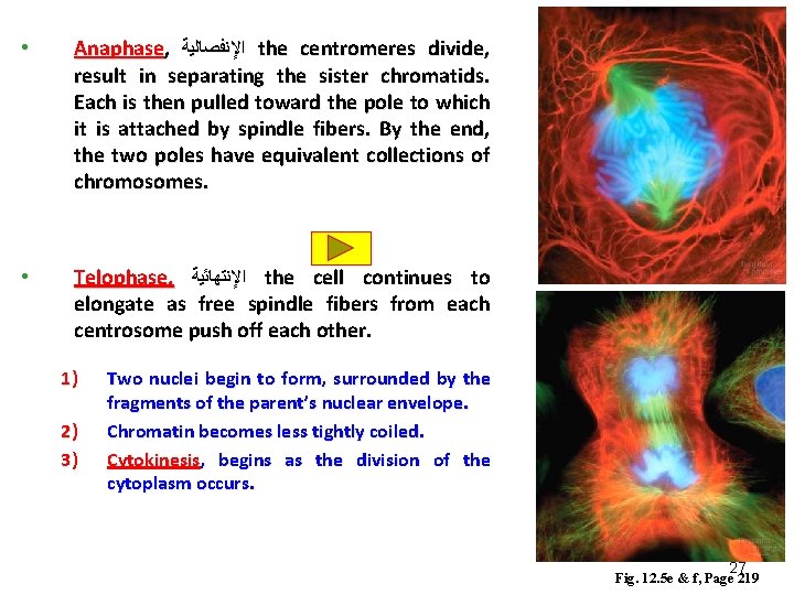
• Anaphase, ﺍﻹﻧﻔﺼﺎﻟﻴﺔ the centromeres divide, result in separating the sister chromatids. Each is then pulled toward the pole to which it is attached by spindle fibers. By the end, the two poles have equivalent collections of chromosomes. • Telophase, ﺍﻹﻧﺘﻬﺎﺋﻴﺔ the cell continues to elongate as free spindle fibers from each centrosome push off each other. 1) 2) 3) Two nuclei begin to form, surrounded by the fragments of the parent’s nuclear envelope. Chromatin becomes less tightly coiled. Cytokinesis, begins as the division of the cytoplasm occurs. 27 Fig. 12. 5 e & f, Page 219
Fig. 12. 5 left 28
Fig. 12. 5 right 29
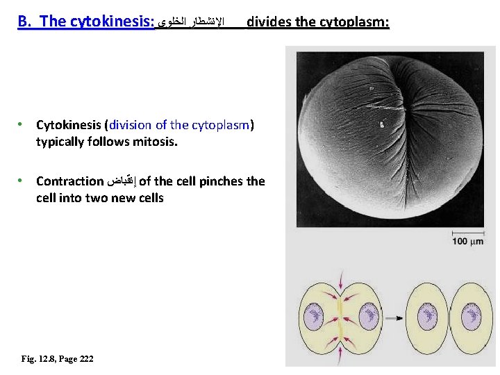
B. The cytokinesis: ﺍﻹﻧﺸﻄﺎﺭ ﺍﻟﺨﻠﻮﻯ divides the cytoplasm: • Cytokinesis (division of the cytoplasm) typically follows mitosis. • Contraction ﺇﻧﻘﺒﺎﺽ of the cell pinches the cell into two new cells Fig. 12. 8, Page 222 30
Fig. 12. 9, Page 223 31
Cell Cycle Interphase Division process Cytokinesis Mitosis G 1 S Prophase ﺍﻟﺘﻤﻬﻴﺪﻳﺔ G 2 Prometaphase ﻗﺒﻞ ﺍﻹﺳﺘﻮﺍﺋﻴﺔ Metaphase ﺍﻹﺳﺘﻮﺍﺋﻴﺔ Anaphase ﺍﻹﻧﻔﺼﺎﻟﻴﺔ Telophase ﺍﻹﻧﺘﻬﺎﺋﻴﺔ 32
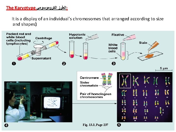
Fertilization and meiosis alternate ﻳﺘﻌﺎﻗﺒﺎ in sexual life cycles v A life cycle of an organism is the generation-to-generation sequence of stages in its reproductive history. v It starts at the conception of an organism until it produces its own offspring ﻧﺴﻞ. v In humans, each somatic cell (all ( cells other than sperm or ovum) has 46 chromosomes. v These homologous chromosome ﺍﻟﻜﺮﻭﻣﻮﺳﻮﻣﺎﺕ ﺍﻟﻤﺘﻤﺎﺛﻠﺔ pairs carry genes that control the same inherited characters. v A karyotype ﺍﻟﻄﺮﺯ ﺍﻟﻜﺮﻭﻣﻮﺳﻮﻣﻰ display of the 46 chromosomes shows 23 pairs of chromosomes, each pair with the same length, centromere position, and staining pattern. 33
The Karyotype ﺍﻟﻄﺮﺯ ﺍﻟﻜﺮﻭﻣﻮﺳﻮﻣﻰ : It is a display of an individual’s chromosomes that arranged according to size and shapes) Fig. 13. 3, Page 237 34
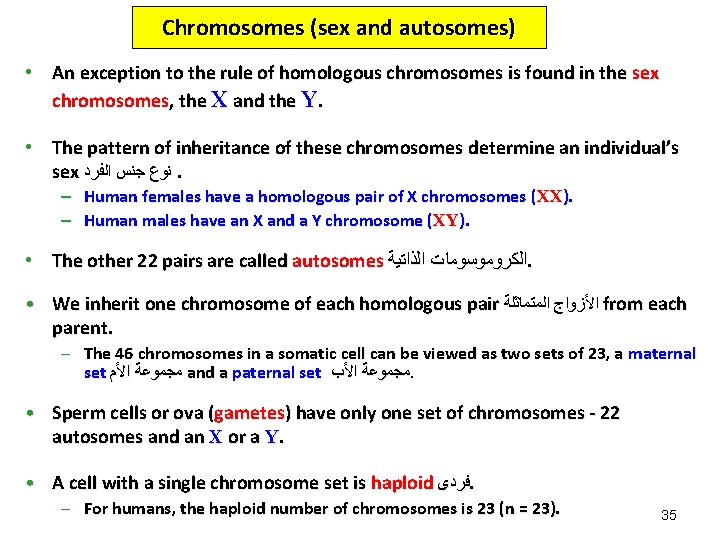
Chromosomes (sex and autosomes) • An exception to the rule of homologous chromosomes is found in the sex chromosomes, the X and the Y. • The pattern of inheritance of these chromosomes determine an individual’s sex ﻧﻮﻉ ﺟﻨﺲ ﺍﻟﻔﺮﺩ. – Human females have a homologous pair of X chromosomes (XX). – Human males have an X and a Y chromosome (XY). • The other 22 pairs are called autosomes ﺍﻟﻜﺮﻭﻣﻮﺳﻮﻣﺎﺕ ﺍﻟﺬﺍﺗﻴﺔ. • We inherit one chromosome of each homologous pair ﺍﻷﺰﻭﺍﺝ ﺍﻟﻤﺘﻤﺎﺛﻠﺔ from each parent. – The 46 chromosomes in a somatic cell can be viewed as two sets of 23, a maternal set ﻣﺠﻤﻮﻋﺔ ﺍﻷﻢ and a paternal set ﻣﺠﻤﻮﻋﺔ ﺍﻷﺐ. • Sperm cells or ova (gametes) have only one set of chromosomes - 22 autosomes and an X or a Y. • A cell with a single chromosome set is haploid ﻓﺮﺩﻯ. – For humans, the haploid number of chromosomes is 23 (n = 23). 35
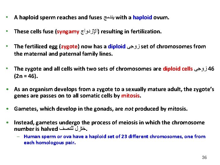
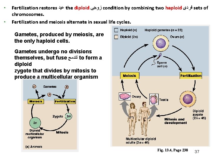
• A haploid sperm reaches and fuses ﻳﻨﺪﻣﺞ with a haploid ovum. • These cells fuse (syngamy )ﺍﻹﺯﺩﻭﺍﺝ resulting in fertilization. • The fertilized egg (zygote) now has a diploid ﺯﻭﺟﻰ set of chromosomes from the maternal and paternal family lines. • The zygote and all cells with two sets of chromosomes are diploid cells ﺯﻭﺟﻰ 46 (2 n = 46). • As an organism develops from a zygote to a sexually mature adult, the zygote’s genes are passes on to all somatic cells by mitosis. • Gametes, which develop in the gonads, are not produced by mitosis. • Instead, gametes undergo the process of meiosis in which the chromosome number is halved ﺧﺘﺰﻝ ﻟﻠﻨﺼﻒ. – Human sperm or ova have a haploid set of 23 different chromosomes, one from each homologous pair. 36
• • Fertilization restores ﻋﻴﺪ the diploid ﺯﻭﺟﻰ condition by combining two haploid ﻓﺮﺩﻯ sets of chromosomes. Fertilization and meiosis alternate in sexual life cycles. Gametes, produced by meiosis, are the only haploid cells. Gametes undergo no divisions themselves, but fuse ﺗﻨﺪﻣﺞ to form a diploid zygote that divides by mitosis to produce a multicellular organism Fig. 13. 4, Page 238 37
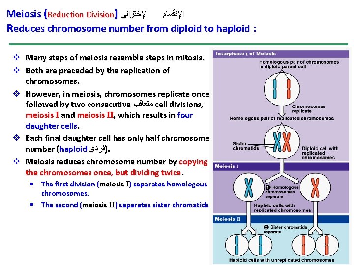
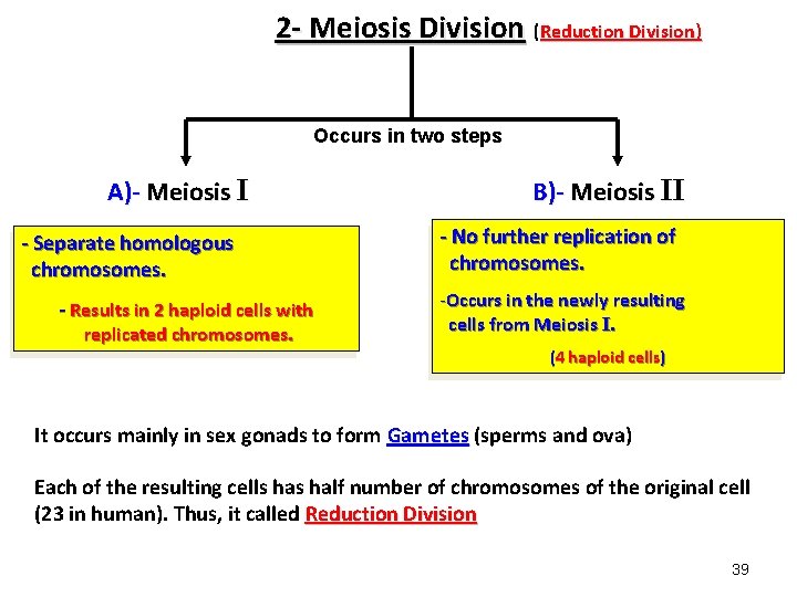
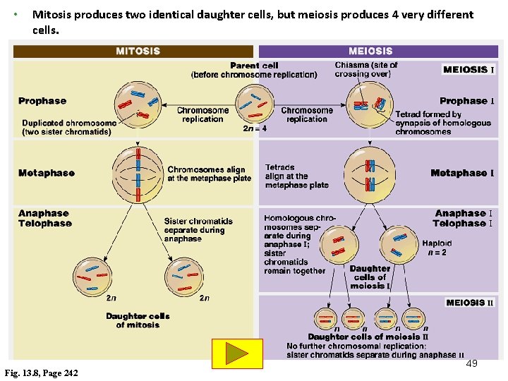
Meiosis (Reduction Division) ﺍﻹﻧﻘﺴﺎﻡ ﺍﻹﺧﺘﺰﺍﻟﻰ Reduces chromosome number from diploid to haploid : v Many steps of meiosis resemble steps in mitosis. v Both are preceded by the replication of chromosomes. v However, in meiosis, chromosomes replicate once followed by two consecutive ﻣﺘﻌﺎﻗﺐ cell divisions, meiosis I and meiosis II, which results in four daughter cells. v Each final daughter cell has only half chromosomes number (haploid )ﻓﺮﺩﻯ. v Meiosis reduces chromosome number by copying the chromosomes once, but dividing twice. § The first division (meiosis I) separates homologous chromosomes. § The second (meiosis II) separates sister chromatids. 38
2 - Meiosis Division (Reduction Division) Occurs in two steps A)- Meiosis I - Separate homologous chromosomes. - Results in 2 haploid cells with replicated chromosomes. B)- Meiosis II - No further replication of chromosomes. -Occurs in the newly resulting cells from Meiosis I. (4 haploid cells) It occurs mainly in sex gonads to form Gametes (sperms and ova) Each of the resulting cells half number of chromosomes of the original cell (23 in human). Thus, it called Reduction Division 39
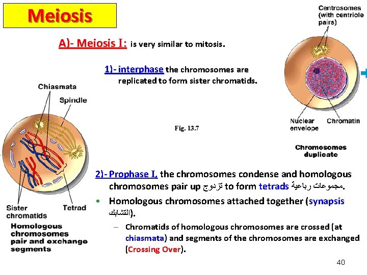
Meiosis A)- Meiosis I: is very similar to mitosis. 1)- interphase the chromosomes are replicated to form sister chromatids. Fig. 13. 7 2)- Prophase I, the chromosomes condense and homologous chromosomes pair up ﺗﺰﺩﻭﺝ to form tetrads ﻣﺠﻤﻮﻋﺎﺕ ﺭﺑﺎﻋﻴﺔ. • Homologous chromosomes attached together (synapsis )ﺍﻟﺘﺸﺎﺑﻚ. – Chromatids of homologous chromosomes are crossed (at chiasmata) and segments of the chromosomes are exchanged (Crossing Over). Over 40
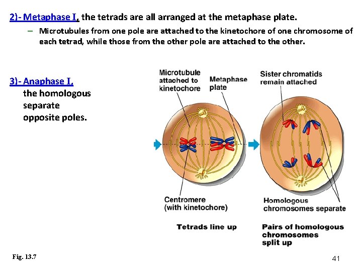
2)- Metaphase I, the tetrads are all arranged at the metaphase plate. – Microtubules from one pole are attached to the kinetochore of one chromosome of each tetrad, while those from the other pole are attached to the other. 3)- Anaphase I, the homologous separate opposite poles. Fig. 13. 7 chromosomes and are pulled toward 41
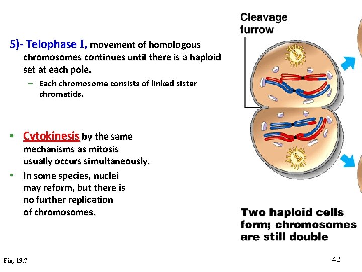
5)- Telophase I, movement of homologous chromosomes continues until there is a haploid set at each pole. – Each chromosome consists of linked sister chromatids. • Cytokinesis by the same mechanisms as mitosis usually occurs simultaneously. • In some species, nuclei may reform, but there is no further replication of chromosomes. Fig. 13. 7 42
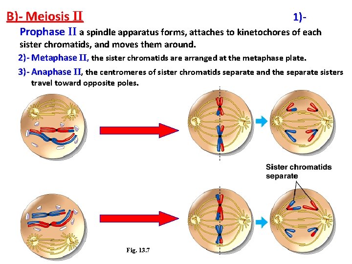
B)- Meiosis II 1)- Prophase II a spindle apparatus forms, attaches to kinetochores of each sister chromatids, and moves them around. 2)- Metaphase II, the sister chromatids are arranged at the metaphase plate. 3)- Anaphase II, the centromeres of sister chromatids separate and the separate sisters travel toward opposite poles. Fig. 13. 7 43
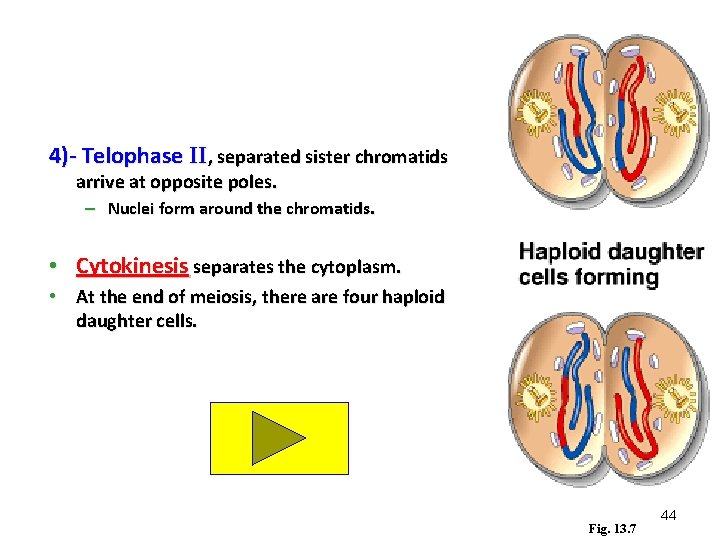
4)- Telophase II, separated sister chromatids arrive at opposite poles. – Nuclei form around the chromatids. • Cytokinesis separates the cytoplasm. • At the end of meiosis, there are four haploid daughter cells. Fig. 13. 7 44
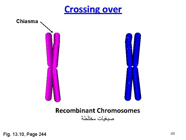
Crossing over Chiasma Recombinant Chromosomes ﺻﺒﻐﻴﺎﺕ ﻣﺨﺘﻠﻄﺔ Fig. 13. 10, Page 244 45
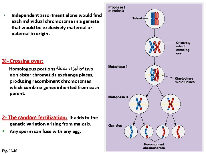
Crossing over Page 244 -Occurs during prophase I. -The two homologous chromosomes joint together very closely. -Two non-sister chromatids of the homologous chromosomes are crossed over at a chiasma point and exchange corresponding segments. -The resulting chromosomes are called “recombinant chromosomes”. -It is important in genetic variation in sexual life cycle. 46
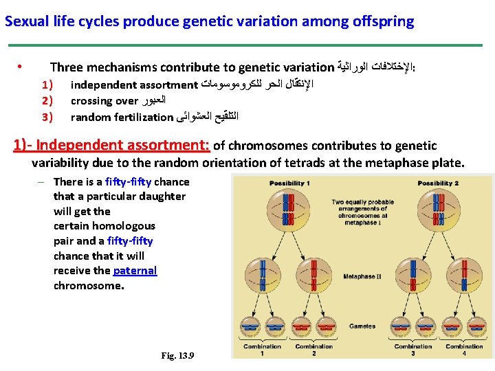
Sexual life cycles produce genetic variation among offspring • Three mechanisms contribute to genetic variation ﺍﻹﺧﺘﻼﻓﺎﺕ ﺍﻟﻮﺭﺍﺛﻴﺔ : 1) 2) 3) independent assortment ﺍﻹﻧﺘﻘﺎﻝ ﺍﻟﺤﺮ ﻟﻠﻜﺮﻭﻣﻮﺳﻮﻣﺎﺕ crossing over ﺍﻟﻌﺒﻮﺭ random fertilization ﺍﻟﺘﻠﻘﻴﺢ ﺍﻟﻌﺸﻮﺍﺋﻰ 1)- Independent assortment: of chromosomes contributes to genetic variability due to the random orientation of tetrads at the metaphase plate. – There is a fifty-fifty chance that a particular daughter will get the certain homologous pair and a fifty-fifty chance that it will receive the paternal chromosome. Fig. 13. 9 cell of meiosis I maternal chromosome of a 47
• Independent assortment alone would find each individual chromosome in a gamete that would be exclusively maternal or paternal in origin. 3)- Crossing over: Homologous portions ﺃﺠﺰﺍﺀ ﻣﺘﻤﺎﺛﻠﺔ of two non-sister chromatids exchange places, producing recombinant chromosomes which combine genes inherited from each parent. 2 - The random fertilization: it adds to the genetic variation arising from meiosis. • Any sperm can fuse with any egg. Fig. 13. 10 48
• Mitosis produces two identical daughter cells, but meiosis produces 4 very different cells. Fig. 13. 8, Page 242 49
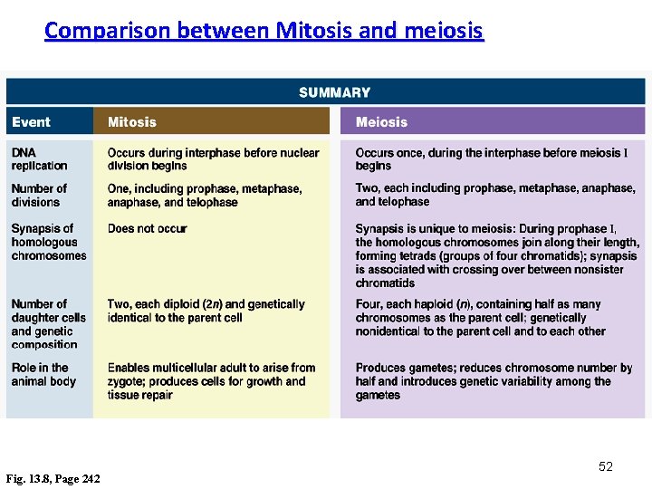
Comparison between Mitosis and meiosis • The chromosome number is reduced by half in meiosis, but not in mitosis. – Mitosis produces daughter cells that are genetically identical to the parent and to each other. – Meiosis produces cells that differ from the parent and each other. • Three events, unique to meiosis, occur during the first division cycle. 1. During prophase I, homologous chromosomes pair up in a process called synapsis. – Later in prophase I, the joined homologous chromosomes are visible as a tetrad. – At X-shaped regions called chiasmata, sections of nonsister chromatids are exchanged. – Chiasmata is the physical manifestation of crossing over, a form of genetic rearrangement. 50
2. At metaphase I homologous pairs of chromosomes, not individual chromosomes are aligned along the metaphase plate. • In humans, you would see 23 tetrads. 3. At anaphase I, it is homologous chromosomes, not sister chromatids, that separate and are carried to opposite poles of the cell. – Sister chromatids remain attached at the centromere until anaphase II. • The processes during the second meiotic division are virtually identical to those of mitosis. 51
Comparison between Mitosis and meiosis Fig. 13. 8, Page 242 52
Sexual life cycles produce genetic variation among offspring • The behavior of chromosomes during meiosis and fertilization is responsible for most of the variation that arises each generation during sexual reproduction. • Three mechanisms contribute to genetic variation: – independent assortment – crossing over – random fertilization
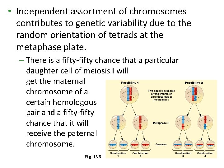
• Independent assortment of chromosomes contributes to genetic variability due to the random orientation of tetrads at the metaphase plate. – There is a fifty-fifty chance that a particular daughter cell of meiosis I will get the maternal chromosome of a certain homologous pair and a fifty-fifty chance that it will receive the paternal chromosome. Fig. 13. 9
• Each homologous pair of chromosomes is positioned independently of the other pairs at metaphase I. • Therefore, the first meiotic division results in independent assortment of maternal and paternal chromosomes into daughter cells. • The number of combinations possible when chromosomes assort independently into gametes is 2 n, where n is the haploid number of the organism. – If n = 3, there are eight possible combinations. – For humans with n = 23, there are 223 or about 8 million possible combinations of chromosomes.
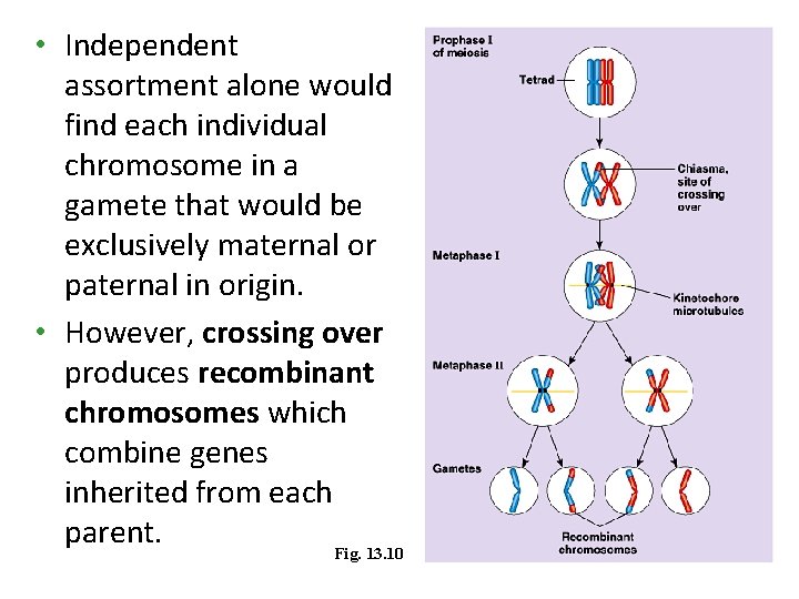
• Independent assortment alone would find each individual chromosome in a gamete that would be exclusively maternal or paternal in origin. • However, crossing over produces recombinant chromosomes which combine genes inherited from each parent. Fig. 13. 10
• Crossing over begins very early in prophase I as homologous chromosomes pair up gene by gene. • In crossing over, homologous portions of two nonsister chromatids trade places. – For humans, this occurs two to three times per chromosome pair. • One sister chromatid may undergo different patterns of crossing over than its match. • Independent assortment of these nonidentical sister chromatids during meiosis II increases still more the number of genetic types of gametes that can result from meiosis.
• The random nature of fertilization adds to the genetic variation arising from meiosis. • Any sperm can fuse with any egg. – A zygote produced by mating of a woman and man has a unique genetic identity. – An ovum is one of approximately 8 million possible chromosome combinations (actually 223). – The successful sperm represents one of 8 million different possibilities (actually 223). – The resulting zygote is composed of 1 in 70 trillion (223 x 223) possible combinations of chromosomes. – Crossing over adds even more variation to this.
• The three sources of genetic variability in a sexually reproducing organism are: – Independent assortment of homologous chromosomes during meiosis I and of nonidentical sister chromatids during meiosis II. – Crossing over between homologous chromosomes during prophase I. – Random fertilization of an ovum by a sperm. • All three mechanisms reshuffle the various genes carried by individual members of a population. • Mutations, still to be discussed, are what ultimately create a population’s diversity of genes.