Control of cell divisionCancer Figure 12 1 The


 – Adenoma-benign – Adenocarcinoma-malignant glandular •")
")


. Cause cancer by adding oncogenes to cells.")

. Cause cancer by adding oncogenes to cells.")
")

- Slides: 63
Control of cell division/Cancer
Figure 12. 1 The functions of cell division • Reproduction • Growth and development • Tissue repair
Figure 12. 5 The cell cycle
Figure 12. 4 Chromosome duplication and distribution during mitosis
Figure 12. 5 The cell cycle
Figure 16. 12 Origins of replication in eukaryotes
Figure 12. 5 The cell cycle
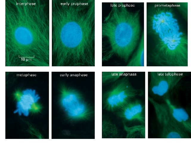
Figure 12. 6 The stages of mitotic cell division in an animal cell: G 2 phase; prometaphase
Figure 12. 6 The stages of mitotic cell division in an animal cell: metaphase; anaphase; telophase and cytokinesis.
Cytokinesis in plants.
Cell Cycle control • The interesting stuff! – How do cells “decide” whether and when to divide? – Checkpoints – What happens when cell cycle control is lost?
Figure 12. 5 The cell cycle
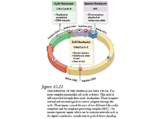
Figure 12. 14 Mechanical analogy for the cell cycle control system Figure 12. 15 The G 1 checkpoint
Cell and Organismal Biology 2009
Figure 12. 13 Evidence for cytoplasmic chemical signals in cell cycle regulation
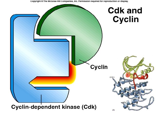
Figure 12. 16 Molecular control of the cell cycle at the G 2 checkpoint MPF-Mitosis promoting factor
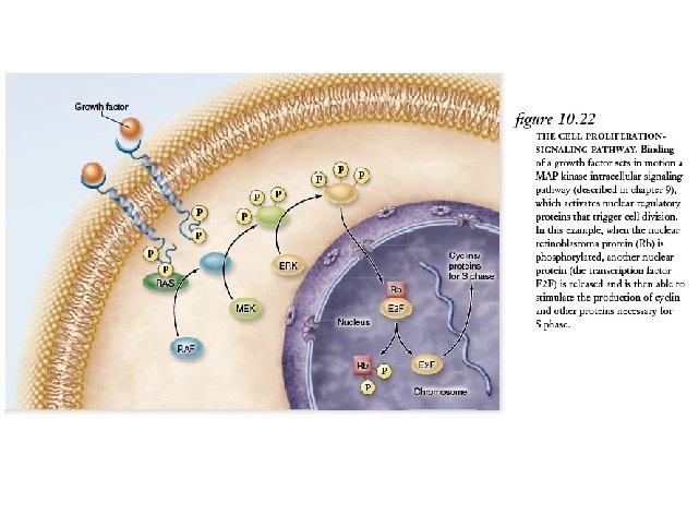
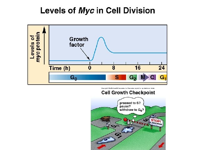
Figure 12. 17 The effect of a growth factor on cell division Fetal Calf Serum Experiment Results
Figure 12. 18 Density-dependent inhibition of cell division
Figure 12. 18 Density-dependent inhibition of cell division
Figure 12. 18 Density-dependent inhibition of cell division
Loss of control of the cell cycle: Cancer
Types of Cancer • Epithelial cell tumors (Carcinoma) – Adenoma-benign – Adenocarcinoma-malignant glandular • Connective tissue or muscle cell tumors (Sarcoma) – Chondroma-benign – Chondrosarcoma-malignant cartilage tumor • Others – Leukemias and nervous system cancers
Progression of tumor size. Breast cancer doubling time is 100 days Mammogram: normal (left) and cancerous (right)
Cancers are progressive Epithelial cell tumor
Steps in the process of metastasis
Papanicolaou test “Pap smear” A. Normal cells- well differentiated B. Precancerous-abnormal differentiation C. Invasive carcinoma-undifferentiated
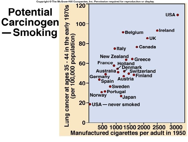
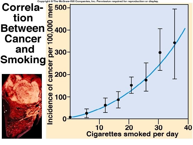
Cancer incidence as a function of age • • Colon cancer in women in England Wales Suggests multiple mutations required to induce cancer
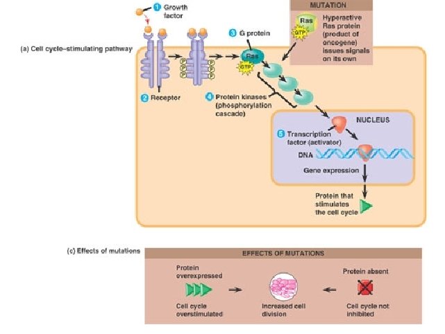
So how is cancer caused? Genes involved in cell cycle regulation are mutated
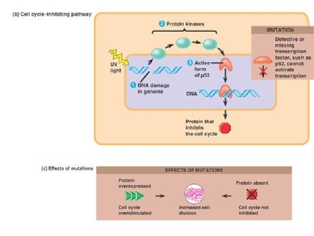
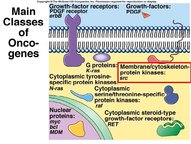
Genes controlling cell division that can cause cancer • • Oncogenes- these are mutated proto-oncogenes which push cells towards cell division (GF receptors, myc, ras etc) Tumor-suppressor genes-these are genes that stop cells from dividing (retinoblastoma, p 53 APC etc)
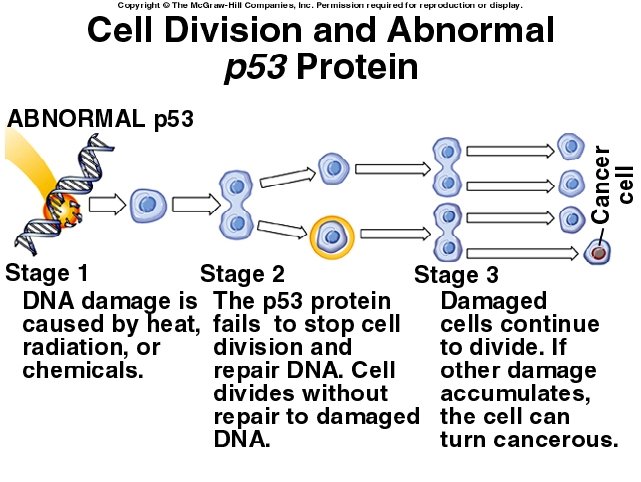
Importance of Structure-an example • P 53 • Mutated in 50% of all human cancers
Cell and Organismal Biology 2009
Figure 19. 11 Genetic changes that can turn proto-oncogenes into oncogenes
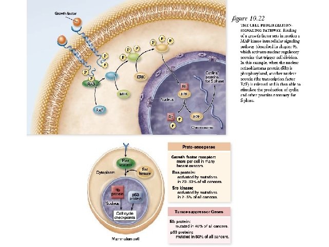
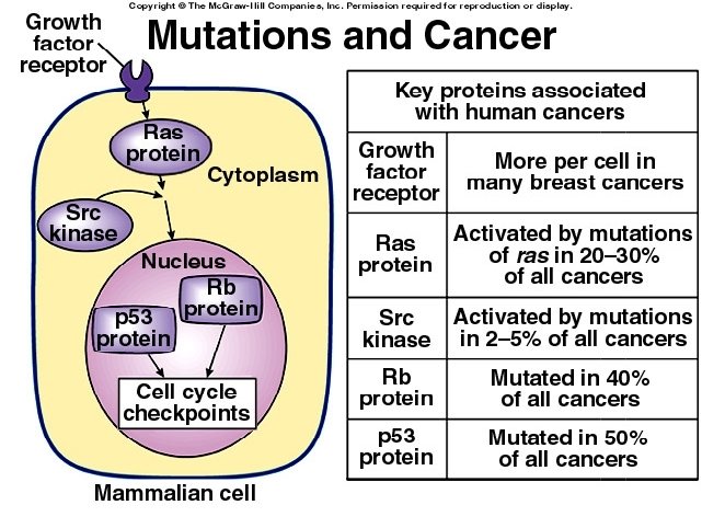
Genes controlling cell division that can cause cancer • Oncogenes- these are mutated proto-oncogenes which push cells towards cell division (GF receptors, myc, ras etc) – ras-mutated in 20 -30% of all cancers – GF receptors-increased in number in many breast cancers – src kinasemutated/affected in 2 -5% of cancers • Tumor-suppressor genesthese are genes that stop cells from dividing – P 53 -mutated in 50% of cancers – Rb-mutated in 40% of cancers
Genes controlling cell division that can cause cancer • Oncogenes- these are mutated proto-oncogenes which push cells towards cell division (GF receptors, myc, ras etc) – ras-mutated in 20 -30% of all cancers – GF receptors-increased in number in many breast cancers – src kinasemutated/affected in 2 -5% of cancers • Tumor-suppressor genesthese are genes that stop cells from dividing – P 53 -mutated in 50% of cancers – Rb-mutated in 40% of cancers
• Science, Vol 274. Oct 18, 1996. p 430
Types and causes of mutations • Rearrangements – Ionizing radiation- forms free radicals that damage DNA • • Translocation Duplications Inversions deletions – Spontaneous mutagenesis- slipped mispairing • Point mutations – UV light- pyrimidine dimers – Chemical mutagenesis
Figure 16. 17 Nucleotide excision repair of DNA damage Xeroderma pigmentosum
Chemical mutagenesis
Chemical mutagenesis • Ames test • Aflatoxin-from the fungus aspergillus that grows on rotting peanuts
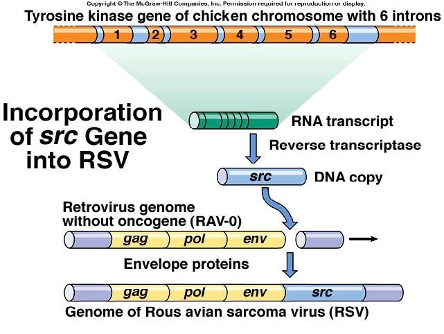
Viruses cause cancer • RNA viruses (retroviruses). Cause cancer by adding oncogenes to cells. – Pick up RNA copies of proto-oncogenes and transfer them to other cells by infection • DNA viruses. Cause cancer by blocking tumor-suppressor proteins.
Figure 19. 13 Genetic changes that can turn proto-ocogenes into oncogenes
Viruses cause cancer • RNA viruses (retroviruses). Cause cancer by adding oncogenes to cells. • DNA viruses. Cause cancer by blocking tumor-suppressor proteins. – Viruses produce proteins that bind to p 53 and RB
DNA viruses produce proteins that block tumorsuppressor action. Normal Virus infected (SV 40/papillomavirus)
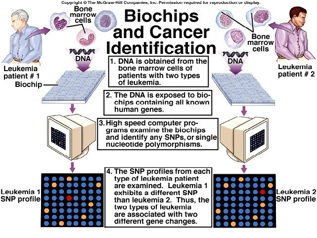
Not all cancers are alike!