Control and Regulation Chapters 15 16 Introduction Two


, the brain")


- Slides: 28
Control and Regulation Chapters 15 &16
Introduction • Two major systems regulate all of the bodies activities. – The Endocrine and Nervous systems • These systems work together to coordinate responses to internal and external stimuli. – Both systems work together at some level to organize responses to all sensory information.
Endocrine System • Composed of organs/tissues called glands and their target organs. • Endocrine Glands produce hormones; – Chemical signals that affect the function of target organs. • Hormones are carried by the blood, but only affect certain organs. – They are specific in their functions
Glands • Endocrine glands are as varied as the organs they control. • They assist in: – Reproduction – Metabolism – Osmoregulation – Embryonic Development – Growth – Metamorphosis – Digestion
Examples of Endocrine Control • Thyroid Gland-Located in the throat of almost all vertebrates – Directs metabolic rate, metamorphosis, growth, and assists in reproductive control • Adrenal Gland-A composite organ composed of 2 tissues – Adrenocortical Gland-Regulates salt absorption in kidneys – Chromafin tissues-Regulate body’s response to stress (Fight-or-Flight)
• Pituitary Gland-Located beneath the brain, has 2 parts – Neurohypophysis-regulates water reabsorption in the kidneys & uterine contractions during labor – Adenohypophysis- regulates growth, lactation, and sperm/egg maturation • Gonads (Ovary/Testis)- Locations varies slightly among groups of vertebrates – Regulate the production of gametes and control development of sexually dimorphic traits
Nervous System • Most of the rapid changes in a vertebrate’s internal or external environments are mediated by the nervous system. • Activation of the correct combination of effectors so that the appropriate responses are made requires a processing of signals within the nervous system that is called coordination or integration. • The nervous system is constantly inundated with nervous signals – many are filtered as they pass to the higher centers so that some are suppressed and others enhanced.
• Often, sensory inputs from different types of receptors are combined to provide a more complete picture of the changes taking place. – These inputs are combined with memories of past events before a response is determined • Information about the outcome of the response is fed back into the nervous system – Thus, reinforcing or altering the past memories. – Although much of vertebrate behavior is based on inherited neuronal connections, vertebrates learn from their experiences.
Components of the Nervous System • The primary functional units of the nervous system are the neurons, or nerve cells. – Transmit signals between neurons or from neurons to organs • Glial Cells – Provide the structural and functional support of neurons cells.
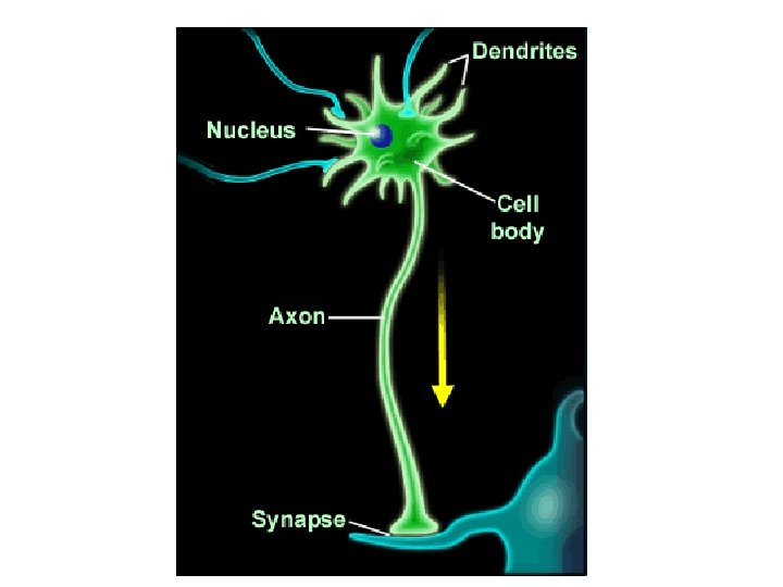
• Neurons are arranges into the: – Central Nervous System (CNS), the brain and spinal cord, and – Peripheral Nervous System (PNS), containing of the nerves between the CNS and the receptors and effectors. • Many morphological types of neurons are known, but all contain the same 4 essential parts: 1. 2. 3. 4. A cell body, or trophic segment Dendrites, or the receptive segment An axon, or conductive segment A terminal arborization, or transmissive segment
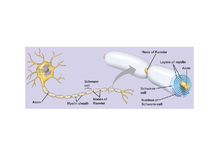
Schwann Cells • Schwann cells associated with many peripheral axons, especially those of autonomic nerves – They envelop the axon but do not continue to grow into a sheath, these cells are referred to an non-myelinated. • In gnathostomes, all axons are surrounded during their embryonic development by glial components called Schwann cells. – The inner tongue-like process of these cells grow around the axon many times so that multiple layers of the Schwann cell’s plasma membrane are laid down, forming the myelin sheath. – Gaps between successive Schwann cells interrupt the myelin sheath at areas called Nodes of Ranvier – Because Nodes of Ranvier are the only regions of the axon accessible to the aqueous extracellular fluids, the waves of depolarization jump from node to node, increasing the speed of transmission of the nerve impulse.
Organization of the Nervous System • Because of the way they develop; the spinal cord and brain of a vertebrate are hollow and lined by a non-nervous and partially ciliated ependymal epithelium. – The adult spinal cord has a small central canal, which enlarges in parts of the brain to form a series of interconnected ventricles. • Cerebrospinal fluid slowly circulates within these spaces.
• Bilateral features of the nervous system are often referred to by the contrasting terms ipsilateral and contralateral. – These terms are especially important in describing the targets of the particular tracts of nerve cells, • Typical spinal nerves attach to the cord at segmental intervals by dorsal and ventral roots. – In most vertebrates, the dorsal and ventral roots of a body segment unite slightly peripheral to the spinal cord to form a spinal – Most of the cranial nerves appear to be serially homologous to either a ventral or dorsal root of a typical spinal nerve.
The Three Connectional Categories of Neurons and their Distribution • Regardless of the differences in their morphologies, all neurons belong to one of three categories: 1. Primary sensory, or afferent neurons • Carry impulses from free nerve endings or specialized receptor cells into the CNS 2. Motor, or efferent neurons • Carry signals from the CNS to the effectors, such as muscles or glands. 3. Interneurons which are confined to the CNS • Receive input from afferent neurons, integrate this information, and send it to the periphery through efferent neurons.
Basic Neuronal Circuitry • There are three basic neuronal pathways common to all vertebrates; – reflexes, – ascending pathways, – descending pathways. • Much of the activity is controlled by reflexes, mediated by cells located in the spinal cord.
• Touching a hot stove initiates a reflex termed a 3 -neuron reflex arc – Because it includes three types of neurons: A sensory neuron, an interneuron, and a motor neuron. – In contrast, the knee-jerk reflex requires only two types of neurons because the sensory neurons synapse directly with motor neurons. • Reflexes like the 2 - and 3 -neuron reflex arc involve neurons that are only on one side of the body and one body segment. – Other reflexes include neurons that cross the midline of the body and affect many body segments. • Axons that cross the midline are called commisures, or decussations.
• Many reflexes in the brain and spinal cord are products of long evolutionary history, – They are innate reflexes. • Others develop during thee lifetime of the animal’s repetitive experiences. – They are conditioned reflexes • Conditioned reflexes are not inherited directly, although the capacity for them to develop is.
• Many sensory impulses ascend from the spinal cord or brainstem to higher centers in the brain. • Groups of axons with similar functions terminate in the same part of the brain and ascend together in common tracts that are described by their points of origin and termination. • Most sensory impulses decussate on their way to higher centers so that impulses originating on the left side of the body terminate on the right side of the brain, but the decussation is not always complete.
Spinal Cord • The spinal cord lies between the brain and spinal nerves, but the cord is far more than a simple passage way for nerve impulses between receptors and effectors and the brain. • Some interneurons of the spinal cord span from one segment of the trunk to one or more other segments and thus are known as intersegmental neurons. – Their presence makes possible considerable sensory-motor integration within the spinal cord. • The rhythmic and coordinated movements of swimming and walking are controlled and integrated reflexively by groups of intersegmental and decussating spinal nerves, – these are central pattern generators.
• The complexity of the spinal cord, and the degree to which the brain exerts control over spinal activity, increases during the course of vertebrate evolution. – Hagfishes and lamprey have relative simple spinal cords; none of their axons are myelinated. – In gnathostomes, the spinal cord is larger, well vascularized, and more rounded. • The evolution of a highly vascular nervous systems was an important part of vertebrate evolution allowing for more complex nervous systems to evolve.
• The brains of birds and mammals exert more control over body activities than do the brains of other vertebrates – Tracts to and from the brain are correspondingly more numerous. • The degree of vascularization of the spinal cord also increases. • As do the number of neuroglial cells that service the neurons in birds and mammals.
Embryonic Development and Regions of the Brain • The brain develops by an enlargement of the cranial end of the neural tube. • First, three brain expansions can be recognized: – a forebrain, or prosencephalon, – a midbrain, or mesencephalon, – and the hindbrain, or rhombencephalon.
Major Trends in Tetrapod Brain Evolution • The brain of vertebrates vary enormously in size and complexity. • But the brain regions of tetrapods, the structures they contain, and their basic organizational features are the same as in fish. – The cerebrum, thalamus, mesencephalic tectum, and cerebellum were the regions most affected as vertebrates adapted to a terrestrial life and became more active with a wider range of behavioral responses.
• As mammals increase in size the isocortex becomes highly convoluted, forming surface folds called gyri, which are separated by grooves called sulci. – A convoluted surface accommodates the increased number of neuron cell bodies needed to process the increased sensory input and motor output of these generally larger mammals.
The Cerebellum • The cerebellum originally served to integrate sensory-motor information and process information from the lateral line. – Lateral lines have been lost in all terrestrial vertebrates. • Amniotes have developed a great range of motion and coordination is essential in these groups – The cerebellum is especially enlarged in birds and mammals.
End