Contraction of Skeletal Muscle and Neuromuscular Transmission Textbook

Contraction of Skeletal Muscle and Neuromuscular Transmission Textbook of medical physiology Guyton & Hall (13 th edition) UNIT II CHAPTER 6 & 7 Pages 75 -95 Dr. Mohammed Alotaibi

Objectives of the lecture At the end of the lecture the student should be able to: Know and describe the followings: � The physiologic anatomy of the skeletal muscle and NM junction. � The general mechanism of skeletal muscle contraction. � Motor End Plate potential and how action potential and excitation- contraction coupling are generated in skeletal muscle. � The molecular mechanism of skeletal muscle contraction & relaxation. � Sliding filament mechanism. � Drugs/ diseases affecting the neuromuscular transmission.

Motor Unit What is a Motor Unit? It is the Motor Neuron (Anterior Horn Cell , Axon) and all the muscle fibers it innervates.

40% of the body are skeletal muscles Sarcolemma

Physiological Anatomy of Skeletal Muscle fiber

3000 Actin 1500 Myosin Responsible for the actual muscle contraction

Myosin Actin M line Myofibril The light and dark bands give skeletal and cardiac muscle their striated appearance.

Fig. 6. 3 Organization of proteins in a sarcomere Ø Titin filaments keep the myosin and actin filaments in place.
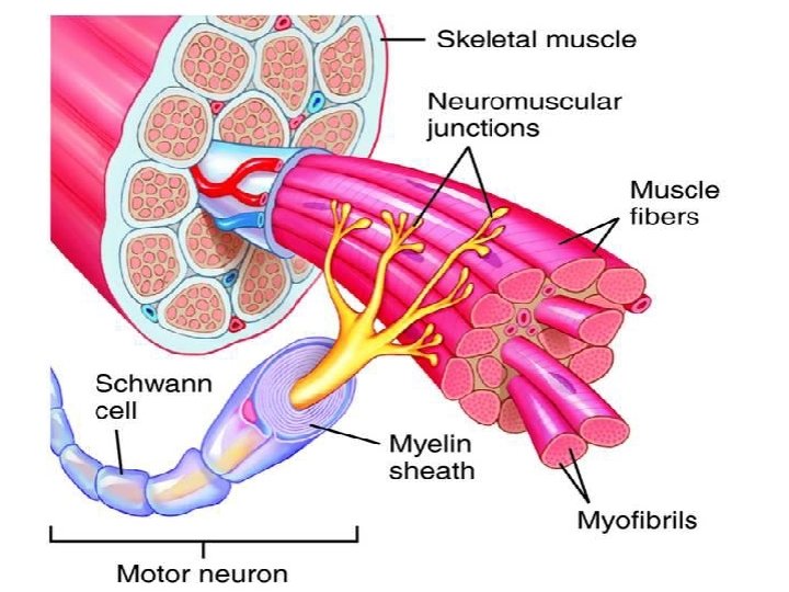

Transmission of impulses from nerve endings to skeletal muscle fibers occurs via: THE NEUROMUSCULAR JUNCTION (NMJ)
 • Synaptic trough/")
Physiologic Anatomy of the Neuromuscular Junction • Motor End Plate (MEP) • Synaptic trough/ gutter • Presynaptic terminal • Postsynaptic terminal • Synaptic space/cleft 125 • Subneural cleft • Acetylcholine (Ach) • Synaptic vesicles • Acetylcholinesterase (Ach-gated ion channels)

Effect of Ach on the Postsynaptic Muscle Membrane Ø Two molecules of Ach must attach to the receptor. Ø Ach-channels open and allow Na+, Ca+, or K+ ions to move through easily; but not negative ions such as Cl- Ø More Na+ ions will pass through which creates a local positive potential change inside the muscle fiber membrane, called the end plate potential (EPP). Ø EPP spreads along the muscle fiber membrane Fig. 7. 3 Acetylcholine gated channels A. Closed B. After Ach attaches

Na + Synapsin Na + Cytoskeleton P Ca 2+- Cal-PK + AP spreads + along a +N the T Na tubules When Ach-gated channels open, sudden influx of Na+ will increase electrical potential in the positive direction as much as 50 -75 m. V and creates a local EPP. This will open voltage gated Na+ channels.

Release of Calcium Ions by the Sarcoplasmic Reticulum Ø As the AP reaches the T-tubule, the voltage change is sensed by [voltage-gated calcium channel dihydropyridine receptors (DHP)] linked to calcium release At rest channels (Ryanodine receptors) which triggers the release of Ca++ initiating contraction. Ø Calcium pump removes calcium ions after contraction occurs. Ø Calcium binds to calsequestrin. Fig. 7. 6 Excitation-contraction coupling in skeletal muscle.

EXCITATION-CONTRACTION COUPLING
 Skeletal Muscle Large Nerves Resting Membrane Potential -80 to -90")
Muscle Action Potential (AP) Skeletal Muscle Large Nerves Resting Membrane Potential -80 to -90 m. V Duration of the Action Potential Lasts 1 - 5 msec Lasts 0. 2 - 1 msec Velocity of Conduction 3 -5 m/sec 39 -65 m/sec

Molecular Mechanism of Muscle Contraction Occurs by a Sliding Filament Mechanism Figure 6 -5. Relaxed and contracted states of a myofibril.

• Molecular Characteristics of the Contractile Filaments Myosin filaments are composed of multiple myosin molecules. Each Myosin molecule has: (1) Head ( 2 ) Tail (3) Hinge (joint ) Each myosin head contains: (1) Actin binding site (2) Myosin ATPase site

• Molecular Characteristics of the Contractile Filaments Actin filaments are composed of actin, tropomyosin and troponin (I, T, C) Double-Stranded: backbone of the actin filament Fig. 6. 7 Actin filament

Molecular Mechanism Fig. 6. 8 “Walk-along” mechanism for muscle contraction. The heads of the cross-bridges bend back and forth and step by step walk along the actin filament, pulling the ends of two successive actin filaments toward the center of the myosin filament.

Sliding Filament Mechanism But what causes the actin filaments to slide inward among the myosin filaments? Forces generated by interaction of the cross-bridges from the myosin filaments with the actin filaments

Forces are inactive The heads of the cross bridges bind with ATP. The ATPase activity of the myosin head immediately cleaves the ATP but leaves the cleavage products, ADP plus phosphate ion, bound to

When the troponin-tropomyosin complex binds with calcium ions, active sites on the actin filament are uncovered and the myosin heads then bind with these sites.

With the active sites on the actin exposed, the myosin heads bind to the actin, forming cross-bridges.

Binding the head of the cross-bridge with the active site causes a conformational change in the head, prompting the head to tilt toward the arm of the crossbridge and providing the power stroke for pulling the actin filament.

Once the head of the cross-bridge tilts, ADP and phosphate ion are released and new molecule of ATP binds. This binding of new ATP causes detachment of the head from the actin.

The new ATP is cleaved to begin the next cycle which “cocks” the head back to its perpendicular condition, ready to begin the new power stroke cycle.

What is Rigor Mortis ? The contracture of skeletal muscles that begins several hours after death due to the loss of ATP.

Drugs That Enhance Transmission at the Neuromuscular Junction Drugs That Stimulate the Muscle Fiber by Ach-Like Action: �Methacholine, Carbachol, and Nicotine. They act for minutes or hours—are not destructed by cholinesterase. Drugs That Stimulate the NMJ by Inactivating Acetylcholinesterase: �Neostigmine, Physostigmine [inactivate acetylcholinesterase for several hours] �Diisopropyl fluorophosphate (nerve gas poison) [inactivates acetylcholinesterase for weeks -------can cause death because of respiratory muscle spasm]

Drugs That Block Transmission at the Neuromuscular Junction Drugs That Block Transmission at the NMJ �Curare & Curariform like-drugs. Prevent passage of impulses from the nerve ending into the muscle by blocking the action of Ach on its receptors on MEP. �Botulinum Toxin. Bacterial poison that decreases the quantity of Ach release by the nerve presynaptic terminals.

Myasthenia Gravis Ø Disease of adult females affects eyelid, extra ocular bulbar and proximal limb muscles. Ø Presents with ptosis, dysarthria, dysphagia, and proximal limb weakness in hands& feet.

Myasthenia Gravis Autoimmune disorder [patients develop antibodies which block or destroy their own Ach receptors]. Ø Occurs in about 1 in every 20, 000 persons. Ø Causes muscle weakness because of the inability of the NMJ to transmit enough signals from the nerve fibers to the muscle fibers. Ø The EPP that occur in the muscle fibers is mostly too weak to initiate opening of the voltage-gated sodium channels. Ø Patient may die of respiratory failure.

Myasthenia Gravis Treatment: Ø Administration of anticholinesterase drugs such as Neostigmine which allows larger than normal amounts of Ach to accumulate in the synaptic space. Ø Corticosteroids and Immunosuppressant drugs to inhibit the immune system, limiting antibody production.

�The End
- Slides: 34