Contents Nasopharynx n Oral cavity n Ear external

 n Orbit n")


")
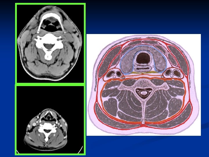
 n n Fat Branches of cranial nerve V 3 Ascending pharyngeal")
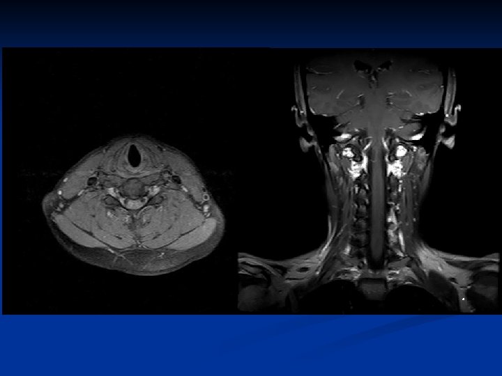
 Fat n Lateral retropharyngeal nodes (of Rouviere) n Medial retropharyngeal nodes")


 & Oral cavity n Keywords: -- Hard & Soft palate -- Tongue")
 -- Facial")


 space --")

 n Infrahyoid epiglottis n False vocal")


- Slides: 68
Contents Nasopharynx n Oral cavity n Ear (external, middle, inner ear) n Orbit n Larynx n Salivary glands n Neck space & vessels n
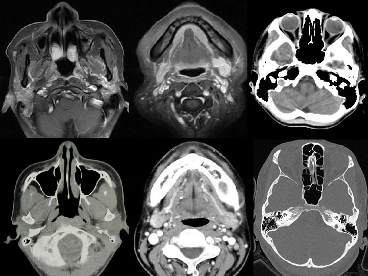
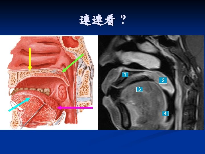
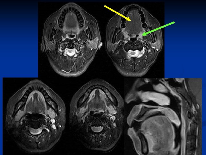
Nasopharynx & Oropharynx
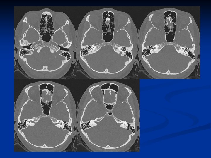
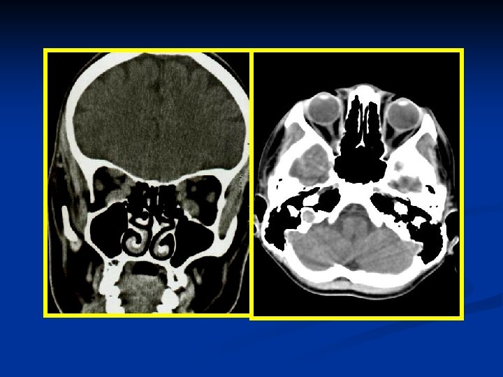
Nasopharynx
Nasopharynx
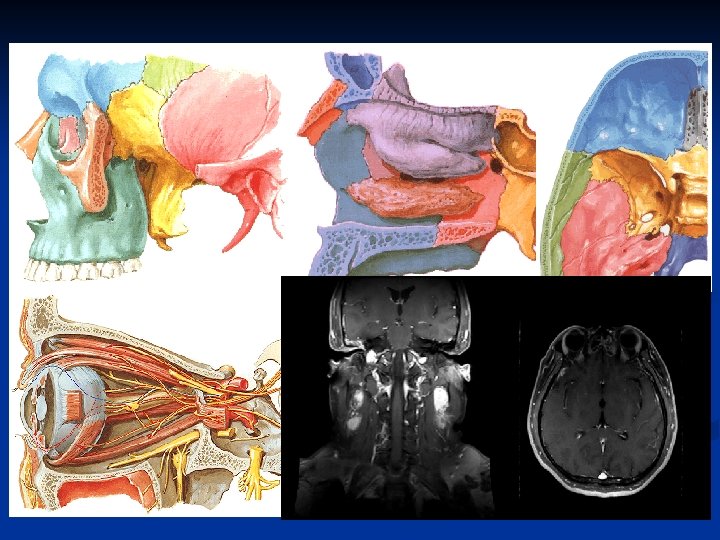
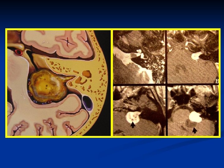
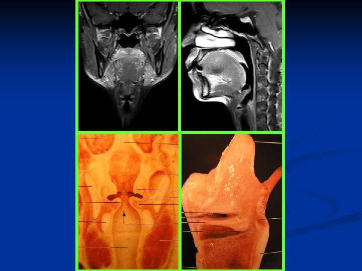
Nasopharynx n Keywords: -- Rosenmüller fossa, Torus tubarius , Eustachian tube orifice -- Pterygopalatine fossa (PPF) -- Parapharyngeal space (PPS) -- Retropharyngeal space (RPS) -- Prevertebral musculature -- Foramen lacerum-ICA (perivascular), -- Foramen ovale-V 3 (perineural)
Rosenmüller fossa, Torus tubarius , Eustachian tube orifice
940127 940930
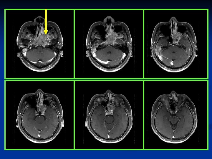
Pterygopalatine fossa (PPF)
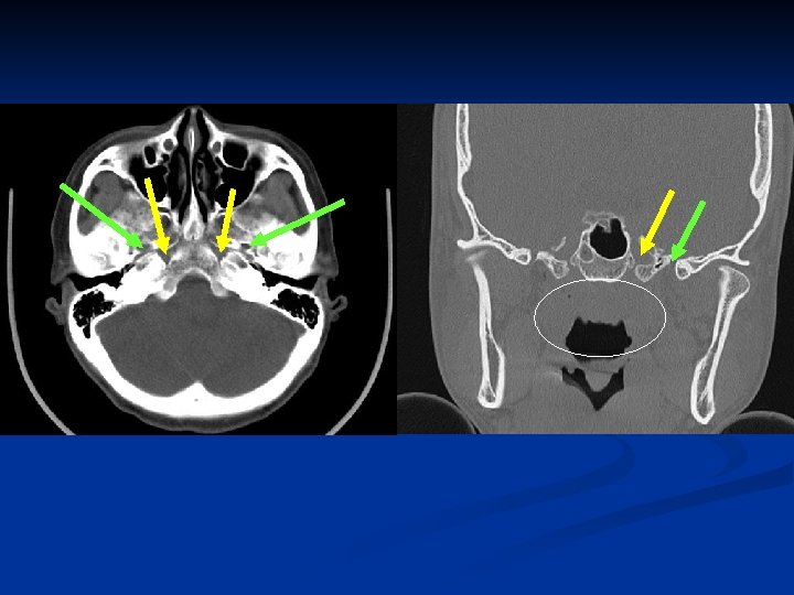
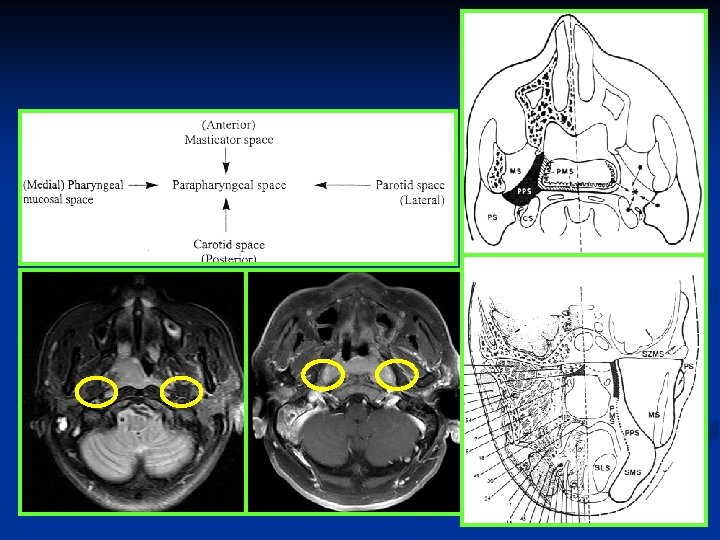
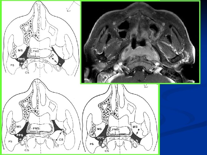
Parapharyngeal space (PPS) n n Fat Branches of cranial nerve V 3 Ascending pharyngeal artery Pharyngeal venous plexus
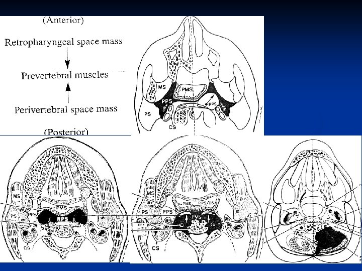
Retropharyngeal space (RPS) Fat n Lateral retropharyngeal nodes (of Rouviere) n Medial retropharyngeal nodes n
Prevertebral musculature
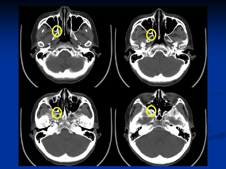
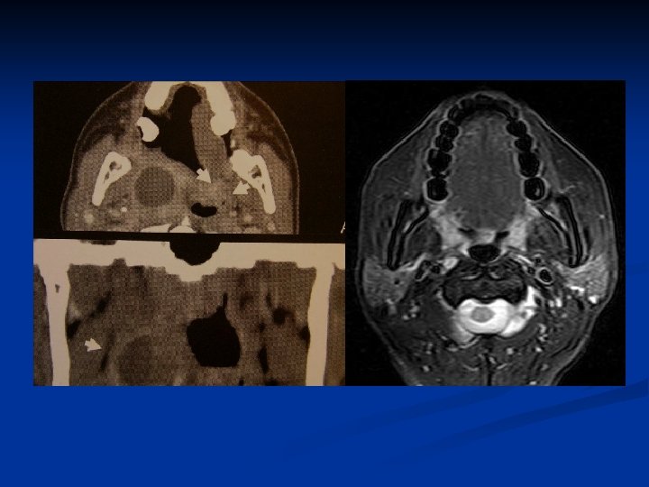
Foramen lacerum-ICA
Foramen ovale-CN V 3 Foramen lacerum-ICA Foramen lacerum Foramen ovale
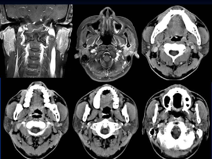
Oropharynx (OP) & Oral cavity n Keywords: -- Hard & Soft palate -- Tongue -- Palatine tonsil; Lingual tonsil (tongue base) -- Posterior OP wall
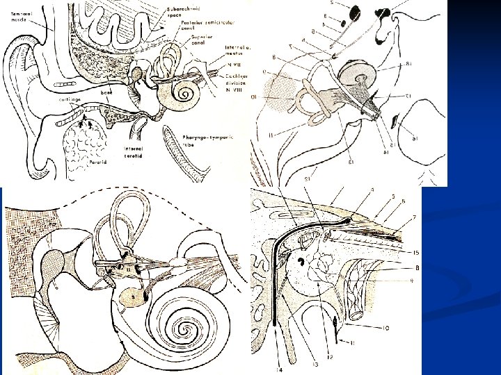
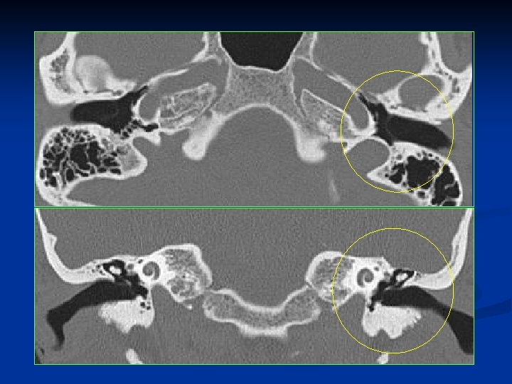
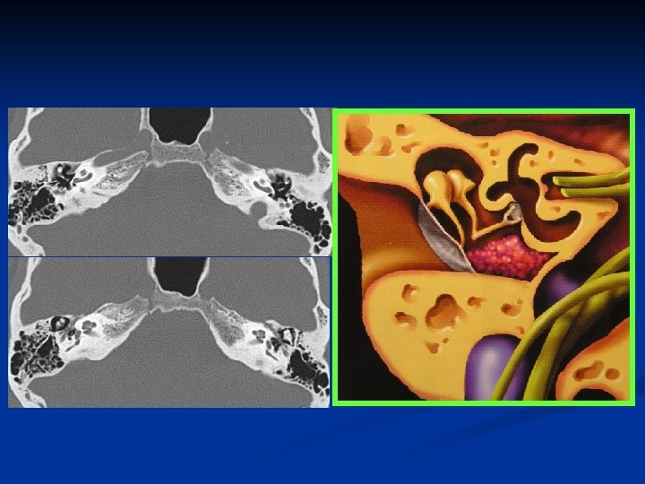
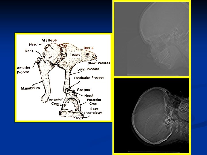
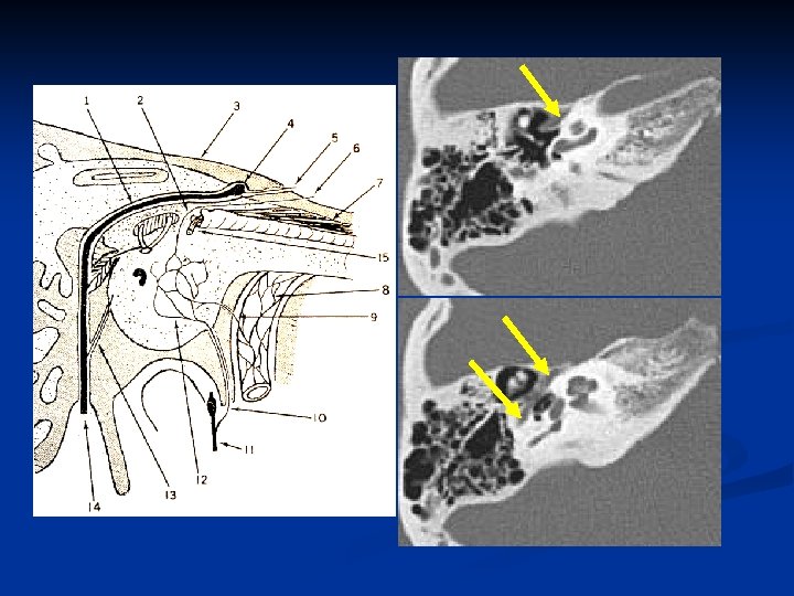
Ear n Keywords: -- Tympanic membrane, -- Ossicular chain (Malleus, Incus, Stapes) -- Facial nerve -- Cochlea, Semicircular canal -- External and internal auditory canals
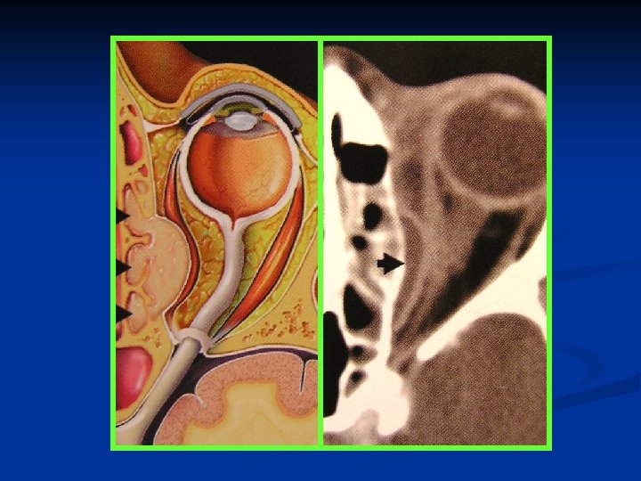
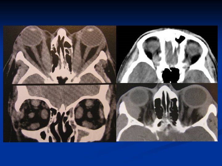
Orbit n Keywords: -- Extraconal & Intraconal -- Lacrimal glands -- Muscles -- Superior & inferior orbital fissures -- Optic canal
Superior & inferior orbital fissures
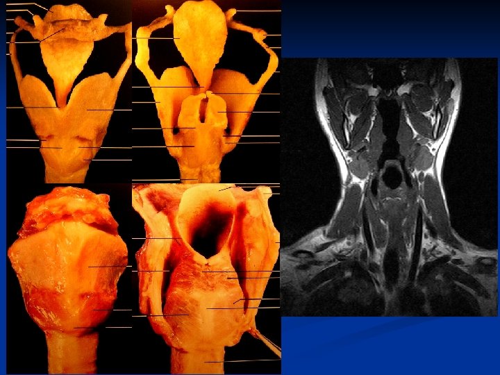
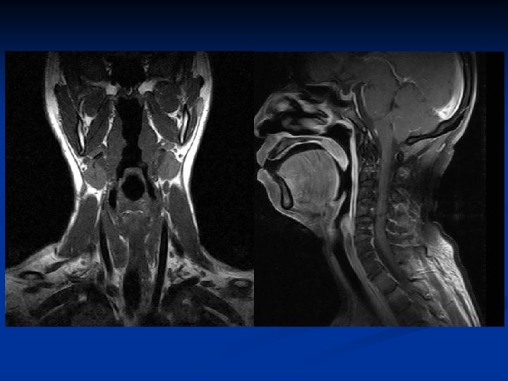
Larynx Composed of a mucosal surface and a supporting cartilaginous skeleton. n Mucosal surface: epiglottis, true and false cords, aryepiglottic folds, and pyriform sinuses n Skeleton: Hyoid cartilage, thyroid cartilage, cricoid cartilage, and arytenoid cartilage n Between mucosa and skeleton: paraglottic and preepiglottic spaces (loose areolar tissues, lymphatics, muscular structures) n
Skeleton n n Hyoid Bone - attachment to epiglottis and strap muscles. Thyroid Cartilage - anterior attachment of vocal folds. Posterior articulation with cricoid cartilage. Cricoid Cartilage - complete ring. Articulates with thyroid and arytenoid cartilages. Arytenoids - two cartilages which glide along the posterior cricoid and attach to posterior ends of vocal folds.
Skeleton Hyoid Bone n Thyroid Cartilage n Cricoid Cartilage n Arytenoids n
Larynx
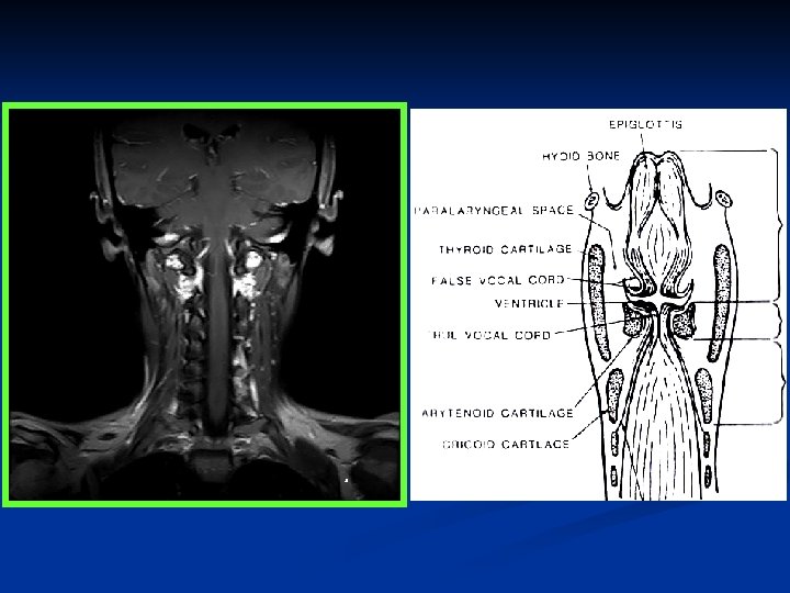
Larynx n Divisions -- Supraglottis -- Glottis -- Subglottis -- Paraglottic (Parapharyngeal) space -- Preepiglottic space
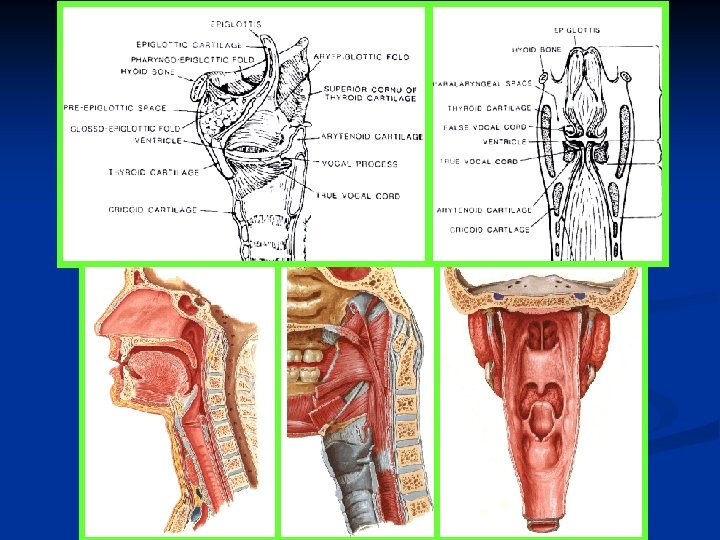
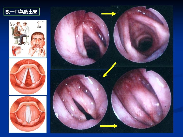
Supraglottis n n n Usually covered with respiratory epithelium containing mucous glands. Epiglottis - leaf-shaped mucosal-covered cartilage, which projects over larynx. Aryepiglottic folds (AE folds) - extend from the lateral epiglottis to the arytenoids. False vocal cords - mucosal folds superior to the true glottis. Separated from true vocal folds by the ventricle. Ventricle - mucosal-lined sac, variable in size which separates the supraglottis from the glottis.
Supraglottis Suprahyoid epiglottis (both lingual and laryngeal aspect) n Infrahyoid epiglottis n False vocal cords (ventricular bands) n Aryepiglottic folds (laryngeal aspects) n Arytenoid cartilages n Ventricles n
Glottis n The true vocal folds attach to the thyroid cartilage at the anterior commissure. The posterior commissure is mobile, as the vocal folds attach to the arytenoids. Motion of the arytenoids effects abduction or adduction of the larynx. The bulk of the vocal fold is made up of muscle covered by mucosa. The free edge is characterized by stratified squamous epithelium. The vocal folds abduct for inspiration and adduct for phonation, cough, and valsalva.
Glottis n True vocal cords, including anterior and posterior commissures
Subglottis n Area from under surface of the true vocal cords to inferior surface of the cricoid cartilage
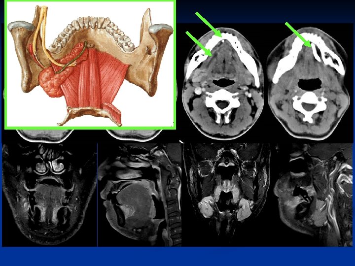
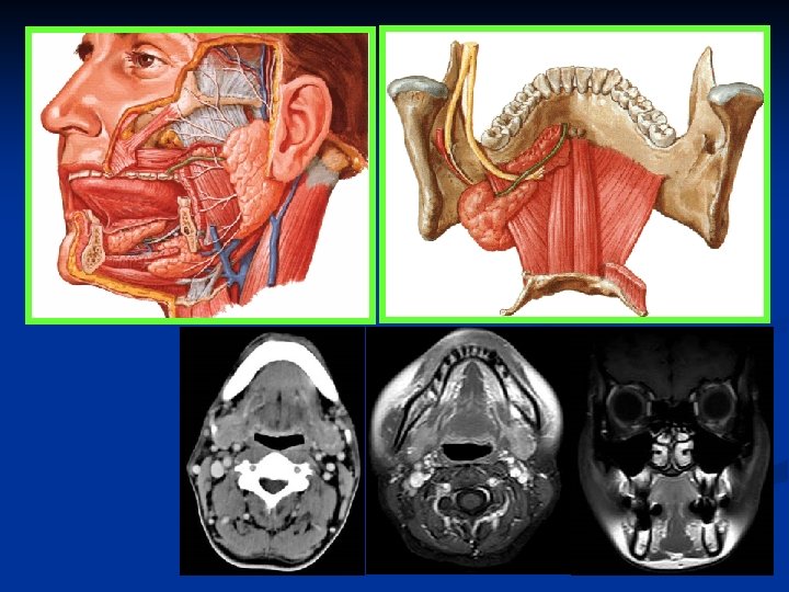
Salivary glands n n Parotid glands Submandibular glands Sublingual glands Minor: tongue, cheeks, lips, palate
Parotid glands The largest of the salivary glands n The duct to this gland (also known as Stensen’s duct) empties within the buccal cavity (the inside of the cheek). n The facial nerve (CNVII) runs through this gland n
Submandibular glands n The Submandibular Gland secretes saliva into ducts called 'Warton's Ducts'. These ducts open on either side of the lingual frenulum.
Sublingual glands n Lie anterior to the submandibular glands under the tongue, beneath the mucous membrane of the floor of the mouth. They are drained by 820 excretory ducts. The largest duct, the sublingual duct (of Bartholin) joins the submandibular duct to drain through the sublingual caruncle.
Neck space & vessels
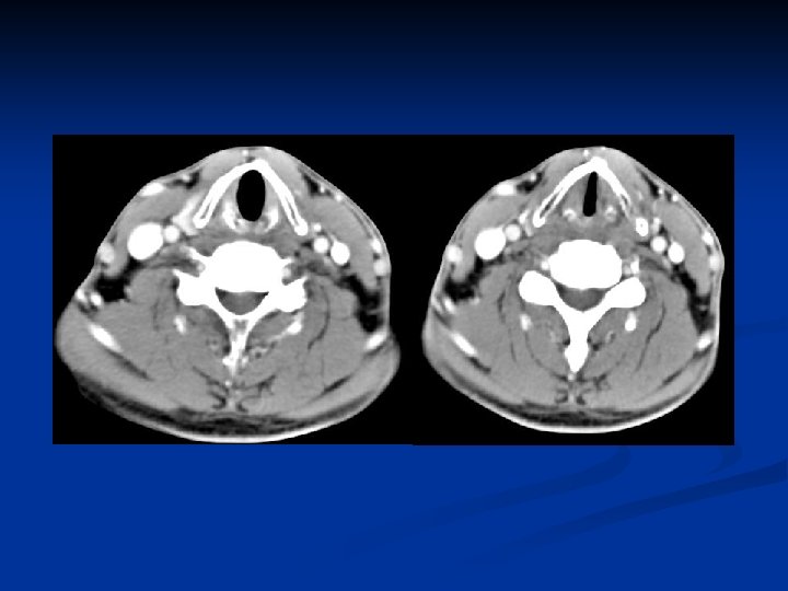
Carotid spaces n Contents: Internal carotid artery Internal jugular vein CN IX, X, XII: nasopharyngeal CS CN X only: oropharyngeal & hypopharyngeal CS Sympathetic plexus Lymph nodes
Thanks for your attention