
CONSORTIUM COGNITION ET VIEILLISSEMENT Traitement des troubles cognitifs
CONSORTIUM COGNITION ET VIEILLISSEMENT Traitement des troubles cognitifs dans les maladies neurodégénératives Structural Changes in Entorhinal Cortex in Mild Cognitive Impairment and Alzheimer’s Disease: An MRI Study Noor Jehan Kabani
Structural Basis of Dementia Cognitive changes have a neural basis. Magnetic resonance imaging is a non-invasive way of studying the neural substrate.
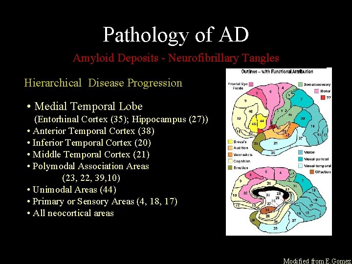
Pathology of AD Amyloid Deposits - Neurofibrillary Tangles Hierarchical Disease Progression • Medial Temporal Lobe (Entorhinal Cortex (35); Hippocampus (27)) • Anterior Temporal Cortex (38) • Inferior Temporal Cortex (20) • Middle Temporal Cortex (21) • Polymodal Association Areas (23, 22, 39, 10) • Unimodal Areas (44) • Primary or Sensory Areas (4, 18, 17) • All neocortical areas Modified from E. Gomez
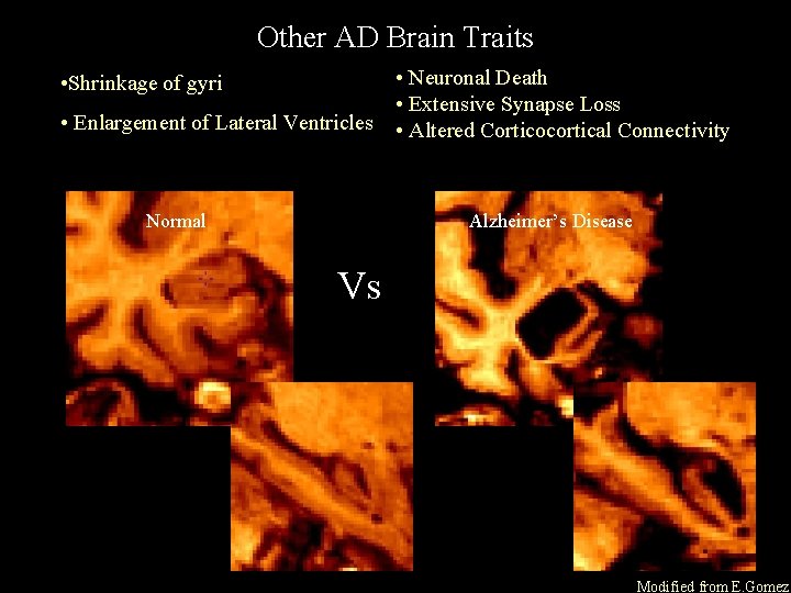
Other AD Brain Traits • Neuronal Death • Extensive Synapse Loss • Enlargement of Lateral Ventricles • Altered Corticocortical Connectivity • Shrinkage of gyri Normal Alzheimer’s Disease Vs Modified from E. Gomez

MRI measures taken to study structural changes in AD
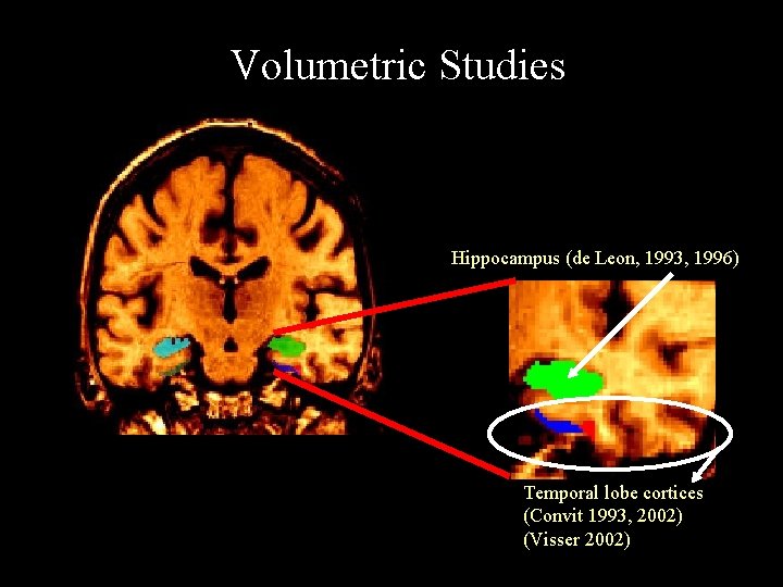
Volumetric Studies Hippocampus (de Leon, 1993, 1996) Temporal lobe cortices (Convit 1993, 2002) (Visser 2002)
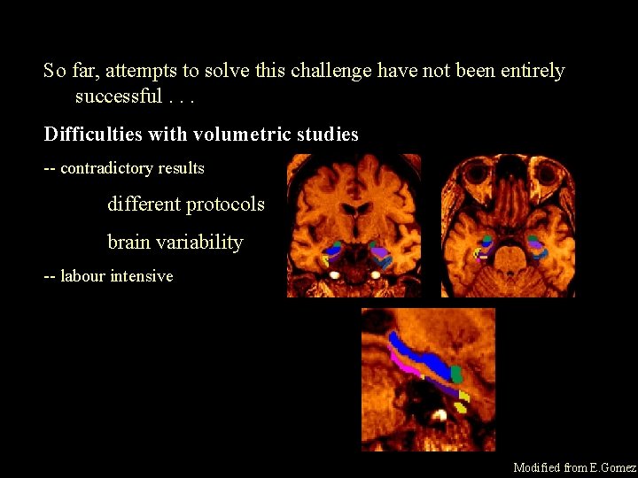
So far, attempts to solve this challenge have not been entirely successful. . . Difficulties with volumetric studies -- contradictory results different protocols brain variability -- labour intensive Modified from E. Gomez
A major weakness volumetric studies depend upon gross morphological changes at the macroscopic level
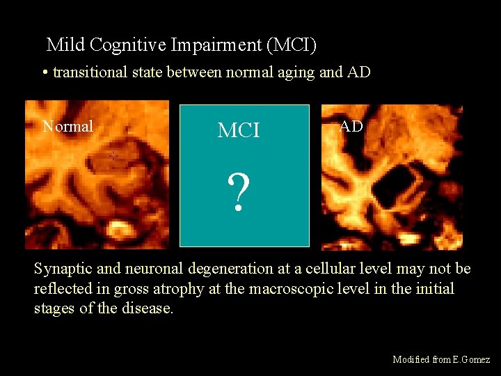
Mild Cognitive Impairment (MCI) • transitional state between normal aging and AD Normal MCI AD ? Synaptic and neuronal degeneration at a cellular level may not be reflected in gross atrophy at the macroscopic level in the initial stages of the disease. Modified from E. Gomez
Rationale For This Study Atrophic changes are not always observed in MCI subjects and even MCI subjects with no medial temporal lobe atrophy may develop AD
Our Approach A different structural imaging parameter MRI Magnetization Transfer (MT) (Wolff and Balaban, 1989)
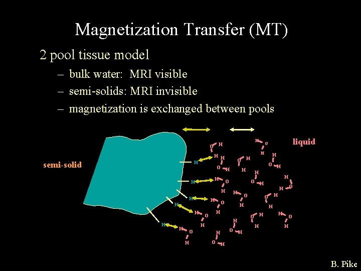
Magnetization Transfer (MT) 2 pool tissue model – bulk water: MRI visible – semi-solids: MRI invisible – magnetization is exchanged between pools H H O H O H H H H O O O H H H O O H H H H O liquid O H H semi-solid H H O B. Pike
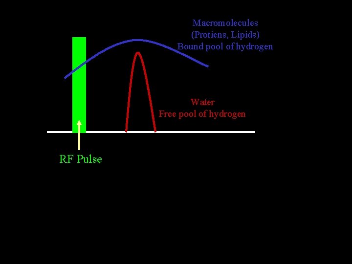
Macromolecules (Protiens, Lipids) Bound pool of hydrogen Water Free pool of hydrogen RF Pulse
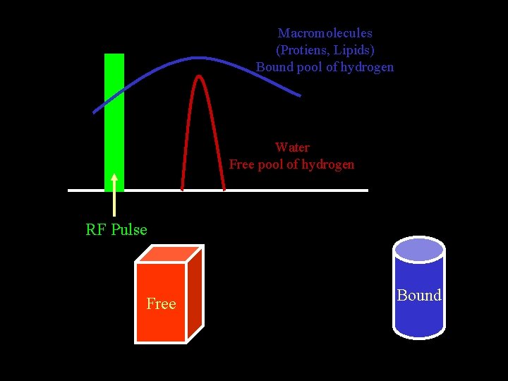
Macromolecules (Protiens, Lipids) Bound pool of hydrogen Water Free pool of hydrogen RF Pulse Free Bound
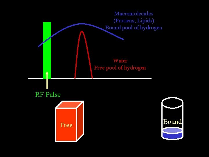
Macromolecules (Protiens, Lipids) Bound pool of hydrogen Water Free pool of hydrogen RF Pulse Free Bound
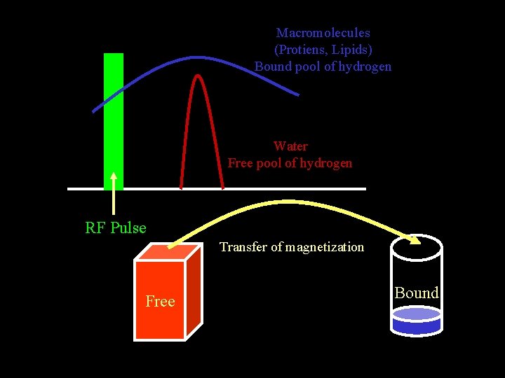
Macromolecules (Protiens, Lipids) Bound pool of hydrogen Water Free pool of hydrogen RF Pulse Transfer of magnetization Free Bound
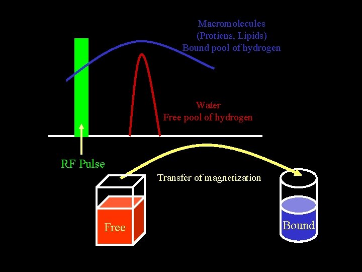
Macromolecules (Protiens, Lipids) Bound pool of hydrogen Water Free pool of hydrogen RF Pulse Transfer of magnetization Free Bound
What does MT measurement yield? MT allows one to indirectly study changes in myelin, protien matrices and cell membranes of the brain that are otherwise not visible using conventional MRI

METHODS
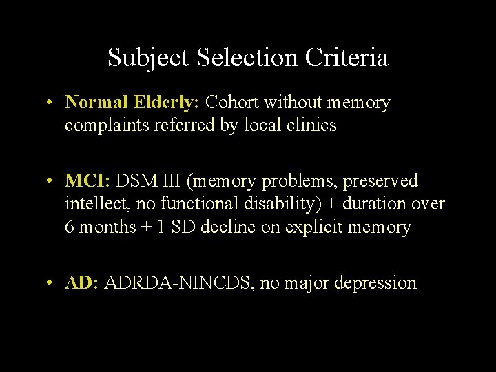
Subject Selection Criteria • Normal Elderly: Cohort without memory complaints referred by local clinics • MCI: DSM III (memory problems, preserved intellect, no functional disability) + duration over 6 months + 1 SD decline on explicit memory • AD: ADRDA-NINCDS, no major depression
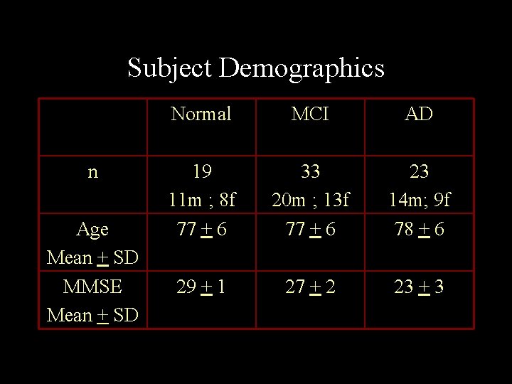
Subject Demographics n Age Mean + SD MMSE Mean + SD Normal MCI AD 19 11 m ; 8 f 77 + 6 33 20 m ; 13 f 77 + 6 23 14 m; 9 f 78 + 6 29 + 1 27 + 2 23 + 3
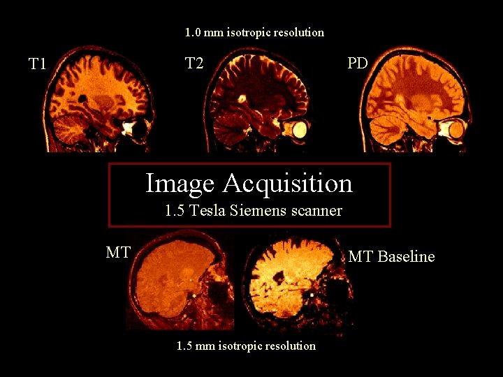
1. 0 mm isotropic resolution T 2 T 1 PD Image Acquisition 1. 5 Tesla Siemens scanner MT MT Baseline 1. 5 mm isotropic resolution

Image Analysis
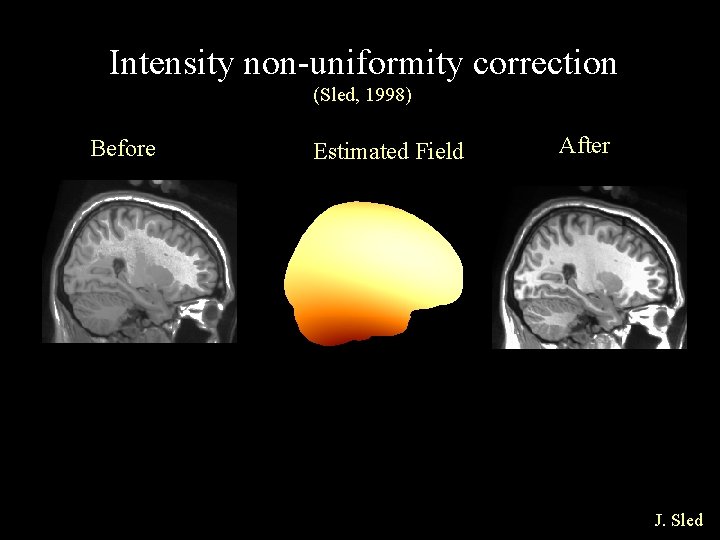
Intensity non-uniformity correction (Sled, 1998) Before Estimated Field After J. Sled
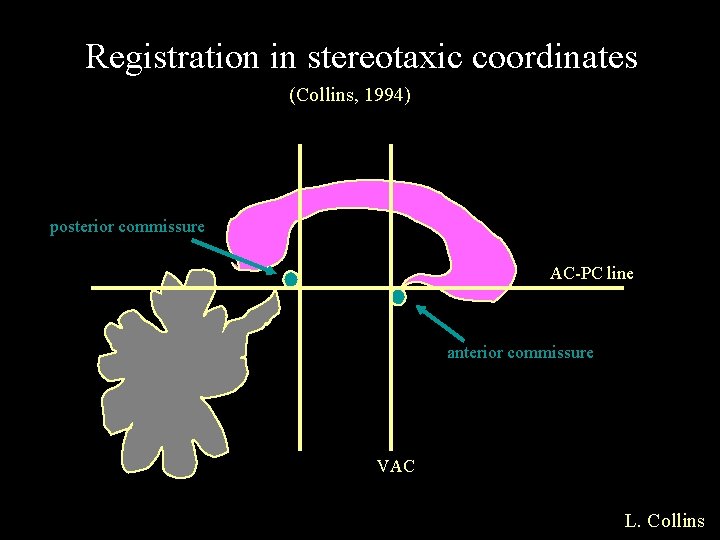
Registration in stereotaxic coordinates (Collins, 1994) posterior commissure AC-PC line anterior commissure VAC L. Collins
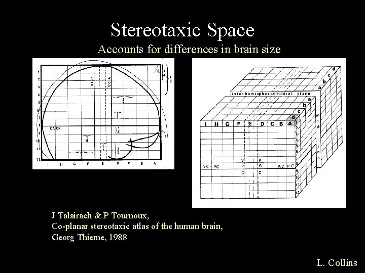
Stereotaxic Space Accounts for differences in brain size J Talairach & P Tournoux, Co-planar stereotaxic atlas of the human brain, Georg Thieme, 1988 L. Collins
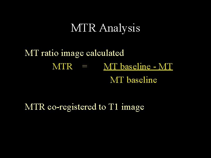
MTR Analysis MT ratio image calculated MTR = MT baseline - MT MT baseline MTR co-registered to T 1 image
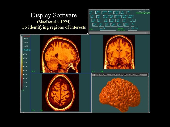
Display Software (Mac. Donald, 1994) To identifying regions of interests
• Labels were painted on T 1 images (1 mm resolution) • Labels were then superimposed on the MTR images • Volume and MTR was calculated

RESULTS PART 1
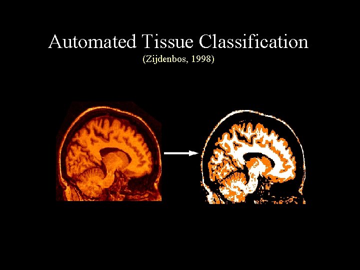
Automated Tissue Classification (Zijdenbos, 1998)
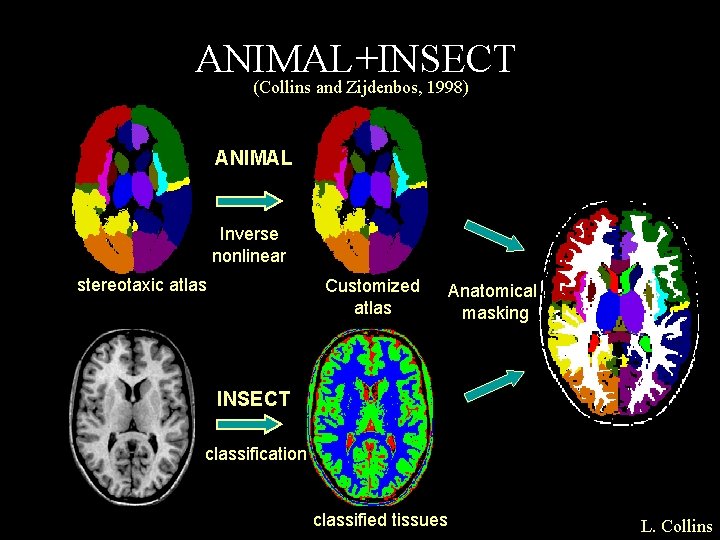
ANIMAL+INSECT (Collins and Zijdenbos, 1998) ANIMAL Inverse nonlinear stereotaxic atlas Customized atlas Anatomical masking INSECT classification classified tissues L. Collins
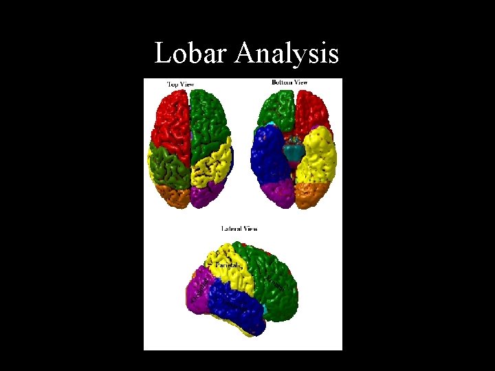
Lobar Analysis

RESULTS PART 2
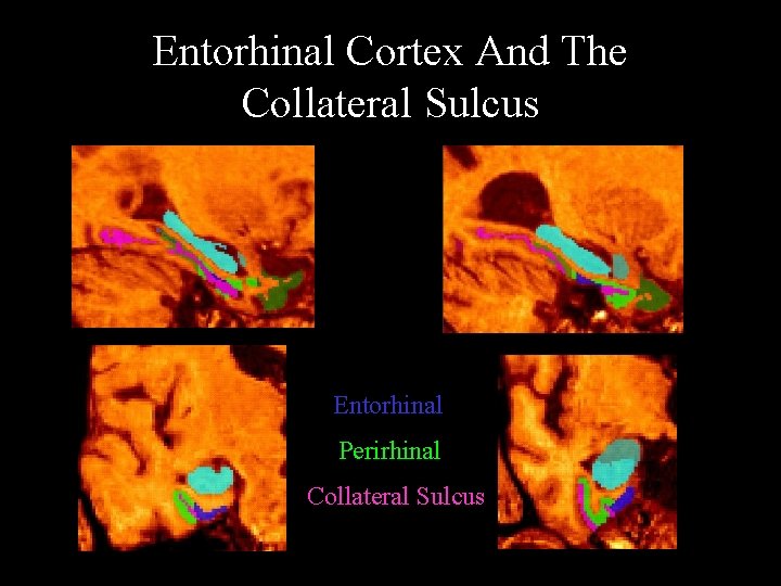
Entorhinal Cortex And The Collateral Sulcus Entorhinal Perirhinal Collateral Sulcus
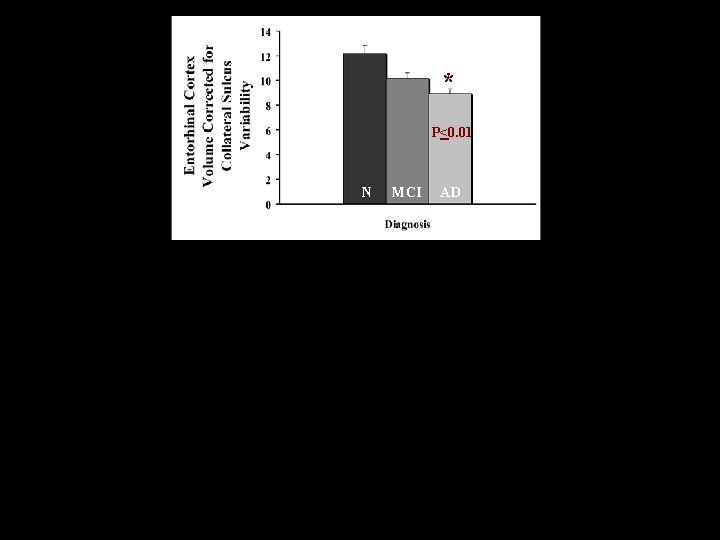
* P<0. 01 N MCI AD
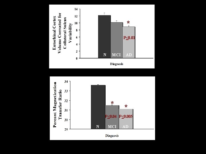
* P<0. 01 N MCI * AD * P<0. 04 P<0. 005 N MCI AD AD
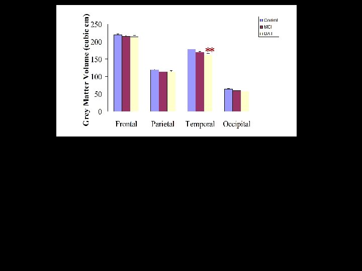
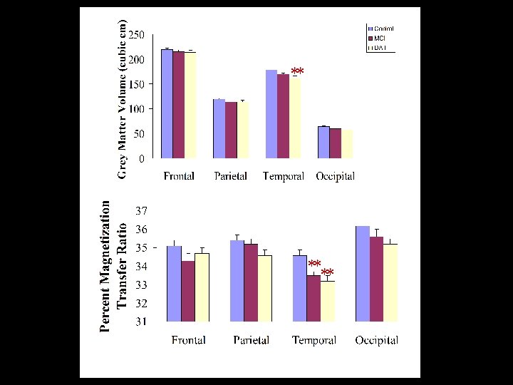
Summary Volumetric measures were lower but no significant difference was found between MCI and normal elderly.
Summary Volumetric measures were lower but no significant difference was found between MCI and normal elderly. MT ratio was significantly lower in the absence of significant volumetric differences.
MTR analysis may well be a sensitive means of detecting early structural changes indicative of incipient dementia before volumetric changes become significant

Acknowledgements • • • Consortium cognition et vieillissement (VRQ) Brain Imaging Center - Montreal Neurological Institute Geriatric unit of the Jewish General Hospital - Montreal Memory Clinic of the Jewish General Hospital - Montreal Alzheimer Society Research Foundation of Canada Canadian Institutes for Health Research • • • Anita Shuper Kate Hanratty Adrienne Dorr Anita Kar Cinzia Gaudelli Kathy De Sousa John Sled Howard Chertkow Shelly Solomon
- Slides: 43