Connective tissue proper Cartilage Bone Origine and function

Connective tissue proper Cartilage Bone

Origine and function of c. t. Origin – embryonic mesenchyme Functions: - nutritive (blood vessels and difusion of nutriens) - protective – immunocompetent cells and antibodies production - connective – between different tissues - mechanical (supporting and mechanical protection of organ – in skull, thorax, pelvis)

Connective tissue types C. t. proper Cartilage Bone __________________ general structure: - cells - intercellular fibers matter ground amorphous substance
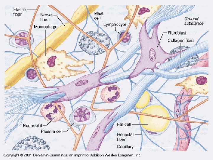

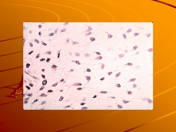
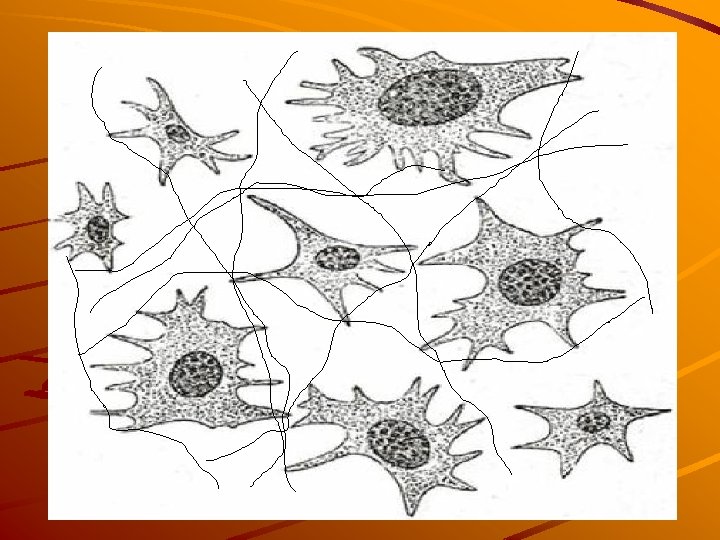
Connective tissue proper - cells FIXED CELLS Fibroblasts, fibrocytes Reticular cells Fat cells (univacuolar, multivacuolar) Pigment cells Undifferentiated cells

Fibroblasts, fibrocytes

Reticular cells Network of cell processes + reticular fibers collagen III
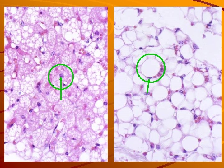
 cells univacuolar multivacuolar (white adip. c. t) (brown adip. c. t. )")
Adipose (fat) cells univacuolar multivacuolar (white adip. c. t) (brown adip. c. t. ) Lipid droplets

Pigment cells neuroectodermal origin

Connective tissue proper - cells MOBILE CELLS Histiocytes macrophages Mast cells Plasma cells Leukocytes
 phagocytosis")
Histiocytes macrophages (belong to monocyte-macrophage system) phagocytosis
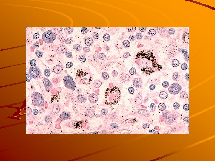

Mast cells heparin, histamin – inflamation mediators
 antibodies (immunoglobulins) production Rough ER")
Plasma cells (B-ly are precursors) antibodies (immunoglobulins) production Rough ER
 ground substance homogenous semifluid material glycosaminoglycans (hyluronic acid, chondroitinsulphate, dermatansulphate, keratansulphate, heparansulphate)")
Intercellular (amorphous) ground substance homogenous semifluid material glycosaminoglycans (hyluronic acid, chondroitinsulphate, dermatansulphate, keratansulphate, heparansulphate) glycoproteins (fibronectin, laminin) – cell adhesive function H 2 O, ions

Fibers collagenous reticular elastic

Collagen fibers „white fibers“, solid and strong, but not elastic 1 – 20 m Ø arranged into bundles collagenase – digestive enzyme in LM – acidophilic (pink in HE)

Collagen fibers 64 nm
 Collagen Type II")
Collagen fiber types Collagen Type I – most ubiquitous (cf. ) Collagen Type II – in cartilage Collagen Type III – reticular fibers Collagen Type IV – "amorphous collagen" without a fiber structure, synthesized by epithelial cel About 40 types of collagen are known
 - a supporting")
Reticular fibers contain type III collagen form a fine meshwork (reticulum) - a supporting mesh in soft tissues such as liver, bone marrow, and lymph organs they are argyrophilic (silver staining) 0. 5 – 2 m Ø
 produced by fibroblasts and smooth")
Elastic fibers „yellow fibers“ - bundles of protein (elastin) produced by fibroblasts and smooth muscle cells in blood vessels. these fibers can stretch up to 1. 5 times their length, and snap back to their original length when relaxed. are branched and wavy spetial staining with orcein or resorcin-fuchsin

C. t. proper classification several types of c. t. proper: classification depends on cells, fibers and ground substance quantity and arrangement abundant ground subst. – „soft tissue“ abundant fibers – „hard tissue“ fibers can be arranged (ir)regularly
 Mesenchyme Jelly-like c. t. (Wharton´s jelly) Collagenous")
CTP classification (types of c. t. ) Mesenchyme Jelly-like c. t. (Wharton´s jelly) Collagenous c. t. areolar (loose) dense irregular Reticular c. t. Elastic c. t. Adipose c. t. white fat brown fat

1. Mesenchyme embryonic c. t. 2. jelly-like c. t. umbilical cord, dental pulp
 c. t.")
3 a. collagenous loose (areolar) c. t.

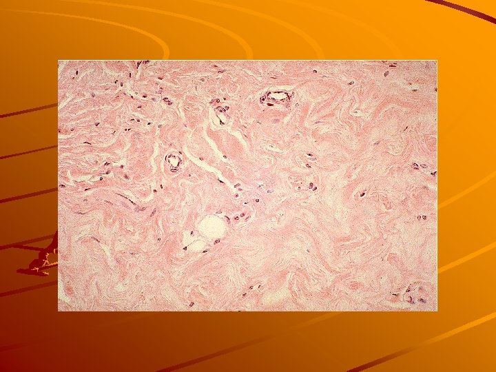
3 b. collagenous dense c. t. irregular tendons, ligaments
 5. elastic. c.")
4. reticular c. t supporting tissue in lymph organs (impregn. ) 5. elastic. c. t. membranes in blood vessels wall(orcein)
 brown fat white fat")
6. adipose tissue fetal adipose tissue (thermoregulation) brown fat white fat

Light microscopy Occular magnif. : 10 x Objectives magnif. 4 x, 10 x, 40 x Total magnif. : 40 x, 100 x, 400 x Don´t use immersion objective! Slide cover slip must be situated up to the lens of objective 10 x

Supporting connective tissue Cartilage Bone
; nutrients are transported to the")
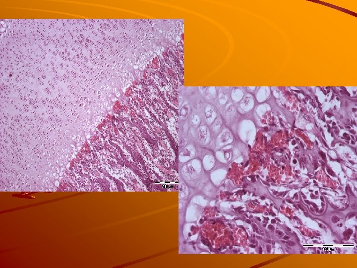
Cartilage - general characteristic Avascular tissue (without blood vessels); nutrients are transported to the cells by diffusion from perichondrium Perichondrium* – dense connective tissue capsule with blood vessels Mesenchymal origin * Is not present on the articular surfaces of cartilage and on the surface of fibrocartilage

General structure of the cartilage Cells – chondrocytes in lacunae Intercellular matter: - ground substance chondroitinsulphates chondronectin - fibers (collagenous or elastic)

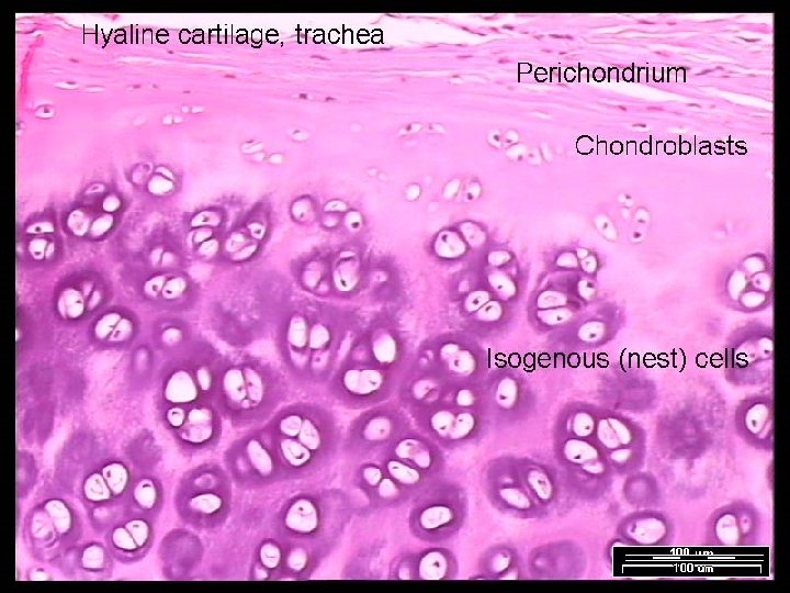
Cartilage cells: chondroblasts, chondrocytes in lacunae produce intercellular matter: - collagen or elastic fibers - ground substance: 1) main glycosamino- glycans are chondroitinsulfates, keratansulfate = acid substances cause basophilia of cartilage, 2) glycoprotein chondronectin (increases adhesion of cells and matrix) cells are involved in cartilage growth: - interstitial growth (isogenic groups) - appositional growth (from perichondrium)
 Chondrocytes form isogenic groups")
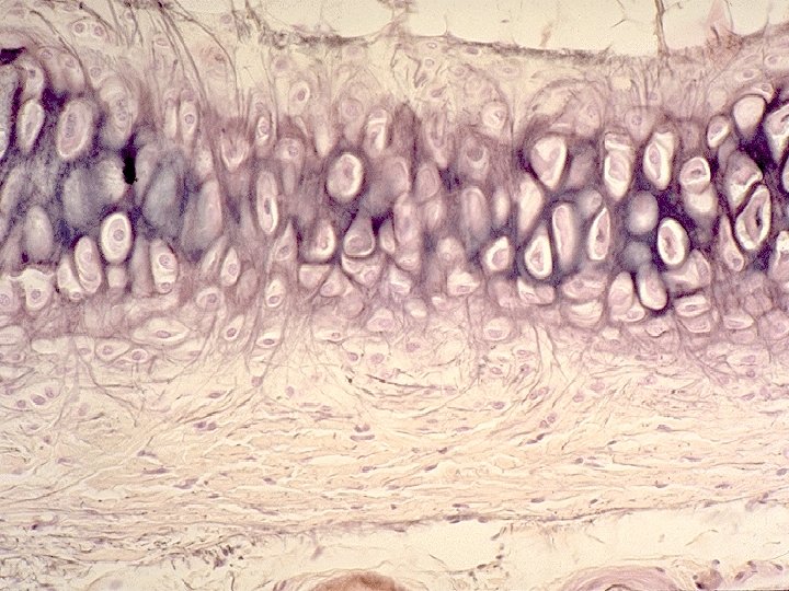
Hyaline cartilage (example: free surfaces of joins, epiphyseal growth plate) Chondrocytes form isogenic groups (in basophilic capsule) Collagenous fibers form bundles Ground substance is abundant and covers the fibers
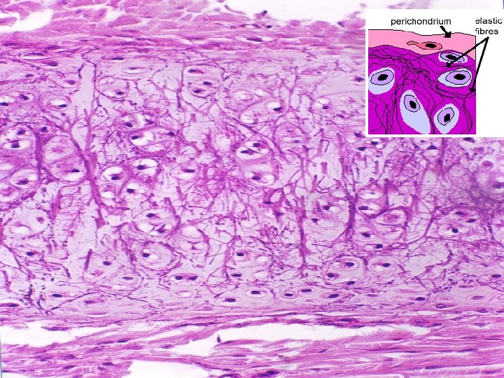
 Chondrocytes in lacunae are dispersed: NO isogenic groups Elastic")
Elastic cartilage (example: auricle, epiglottis) Chondrocytes in lacunae are dispersed: NO isogenic groups Elastic fibers Ground substance
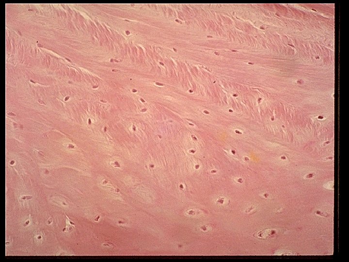
 Small and flattened chondrocytes Thick bundles of collagenous fibers")
Fibrocartilage (example: symphysis, intervertebral discs) Small and flattened chondrocytes Thick bundles of collagenous fibers Small amount of ground substance No perichondrium

Bone tissue Organic component Cells Collagenous fibers Ground substance is mineralized Inorganic component Minerals – hydroxyapatite crystals Ca 10(PO 4)6(OH)2
 - ground substance: 1)")
Intercellular matrix Organic component: - collagen fibers (collagen type I) - ground substance: 1) main glycosaminoglycan are chondroitin-sulfate, keratan-sulfate, 2) fibronectin, osteopontin (increase adhesion of cells and matrix), osteocalcin (binds Ca) Inorganic component: crystals of hydroxyapatite

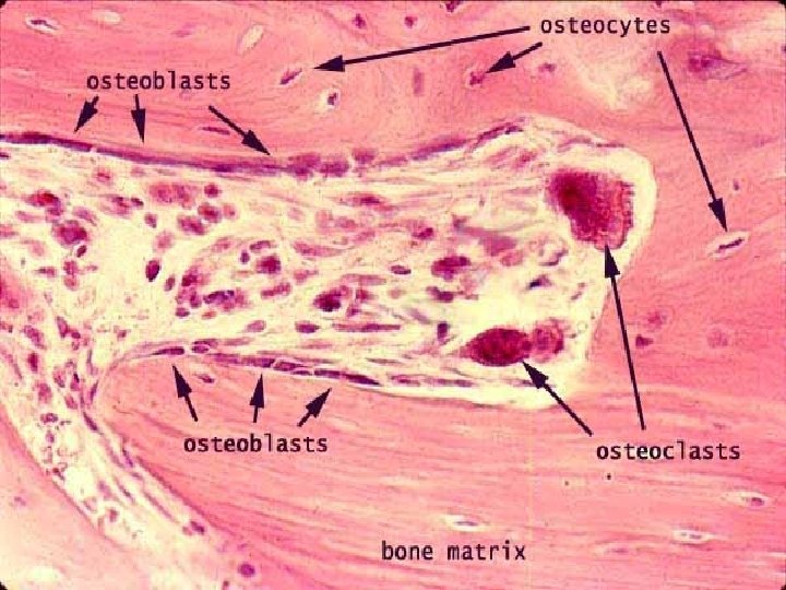
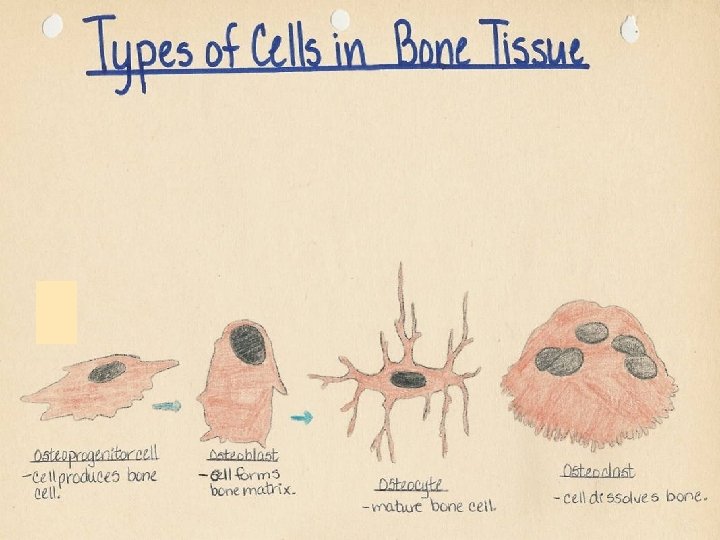
Bone cells osteoprogenitor cells osteoblasts osteocytes osteoclasts
")
on the surface of the bone inside of the bone matrix (in lacunae)
 - synthesize non-mineralized intercellular matrix")
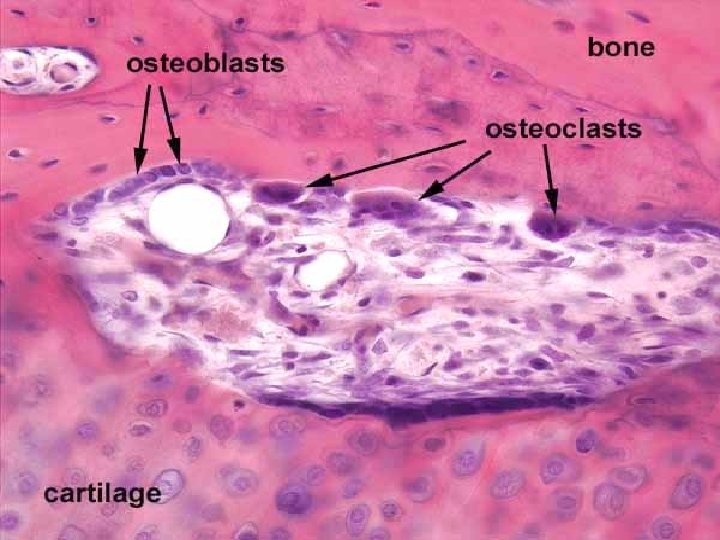
osteoblasts - on the surface of bone (epitheloid arrangement) - synthesize non-mineralized intercellular matrix = osteoid (ground substance + collagen) - periosteal osteoblasts – growth of bone by apposition

osteocytes - osteoblasts entrapped in mineralized bone matrix = osteocytes (cytoplasmic processes in canaliculi ossium striation of bone lamellae) - transport of minerals from blood into the matrix
 –")
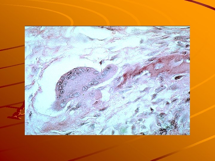
osteoclasts - in Howship´s lacunae - large multinucleated cells (up to 50 nuclei) – arrise by fusion of monocytes - produce acid phosphatase – enzyme involved in bone resorption during ossification, bone remodeling or reparation

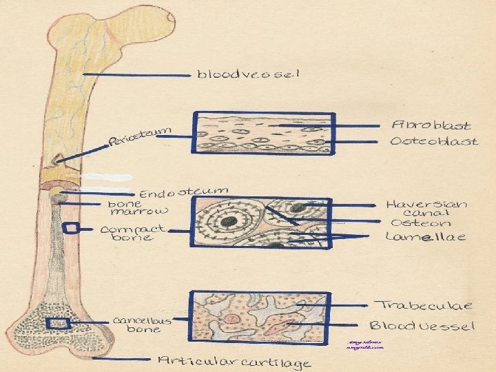
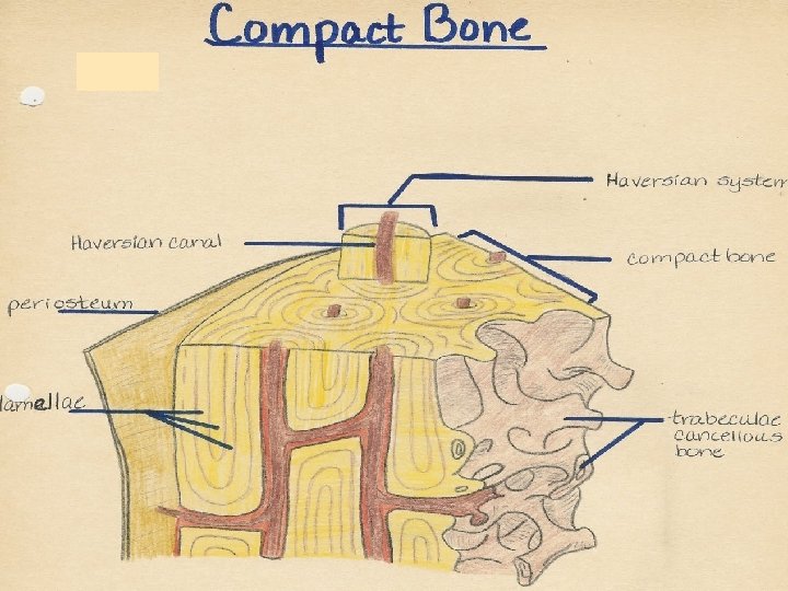
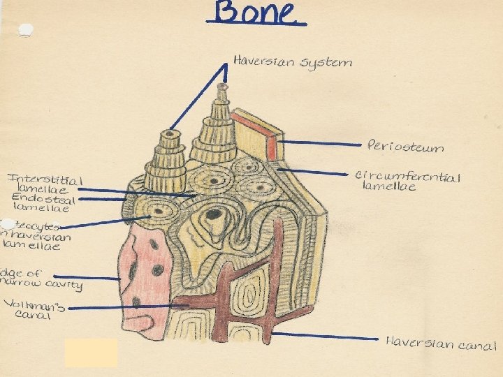
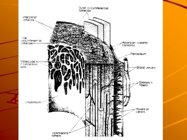
Periosteum – endosteum Periosteum dense c. t. capsule attached to bone by Sharpey's fibers (collagen); is composed of two layers: 1)outer fibrous layer of dense irregular c. t. and 2)inner cellular layer composed of cells that give rise to osteoblasts. Endosteum thin layer of connective tissue containing osteoprogenitor cells and osteoblasts

Periosteum – endosteum
 Lamellar spongy (cancellous) compact")
Types of bone tissue Fibrillar (woven) Lamellar spongy (cancellous) compact
 fibrillar bone – „primitive“ Arrise by primary ossification and is transformed")
Bone types A) fibrillar bone – „primitive“ Arrise by primary ossification and is transformed into the compact bone during secondary ossification Forms tuberositas ossium Bundles of collagen fibers osteocytes
 lamellar (cancellous) bone spongy - compact")
Bone types B) lamellar (cancellous) bone spongy - compact
 bone")
Spongy (trabecular) bone

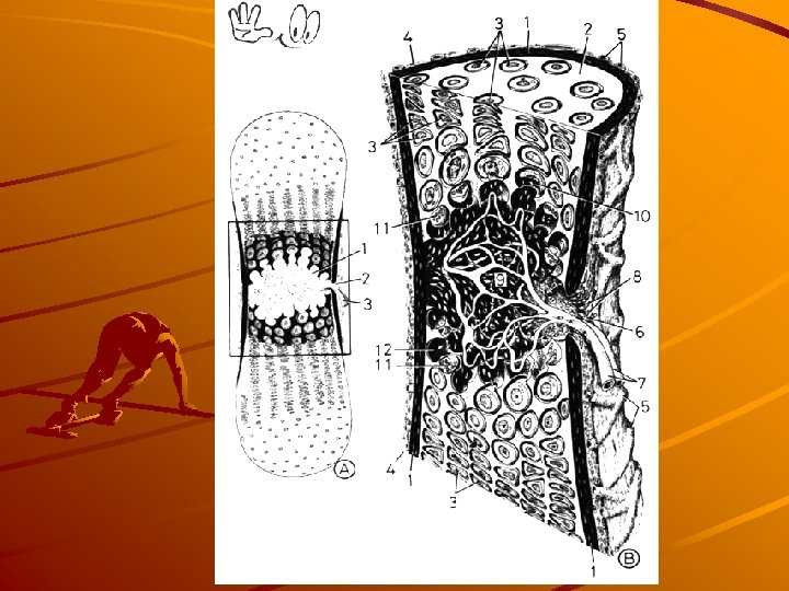
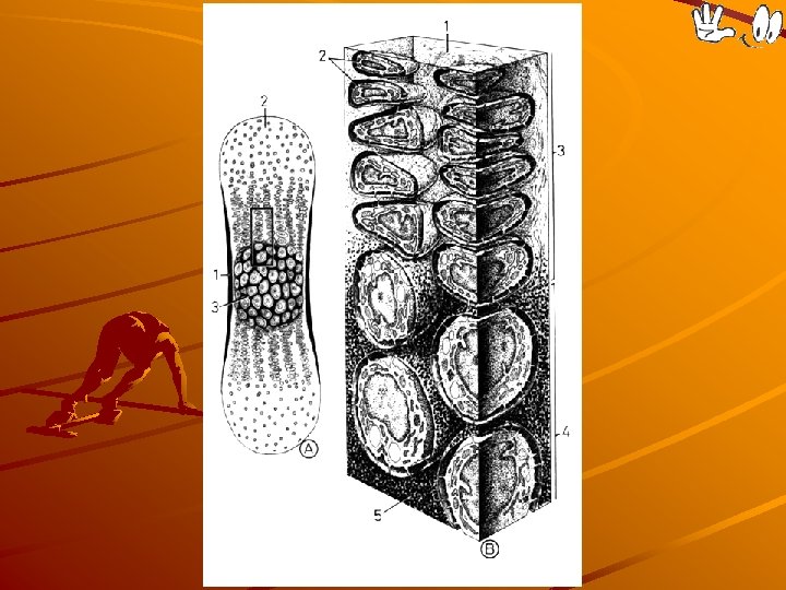
compact bone collagen fibers in mineralized ground substance form lamellae, which are arranged as: Haversian lamellae = osteon interstitial lamellae circumferential lamellae (inner, outer)

OSTEON: 1 – Haversian canal contains 2, 3 – blood vessels, 4 – nerves in loose connective tissue; 5 – lamellae 7 – osteocytes in lacunae (6) 8 – cytoplasmic processes in canaliculi ossium

Osteon

Outer circumferential lamellae Haversian system Inner circumferential lamellae Haversian canal Interstitial lamellae

Ossification – bone formation PERIOSTEUM contains blood vessels and osteoprogenitor cells osteogenic potential osteoblasts (producce intercellular matrix OSTEOID Osteoid + minerals (from blood) = OSSEIN Osteoblasts entrapped in ossein osteocytes, ( osteocytes + ossein = BONE) The result of primary ossification is fibrillar bone Resorption, remodelation of fibrillar bone by osteoclasts The result of secondary ossification is compact bone (are involved in destruction of woven bone)

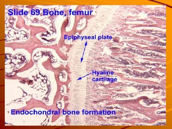
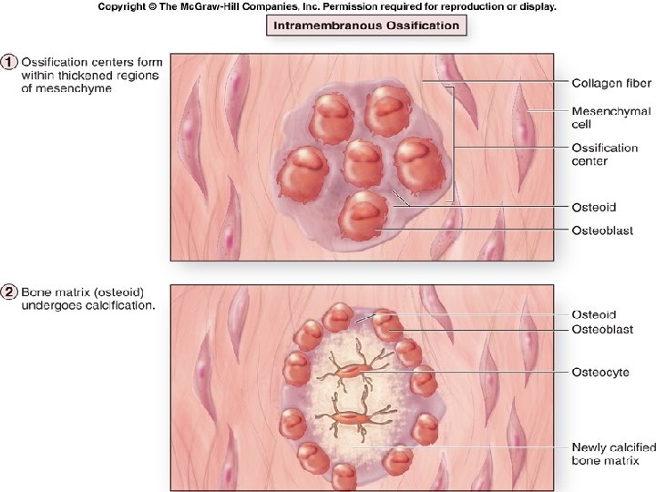
Ossification types Intramembranous ossification - bone develops in mesenchymal membrane flattened bones Endochondral ossification - bone develops on a cartilage model long bones

Intramembranous ossification Formation of bone directly from mesenchyme. Increased vascularity of mesenchyme Mesenchyme cells osteoprogenitor cells osteoblasts Osteoblasts produce osteoid and transform into osteocytes. Periosteum + endosteum (dura mater in skull) NEUROCRANIUM and other flat bones

mezenchymocytes osteocytes Osteoprogenitor cells s o st a l ob ste

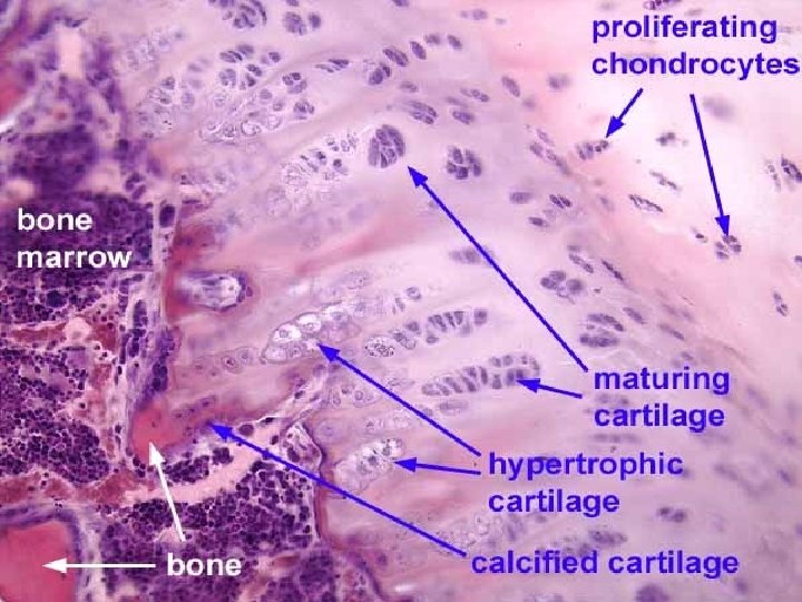
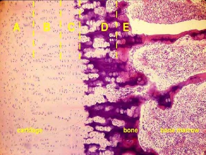
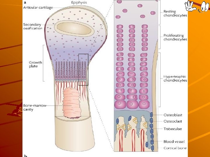
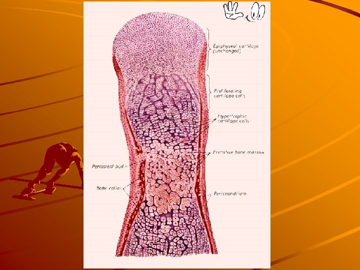
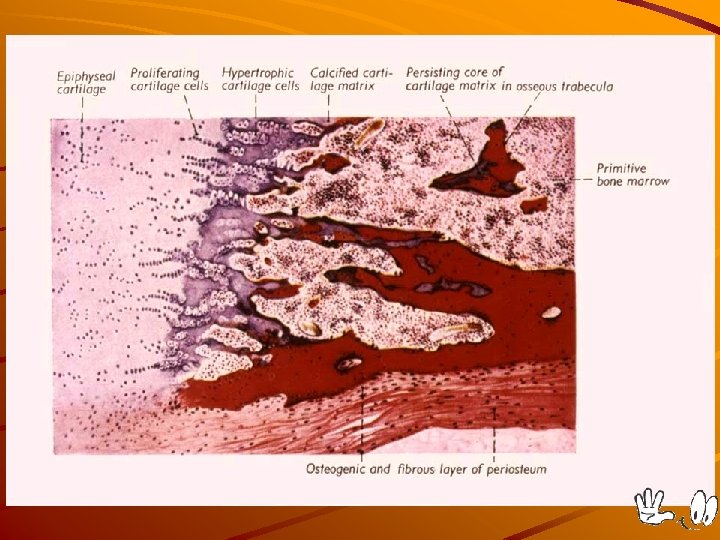
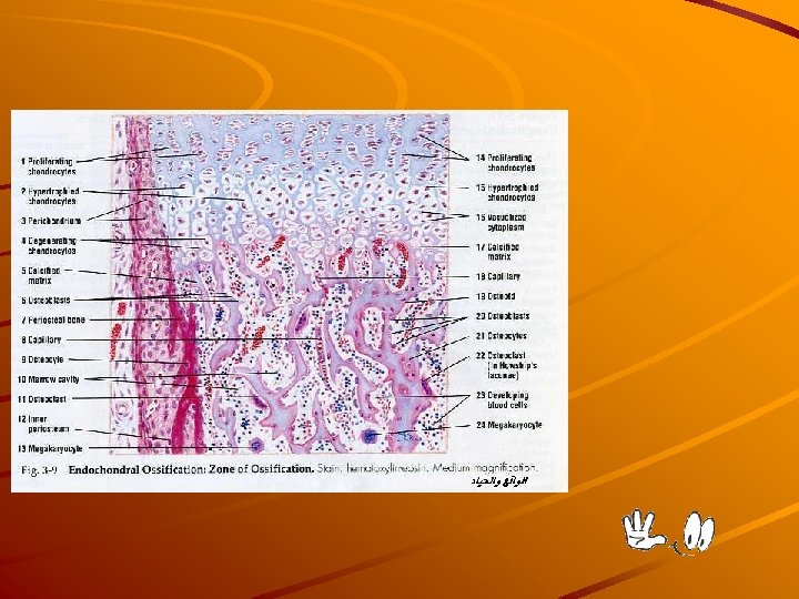
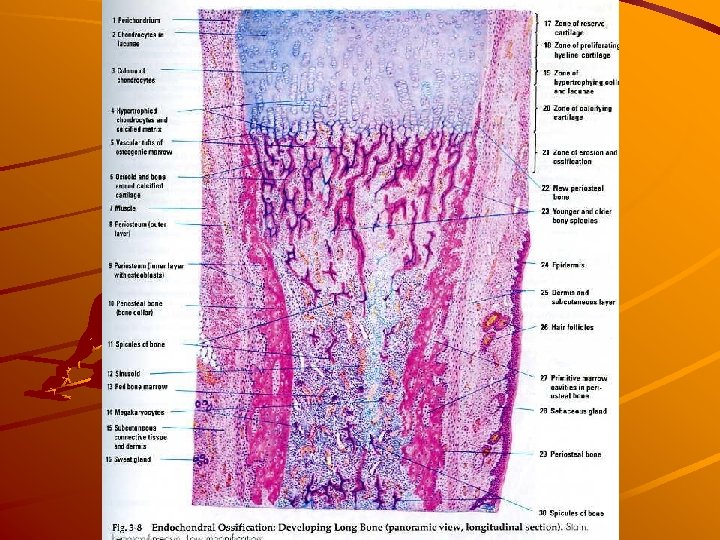
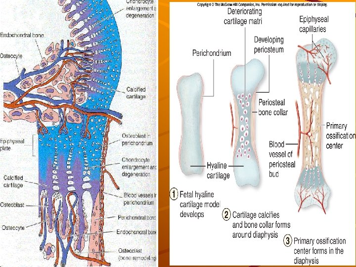
Endochondral ossification PRIMARY OSSIFICATION CENTER: Occurs in the center of the diaphysis, and extends toward both epiphyses. SECONDARY OSSIFICATION CENTER: in the center of the epiphyses. EPIPHYSEAL PLATE: is hyaline cartilage which alllows bone growing in the length.
 Blood vessels invade")
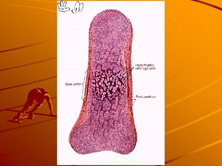
Endochondral ossification Perichondrium arround diaphisis transforms into periosteum (fibroblasts osteoprogenitor cells) Blood vessels invade the cartilage and bring osteoprogenitor cells, blood cells, bone marrow cells, macrophages, endothelial cells. Ca and other ions are transported by blood and intercellular matrix around the chondrocytes is calcified – nutrients cannot difuse to them – cells degenerate and die <to be continued>
 they")
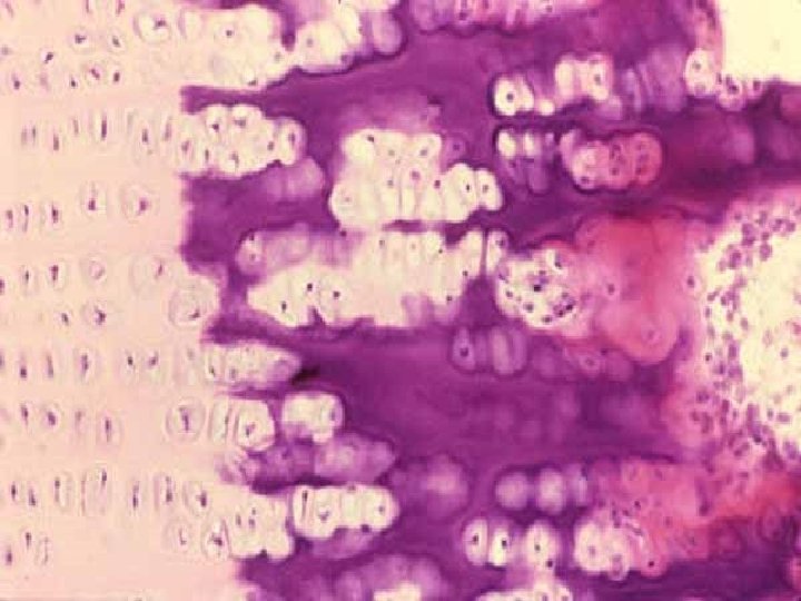
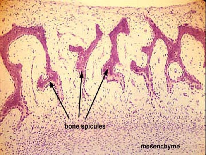
Endochondral ossification after chondrocyte death mineralized intercellular matrix forms spicules (rests of cartilage) they are occupied by osteoblasts producing osteoid surrounds osteoblasts and is mineralized by ions from blood; osteoblasts osteocytes primary ossification = fibrillar bone resorption - osteoclasts secondary ossification = lamellar bone

OSSIFICATION ZONES RESERVE ZONE: normal hyaline cartilage PROLIFERATIVE ZONE: chondrocytes in columns ZONE of HYPERTROPHY: enlarged chondrocytes CALCIFIED ZONE: (different staining of intercellular matter in light microscope) „LINE of erosion“ – imaginary line OSSIFORM ZONE: spicules, osteoid, ossein in regions of bone formation

No rm al Pr ne st eo i d an d of rt ila e er o si os si ca on fo rm zo ne s ca rt of h er tr ed Li O ra tiv e ifi ne ife yp Ca lc al in e ca rt ol Zo hy op h y ila ge
 Elastic cartilage (26. Eoiglottis, HE) (27.")
Cartilage Bone Slides: Hyaline cartilage (28. Trachea, HE) Elastic cartilage (26. Eoiglottis, HE) (27. Elastic cartilage, orcein) Lamellar bone (… Endochondral ossification (… Atlas EM:
 or immobile joints. Syndesmosis: Bones connected")
Bone junctions - joints SYNARTHROSES: Poorly moveable (fibrous) or immobile joints. Syndesmosis: Bones connected by dense fibrous connective tissue, as in SKULL SUTURES Synchondrosis: Bones connected by cartilage, as in the PUBIC SYMPHYSIS. Synostosis: bones connected by bone tissue, as in the PELVIS – DIARTHROSES: Movable joints Articular Cartilage made of hyaline cartilage, without perichondrium, covers the moving bone-ends. Joint Capsule is continuous with the periosteum. Synovial Membrane lines the joint capsule. It secretes synovial fluid into the joint space.

Diarthrosis

Thanks for attention
membranózní osifikace Mezenchym + vaskularizace: mezenchymocyty osteoblasty : produkce osteoprogenitorní bb. osteoidu Osteoblasty +")
(Intra)membranózní osifikace Mezenchym + vaskularizace: mezenchymocyty osteoblasty : produkce osteoprogenitorní bb. osteoidu Osteoblasty + osteoid mineralizace: osteoblasty osteocyty
 A: chrupavčitý model kosti: 1 - epifýza, 2, střed diafýzy B:")
Enchondrální osifikace (chondrogenní) A: chrupavčitý model kosti: 1 - epifýza, 2, střed diafýzy B: periostální manžeta (z perichondria) invaze krevních cév s osteoklasty (na čele ) a osteoprogenitorními bb. (podél cév )
 1 - periost 2 – osteoklasty")
A: hypertrofie a kalcifikace chrupavky B: detail (A) 1 - periost 2 – osteoklasty 3 – osteocyty v kostěné manžetě pod periostem 5 – monocyty 6 – osteoprogen. bb.

Zóna normál. hyalinní chrupavky Zóna proliferující chrupavky Zóna hypertrofické chrupavky Zóna kalcifikující chrupavky 2 4 5 6 9 – – – osteoklasty primární kost s osteocyty osteoblasty osteoid (pod osteoblasty) kapiláry

Lamellar bone

Ossification – bone formation PERIOSTEUM contains blood vessels and osteoprogenitor cells osteogenic potential Osteoprogenitor cells osteoblasts (production of intercellular matrix OSTEOID Osteoid + minerals (from blood) = ossein Osteoblasts entrapped in ossein osteocytes, osteocytes + ossein = BONE The result of primary ossification is woven bone The result of secondary ossification is compact bone (osteoclasts are involved in destruction of woven bone)
- Slides: 106