Connective Tissue General Types of Connective Tissue Loose


- Slides: 51
Connective Tissue
General Types of Connective Tissue ● Loose Connective Tissue - Ground substance - Bundles of collagen, elastin, and watery matrix Cells: fibroblasts, macrophages, mast cells, fat cells
General Types of Connective Tissue ● Dense Connective Tissue - Collagenous Connective Tissue ■ Regularly Arranged (DCCTRA) ■ Irregularly Arranged (DCCTIRA) - Elastic Connective Tissue ■ Regularly Arranged (DECTRA) ■ Irregularly Arranged (DECTIRA)
General Types of Connective Tissue ● Specialized Connective Tissue Cartilage ■ Hyaline ■ Fibrocartilage ■ Elastic Bone ■ Endochondral ossification ■ Intramembranous ossification
Basic Connective Tissue
Areolar Connective Tissue LCT derived from mesenchyme. A A: Fibroblasts- secrete Reticulin, Elastin, Collagen C B: Collagen Fibers B C: Elastic Fibers
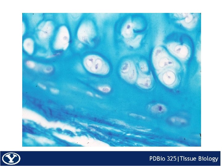
Hyaline Cartilage DCCTRA A A: Chondroblast B: Chondrocytes D B C: Nest cells D: Perichondrium-DCCTRA C
Practice
Elastic Cartilage C DECTRA A: Chondrocytes. A B: Chondroblast layer. C: Perichondrium is stained pink. B
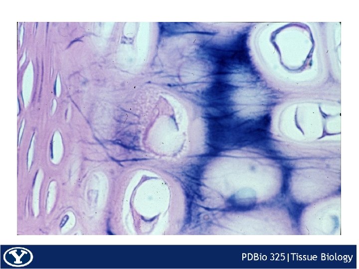
Fibrocartilage A DCCTIRA A: Transverse fibers. B: Longitudinal fibers. C: Fibroblasts. B
Digestive System
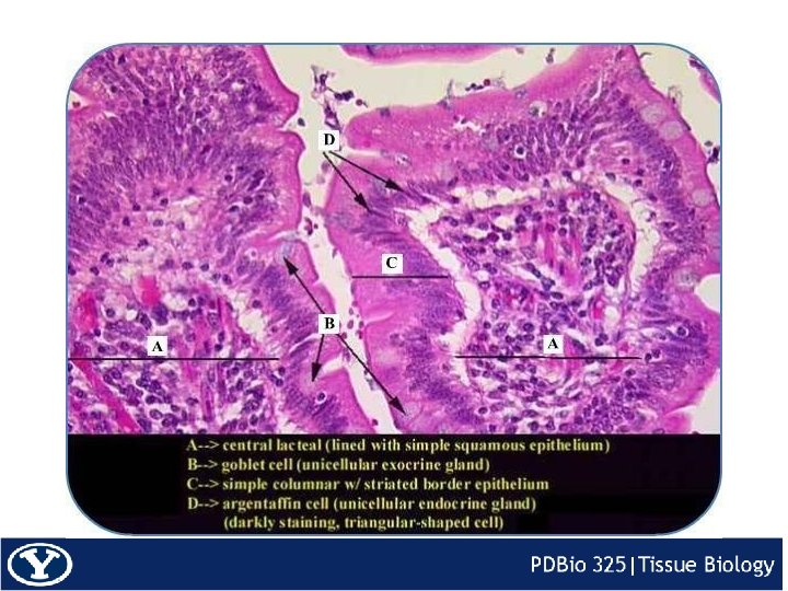
Intestines A: Lamina propria- LCT B: Muscularis mucosa C: Submucosa. A C B
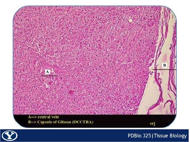
Liver: Hepatic/Glisson’s Capsule A DCCTRA A: Glisson’s Capsule surrounds the liver and is made of DCCTRA.
Musculoskeletal System
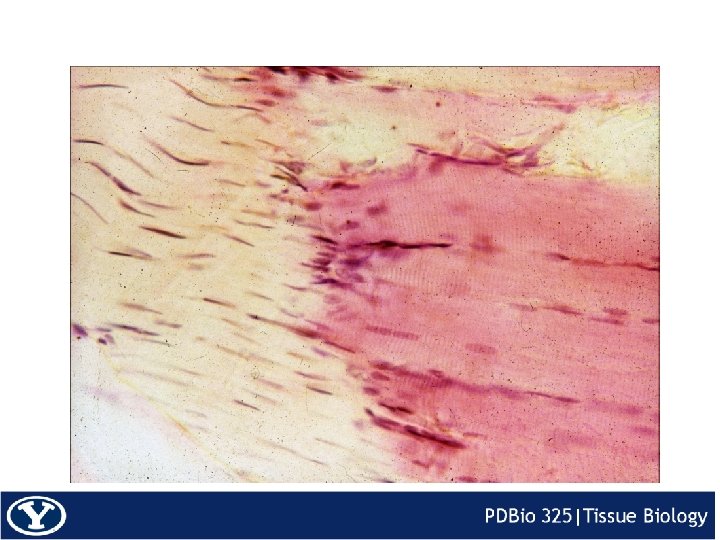
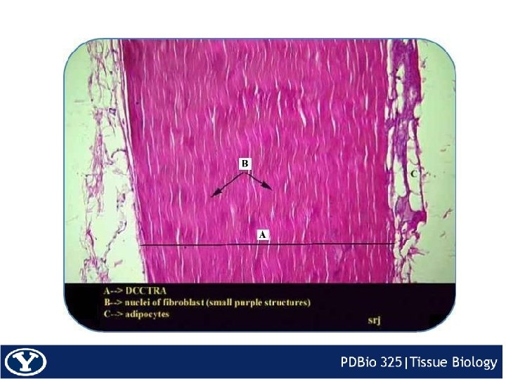
Tendon DCCTRA A: Compacted collagen fibers in regular arrangement. B: Fibroblast nuclei A B
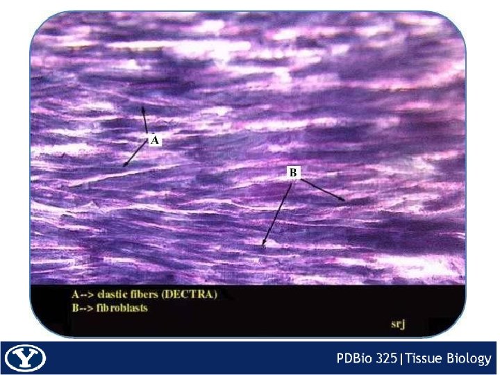
Ligament DECTRA
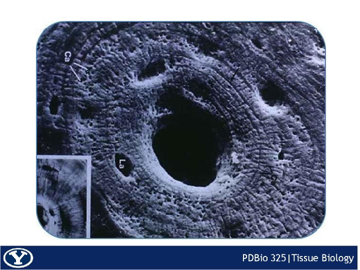
Bone 9 11 B 9. Bone: Longitudinal Cut A: Haversian canals B: Osteocytes in lacunae. A C: Canaliculi C 10. Bone: Transverse Cut A: Haversian canal. B: Concentric lamellae C: Interstitial lamellae 11. SEM of Osteon 10 B A C
Practice A C B A: concentric lamellae B: osteon C: interstitial lamellae
Endochondral Bone Formation A: Zone of Reserve– B: Zone of Proliferation– C: Zone of Hypertrophy– A B D: Zone of Calcification– E C E: Zone of Resorption– F: Perichondrium- DCCTRA G: Periosteum- DCCTRA F G D
Review: Name all the zones from right to left E D G C A B F
Clinical Connection
Osteosarcoma ● Malignant neoplasm of mesenchymal origin ● Common form of bone cancer ● Exhibits osteoblastic differentiation and produces osteoid Sources: http: //radiopaedia. org/images/24107 http: //en. wikipedia. org/wiki/Osteosarcoma
Developing Tooth A: Dental pulp. B: Odontoblast layer is simple columnar epithelium w/ dentin F C: Predentin E D D: Dentin E: Enamel F: Ameloblast layer is simple columnar epithelium w/ enamel. C B A A
Practice
Respiratory System
Trachea A: Chondroblast layer. A B: Perichondrium C: Ciliated pseudostratified columnar epithelium with LCT just below the epithelium. C B
A B C
Dermis
LCT B A: LCT of dermis A B: Keratinized Stratified squamous epithelium of the epidermis. A A
Lymphatic System
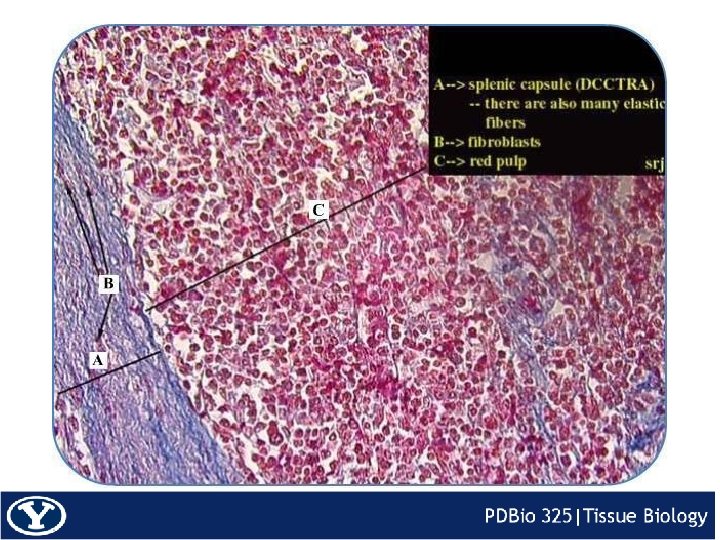
Splenic Capsule C DCCTRA with Elastic Fibers A A: Splenic capsule B B: White pulp (within the lymphatic nodules) C: Red Pulp C
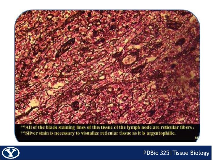
Lymph Node: Reticular Fibers Don’t worry about this too much this week! A: Reticular fibers. A B: Dense lymphatic tissue. B
Cardiovascular System
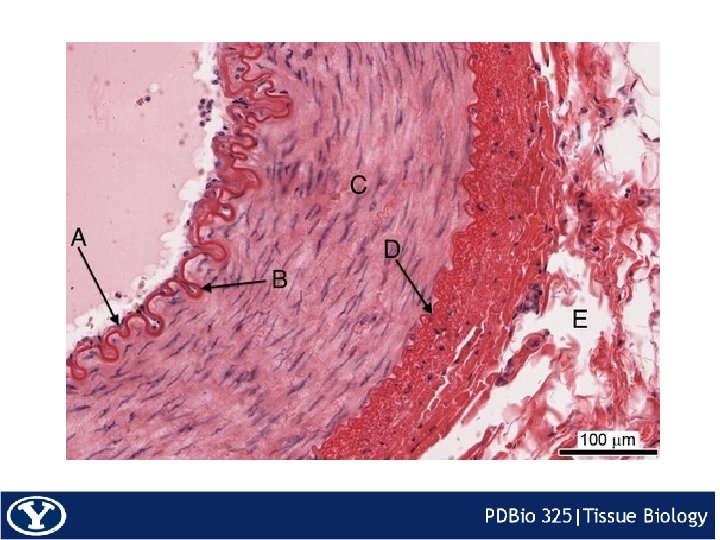
Inner & Outer Elastic Lamina A: Internal elastic lamina B: External elastic lamina B DECTIRA in diastole DECTRA in systole A *****(your slides will always be diastole) so specify the phase when writing on exams! C: Tunica Adventitia. LCT w/ some DCCTRA C
Miscellaneous
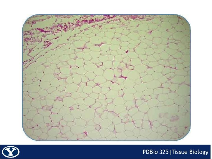
Adipose Tissue Unilocular Adipocytes A: White adipose cells A
Oviduct: Lamina Propria A: Ciliated simple columnar epithelium B: Lamina propria B A
Practice