CONNECTIVE TISSUE Dr Iram Tassaduq INTRODUCTION Connective tissue


 shows q")

 Almost as numerous as fibroblasts q Most abundant in richly vascularized areas")


 and")


- Slides: 33
CONNECTIVE TISSUE Dr Iram Tassaduq
INTRODUCTION ‘Connective tissue’ is the term traditionally applied to a basic type of tissue of mesodermal origin which provides structural and metabolic support for other tissues and organs throughout the body, also known as “Supporting tissue”.
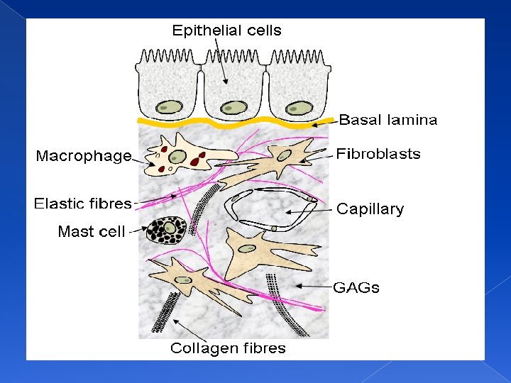
COMPOSITION q Cells q Fibers q Ground substance
FUNCTIONS Establishing a structural framework q Supporting, surrounding and interconnecting tissues q Exchange of nutrients and waste products q Storing energy reserves q Defending the body from microorganisms q Protecting delicate organs q
CLASSIFICATION CONNECTIVE TISSUE Embryonic Adult
EMBRYONIC CONNECTIVE TISSUE MESENCHYMAL MUCOUS
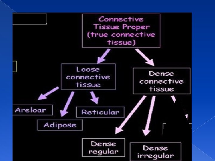
ADULT CONNECTIVE TISSUE PROPER SPECIALIZED CONNECTIVE TISSUE
SPECIALIZED CONNECTIVE TISSUE CARTILAGE ELASTI C HYALINE BONE FIBRO CARTILAGE COMPACT SPONGY
CELLS OF CONNECTIVE TISSUE q q q RESIDENT CELLS Fibroblasts Macrophages Adipose cells Mast cell Plasma cells WANDERING CELLS q Lymphocytes q Plasma cells q Neutrophils q Eosinophils q Basophils q Monocytes
FIBROBLASTS One of the two most numerous cells of C. T. q Large, flat cells with branching processes and appear fusiform in profile. q Young fibroblasts q Mature/inactive fibroblasts q
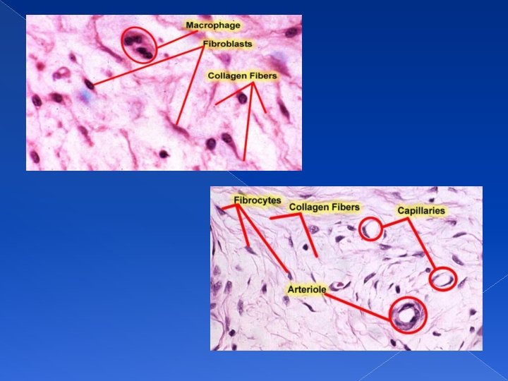
FIBROBLASTS q q Section of rat skin. A connective tissue layer (dermis) shows q several fibroblasts, which are the elongated cells. (H&E) stain. Most common cells Synthesize proteins, such as collagen and elastin that forms collagen, reticular, and elastic fibers Secrete “Ground Substance” Involved in production of “Growth Factors” Wound healing
• Fibroblasts that are actively engaged in synthesis are richer in mitochondria, lipid droplets, Golgi complex, and rough endoplasmic reticulum than are quiescent fibroblasts (fibrocytes).
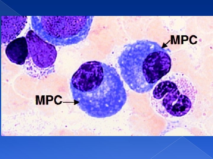
MACROPHAGES (HISTIOCYTES) Almost as numerous as fibroblasts q Most abundant in richly vascularized areas q Fixed/resting macrophages q Free/wandering macrophages q
MACROPHAGES q q q q Size 10 -30 µm Oval or kidney shaped nucleus located eccentrically Monocytes-Marophages Kupffer cells-liver, microglial cells-CNS, osteoclasts- bone tissue Phagocytosis Have well-developed Golgi complex, many lysosomes, and a prominent rough endoplasmic reticulum ‘Foreignbody giant cell’
IN RESTING PHASE Appear as irregular cells with short and blunt processes but sometimes the processes may be long, slender and branching. q Nucleus is rounder, smaller and more heterochromatic than that of fibroblast. q Cytoplasm stains darkly & may contain few small vacuoles q
IN ACTIVE STATE Cell becomes larger with bigger nucleus and prominent nucleolus q Cytoplasm becomes filled with granules and vacuoles, containing ingested material q
FUNCTIONS OF MACROPHAGES Important agent of defense and part of Mononuclear phagocyte system. q These are secretory cells that produces several important substances including enzymes and proteins of complement system q They play very important role in immune system and act as antigen presenting cells. q They are capable of motility and when suitably stimulated grouped around a large foreign body to form multinucleated foreign body giant cells q
ADIPOSE CELLS The large mesenchymal cells after accumulation of fat droplets in their cytoplasm are called adipocytes q Occur singly or in clumps along small blood vessels q
ADIPOSE CELLS q q q Also called “Fat Cells” Size 50 -150 µm Cytoplasm is dispalced to periphery by a single large fat droplet Nucleus is pressed against the cell membrane “Signet ring” Functions in energy reserves, insulation, protection, and support
TYPES OF ADIPOSE TISSUE q White or unilocular q Brown or multilocular
UNILOCULAR FAT q q q Also called white fat Widely distributed in body Main component of adult fat Single fat droplet in cytoplasm Cells show signet ring appearance
MULTILOCULAR FAT q q q Also called brown fat Mainly found in children Multiple small fat droplets Help in heat production Rich blood supply Numerous mitochondria
MAST CELLS Mast means well fed. Their cytoplasm is full of coarse granules so this name is given. q Tend to occur in small groups around blood vessels. q Irregularly oval in outline, have short pseudopodia q Nucleus is small being crowded by large number of prominent granules q
FUNCTIONS OF MAST CELLS q As these granules contain heparin (a powerful anticoagulant) and histamine (a potent vasodilator) so they play very vital role in homeostasis.
METACHROMASIA
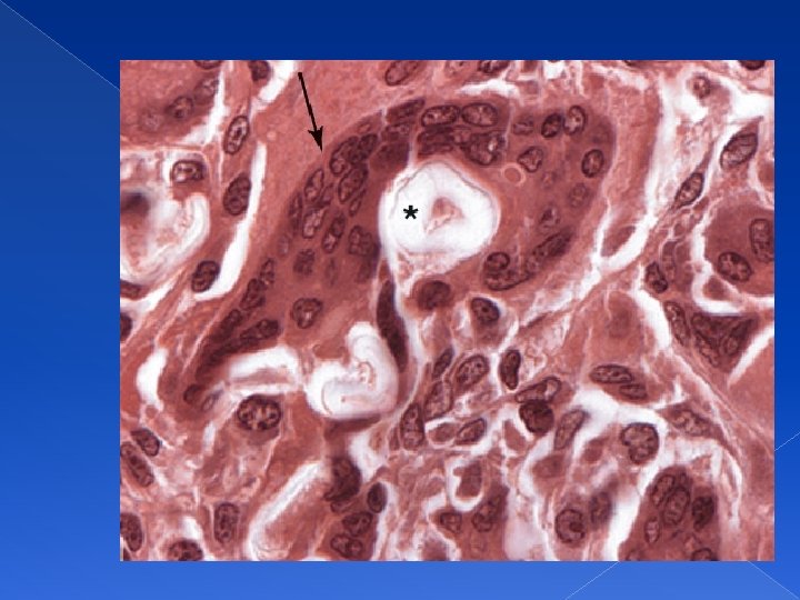
PLASMA CELLS q q q q Portion of a chronically inflamed intestinal villus. PT stain Large, ovoid cells Nucleus is spherical and eccentrically placed ‘cart-wheel appearance’ Basophilic cytoplasm due to abundant RER Rare in most connective tissues Produce antibodies Average life is short, 10– 20 days