Connective Tissue Connective tissue may be defined as

Connective Tissue

§ Connective tissue may be defined as that group of tissues predominantly composed of extracellular or intercellular material (matrix), secreted mainly by its cells, which are, therefore, usually widely spaced. § The extracellular matrix consists of fibers and amorphous ground substance. § So there are three elements of connective tissue (cells, fibers and ground substance). ALLAH Subhanahu develops connective tissue from embryonic mesoderm and, in the head region, largely from neural crest cells.

CLASSIFICATION § Primitive or undifferentiated connective tissue Mesenchymal tissue § Intermediate or maturing connective tissue Mucoid tissue § Adult or Mature connective tissue

§ Mature Connective Tissue is mainly of three types § Soft, § Hard and § Fluid-like

§ Soft Connective tissue is again of two types § Loose connective tissue § Dense connective tissue
 §")
Classification of Loose connective tissue § Ordinary § Areolar (Loose Areolar connective tissue) § Cellular (Cellular connective tissue) § Vascular (Vascular connective tissue) § Special § § Adipose Reticular Lymphoid Pigmented
 § Regular (Dense")
Classification of Dense connective tissue § Irregular (Dense Irregular connective tissue) § Regular (Dense Regular connective tissue) § White fibrous tissue § Yellow elastic tissue
 § § Cartilage Hyaline Bone Non-Articular Elastic Fibrous Compact Spongy")
Hard (Skeletal connective tissue) § § Cartilage Hyaline Bone Non-Articular Elastic Fibrous Compact Spongy or Cancellous
 Bone marrow or Myeloid tissue Blood")
§ Fluid like (Haemopoietic connective tissue) Bone marrow or Myeloid tissue Blood

§ CLASSIFICATION OF CONNECTIVE TISSUE § Primitive or undifferentiated connective tissue Mesenchymal tissue § Intermediate or maturing connective tissue Mucoid tissue § Adult or Mature connective tissue § Mature Connective Tissue is mainly of three types Soft, Hard and Fluid-like § Soft Connective tissue is again of two types § Loose connective tissue § Dense connective tissue § Classification of Loose connective tissue § Ordinary Areolar (Loose Areolar connective tissue) § Cellular (Cellular connective tissue) § Vascular (Vascular connective tissue) § Special Adipose § Reticular § Lymphoid § Pigmented
")
§ § § § Classification of Dense connective tissue Irregular (Dense Irregular connective tissue) Regular (Dense Regular connective tissue) White fibrous tissue Yellow elastic tissue Hard (Skeletal connective tissue) Cartilage Hyaline Non-Articular Elastic Fibrous Bone Compact Spongy or Canccllous Fluid like (Haemopoietic connective tissue) Bone marrow or Myeloid tissue Blood

COMPONENTS OF CONNECTIVE TISSUE § There are three components of connective tissue: 1. 2. 3. Cells Fibers Ground substance

Cells § § § § § Mesenchymal cells Fibroblasts Macrophages Plasma cells Mast cells Fat cells Reticular cells Lymphocytes Pigment cells Fibrocytes

Fibers § Following types of fibers are found in different types of connective tissues. 1. Collagen fibers 2. Reticular fibers 3. Elastic fibers

Mesenchymal connective tissue § It is also called Mesenchymal connective tissue or simply mesenchyme. § In fact it is nothing but embryonic mesoderm, may be extraembryonic or intraembryonic mesoderm. § It is typical unspecialized connective tissue of early weeks of embryonic life. § It is composed of mesenchymal cells and matrix.

§ Mesenchymal cells have branching processes. § They often appear to join those of neighboring cells, although they do not form a true syncytium. § Ground substance is a coagulable fluid in the earliest stages but later contains fine reticular fibers. § So matrix consists of ground substance and fine reticular fibers. § The mesenchymal cells and reticular fibers are gradually replaced by mature types of cells (e. g. fibroblasts) and collagen fibers respectively as the mesenchyme develops and differentiates into the adult connective tissue.

Undifferentiated Mesenchymal cells: § They are small and stellate in shape, usually located along the walls of blood vessels, particularly capillaries, where they are referred to as perivascular or adventitial cells. § They can differentiate into the usual cell types found within loose connective tissue or into smooth muscle cells of blood vessels when required.

Mucoid tissue § It is also called mucoid or mucous connective tissue. § It is an intermediate type of connective tissue, found chiefly as a stage in the development of connective tissue from mesenchyme to the adult type. § It also exists as Wharton's jelly, which form the bulk of umbilical cord, where it does not differentiate further.

§ It consists of a copious matrix and large stellate cells called fibroblasts. § The matrix is largely made up of soft and jelly like ground substance and a fine meshwork of collagen fibers. § Usually few fibers occur in typical mucoid tissue, though at birth the umbilical cord shows a considerable development of perivascular collagen fibers.

§ Fibroblasts have oval nuclei and branching processes often appear to fuse with those of neighboring cells. A few macrophages and wandering lymphocytes are occasionally found. § SITES: In addition to umbilical cord, after birth, mucoid tissue is still to be seen in the pulp of a developing tooth. In the adult the vitreous body of the eye and the nucleus pulposus of the intervertebral disc are persistent forms of mucoid tissue in which the fibers and cells ore very few in number,
 of the umbilical")
§ Umbilical Cord The two arteries and one vein (upper left) of the umbilical cord are surrounded by a large amount of connective tissue. § The proportion of "ground substance" is higher in this tissue, and it is sometimes called "Wharton's jelly".
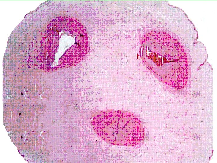

§ Umbilical Cord A closer look shows the preponderance of ground substance (largely glycoproteins and proteoglycans) and relatively few cells.

Loose connective tissues § It is formed by direct differentiation of mesenchyme. § It is loosely arranged and fibroelastic. § It binds other tissues, organs and their components together. § Owing to its flexibility, it allows a considerable degree of mobility between different structures and organs.

§ It contains fibroblasts, macrophages, reticular cells, plasma cells, mast cells, and lymphocytes. § Neutrophils and eosinophils are also seen. § Collagen fibers are most prominent. § Elastic fibers form a continuous branching network. § Reticular fibers are present in the loose connective tissue that borders upon other structures.

§ Loose connective tissue derives its name from the fact that its fibers are loosely arranged. The cells and fibers are embedded in fluid like ground substance. § SITES: Loose connective tissue is widely distributed in the body. In fact it occupies any unoccupied space in the body.

§ Loose connective tissue is of two types: § Ordinary loose connective tissue § Special loose connective tissue

§ Ordinary loose connective tissue: § There are three varieties of ordinary loose connective tissue.

Areolar connective tissue: § The name areolar is descriptive of the general appearance produced by small spaces, which contain only an amorphous ground substance, which is usually not seen because it is washed off in routine preparations. Here the most common cells are fibroblasts and macrophages. Fat cells (adipocytes) are seen in small groups, particularly around blood vessels. Reticular cells, plasma cells and mast, cells are also seen. Lymphocytes are scattered throughout the areolar tissue.

SITES: § It forms all the superficial fascia and part of the deep fascia e. g. cribriform fasia. § It also forms Submucosa and mucosa (lamina propria) of digestive, respiratory and urogenital tracts. § It also covers these tracts. § Here it is in the forms of fibrosa or subserous connective tissue. § Fat free subcutaneous connective tissue as in eyelids, penis, scrotum and labia is also areolar connective tissue.

§ It is found between and around muscles, nerves and blood vessels, particularly endomysium and endoneurium. § Similarly it is also found around and in the tendons, aponeurosis and ligaments. § It is present underneath the endothelium as subendothelial connective tissue and forms the adventitia of the blood vessels.

§ It covers certain organs such as pancreas, kidneys etc. § It divides the organs and glands into lobes and lobules and even present within the lobules. § Here it is called Interlobar, interlobular and intralobular connective tissue.

Cellular connective tissue: § When the cellular component increases the loose areolar connective tissue becomes cellular connective tissue. § SITES: § Cortex of the ovary

Vascular connective tissue: § When the blood vessels increase in a loose areolar connective tissue, the tissue is given the name vascular connective tissue. § SITES: § Medulla of the Ovary § In fact in a loose connective tissue according to the requirements ALLAH subhanahu Taala increases the cellular element or vascular element, human beings name it cellular connective tissue or vascular connective tissue accordingly.

§CELLS OF LOOSE CONNECTIVE TISSUE

Fibroblasts § These are one of the two most numerous cells of areolar connective tissue, the other being macrophages or Histiocytes. Fibroblasts, as their name suggests, are considered to be responsible for the formation of the fibers and also are thought to elaborate most, if not all, of the amorphous component of the matrix (principally glycosaminoglycans ).

§ They are large, flat, branching cells, which appear fusiform, or spindle shaped in profile. § The branching processes are slender. In most histological preparations, the outlines of the cells are indistinct. § The nucleus is deeply stained with basic dyes. § It is oval or elongated with one or two nucleoli and finely granular chromatin.

§ In young fibroblasts, which are actively engaged in protein synthesis the high concentration of granular endoplasmic reticulum. § Mitochondria appear as slender rods and are most numerous near the nucleus. § Golgi apparatus is also present close to the nucleus. § Microtubules are also present and seem to be required for translocation of secretary vesicles.

Fibrocytes § The old and relatively inactive fibroblasts are given the name fibrocytes. § Here the cytoplasm is sparse and only weakly basophilic, the endoplasmic reticulum is scanty. § The nucleus is flattened and heterochromatic (close-faced).

Macrophages § ALLAH Subhanahu Taala has placed important agents of defense in the body called macrophages. § They are almost as numerous as fibroblasts in loose connective tissue and are more abundant in richly vascularized areas.

§ Generally they are irregular shaped cells with processes, which usually are short and blunt. § Occasionally they may exhibit long, slender branching processes. § They are relatively larger cells (15 -20 um in diameter). Their nuclei are ovoid, usually indented and heterochromatic. § Nucleolus is present but not so conspicuous. § The cytoplasm is mildly basophilic and typically has a frothy appearance under the light microscope. § Ultrastructurally, macrophages contain numerous lysosomes, which digest ingested material.

Macrophage

§ The macrophage is derived from the blood-borne monocyte, which migrates into tissue and differentiates into this phagocytic cell. § Here it is stained blue after gobbling up trypan blue stain. § Look for the irregular cell border and numerous phagosomes.

§ ALLAH Subhanahu Ta`ala has enabled macrophages to multiply mitotically to some extent, but he develops them largely from haemopoeitic stem cells in the bone marrow, circulates in the blood as monocytes before sending them to their final extravascular sites through the venule walls.

§ There are two types of macrophages: 1. Stationary or fixed macrophages: Attached to fibers of the matrix. They are irregular with many filopodia. 2. Motile or nomadic macrophages: Free with in the matrix. They are of a more rounded and regular form.

§ Macrophages are important phagocytes, forming part of the macrophage system. They are also able to dispose of dead or moribund cells prior to tissue regeneration. Because of their mobility and phagocytic activity, they are able to act as scavengers, engulfing extravasated blood cells, dead cells, bacteria, and foreign bodies.

Fat cells § There are two types of fat cells: § Unilocular fat cells § Multilocular fat cell.

Mast cells § ALLAH Rab-ur-Ezzat has blessed us with very important type of defense cells called mast cells. § They are widely distributed in loose connective tissues and often preset in the fibrous capsules of certain organs such as liver. § They are characteristically numerous in relations to blood vessels and nerves.

§ Mast cells are irregularly oval in outline, about 12 um in diameter, with short pseudopodia, an indication of their slow mobility. § The nucleus is centrally placed and relatively small. § They are easily identified by large number of prominent Cytoplasmic granules. § Mast cells also contain a well developed Golgi apparatus and scanty endoplasmic reticulum.

§ The Cytoplasmic granules or vesicles are water soluble and exhibit metachromasia (staining a color different from the dye being used) with basic dyes such as toludine blue, methyline blue, alcian blue and azure A. § These basic dyes instead of giving usual basic dyes colors produce. § The normal basic dyes usually stain blue like colors. The acidic dyes stain red like colors. § Instead of giving blue colors the basic dyes here give purple red color to Cytoplasmic granules. § This metachromatic response of these Cytoplasmic granules.
. §")
§ The granules also show a strongly positive reaction with periodic acid-Schiff PAS). § Electron micrographs show that a unit membrane bound the granules and their average diameter is 0. 5 um. § Thegranules usually contain dense osmiophilic material, which may be finely granular, lamellar, or in the form of membranous whorls; these variants may coexist in the same vesicle, which may also present, in places a crystalline substructure. § For these reasons they have sometimes been termed compound granules.

Mast Cells

§ The numerous granules of the mast cells take up the toluidine blue stain and appear more "purple" than other stained cells. § Histamine released from the mast cell plays a key role in inflammation.

Plasma cells § These cells are found frequently in § serous membranes and § lymphoid tissue and § are plentiful in sites of chronic inflammations.

§ ALLAH Subhanahu has made its nucleus rounded and placed it eccentrically in a basophilic cytoplasm. § The chromatin material is arranged like the spokes of a wheel or hours on the clock. § So nucleus presents a cartwheel or clock face appearance.

§ There is a lighter stained area in the cytoplasm close to the nucleus containing Golgi apparatus and centriole. § ALLAH Subhanah has provided these cells an extensive rough endoplasrnic reticulum for the production of antibodies. § Occasionally acidophil inclusions called Russell bodies are seen in the cytoplasm.

Plasma Cells

§ Plasma cells are easily distinguished by their oval shape and large, offset nucleus with chromatin clumped in a "clockface" pattern, and an adjacent pale patch of clear cytoplasm. § Plasma cells are derived from Blymphocytes and secrete antibodies.

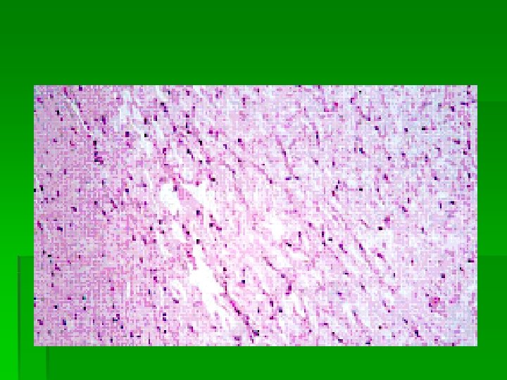
Collagen fibers: § These are found in all types of connective tissue and consist of protein collagen. § In the fresh state, e. g. in tendons, they appear white, and hence also are termed “white” fibers.

§ They are extremely tough and tend to form bundles. § Within a bundle, fibers are held together by a small amount of amorphous cement substance (mucoprotein). § Individual collagen fibers vary from 1 to 12 um in diameter. § The fibers are of indeterminate length and have a straight or wavy course.

§ They are soft and flexible, relatively inelastic, and have high tensile strength. § They are transparent and homogeneous but show a faint longitudinal striation. § It is found under higher magnifications that they are composed of smaller fibers held together by amorphous material. These smaller fibers are called fibrils. There diameter is 0. 2 to 0. 5 um. These fibrils are aligned in a parallel direction, giving the appearance of a longitudinal striation.

§ Collagen is a protein, which stains with most acid dyes. Hence the fibers are stained pink/red with eosin, red by van Gieson’s picrofuchsin and blue by aniline blue of Mallory’s connective tissue. § They are stained green or blue in Masson’s trichrome stain depending upon the modification used I. e. methylene blue or green. § They are stained yellow or brown after silver impregnation (Bielchowsky’s method). § Collagen fivers give a week reaction to periodic acid-Schiff (PAS) technique.

Collagen Fibers
 stain leaves collagen green or blue. § In the skin,")
§ The Masson (trichrome) stain leaves collagen green or blue. § In the skin, Type 1 collagen predominates, as shown by the thick, wavy bundles.

§ Electron micrographs show that each of the collagen fibril is composed of still smaller fibrils called microfibrils. § The later are relatively uniform in diameter in any given connective tissue e. g. in adult human dermis, they are about 100 nm in diameter throughout dermis: but vary in different locations and in different stages of development, ranging from about 20 to 200 nm.

§ Studies using autoradiography, electron micrography and sophisticated biochemical techniques have shown that collagen microfibrils are composed of collagen molecules called tropocollagen. Each molecule is approximately 280 nm long and 1. 5 nm wide. Alignment of these macromolecules is end-toend, but not touching, and with the molecules in adjacent rows arranged in parallel but staggered about one-fourth of their length. This gives rise to the 64 -nm periodicity of collagen commonly seen by electron microscopy.

Elastic Fibers

§ Sheets of elastic fibers, called elastic lamellae, are common in the aorta, shown here. § These lamellae give a distinctive refractive appearance when you focus through them.

Reticular Fibers

§ Reticular fibers are composed of type III collagen, and appear black with a silver stain. § In many organs and basement membranes, they provide a supportive framework.

Loose Connective Tissue
 is a \"space")
§ Loose CT in the duodenum (see the Brunner's glands? ) is a "space filler" that provides the flexibility the GI tract needs. § Many of the nuclei present are probably fibroblasts, but other cell types common in loose CT include plasma cells, mast cells, and lymphocytes.

Loose Connective Tissue

§ Loose CT is also found in the mesentary (surrounding the blood vessels and Pacinian corpuscle), and in addition to the cell types mentioned previously, loose CT may include adiopocytes.

Dense Irregular CT

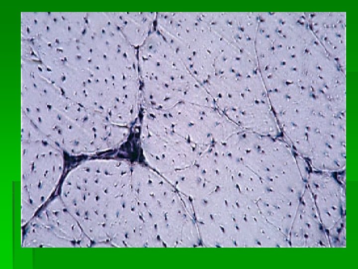
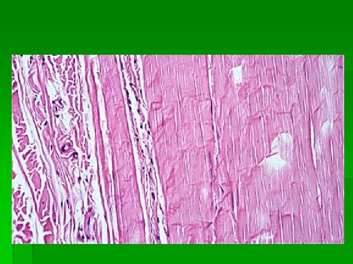
Tendon § In cross section, collagen fibers make up the pale pink background. § The fine lines separate fiber bundles; numerous fibroblast nuclei can be seen.

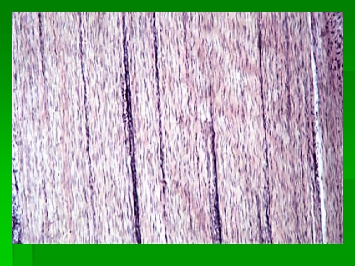
§ Tendon Longitudinal, wavy fibroblasts run along the tendon. Recall that the collagen and elastic fibers of the tendon are extracellular.

§ Tendon Here the tendon has far fewer fibroblasts. Notice the presence of skeletal muscle marks the point of insertion.

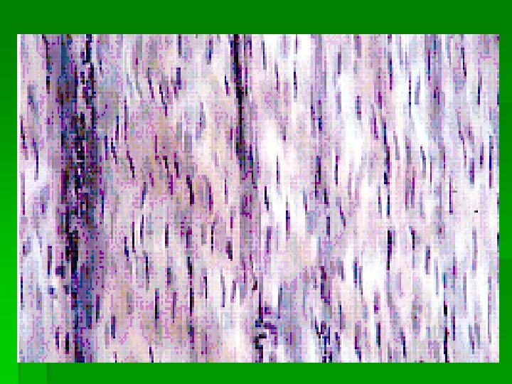
§ Tendon Typically the tendon is characterized by fibroblasts in regular, parallel arrangements, as shown here. § These are often described as "boxcars" in a line (at least by railroad aficionados). Look Alikes: § Smooth Muscle, Skeletal Muscle, Nerve.
- Slides: 84