Connective Tissue 1132020 Dr Shatarat 1 Connective Tissue


.")


 specialized to store fat. 2 types:")


 • contains a resilient gel as ground substance.")


- Slides: 22
Connective Tissue 11/3/2020 Dr. Shatarat ﺍﻣﺠﺪ. ﺩ ﺍﻟﺸﻄﺮﺍﺕ 1
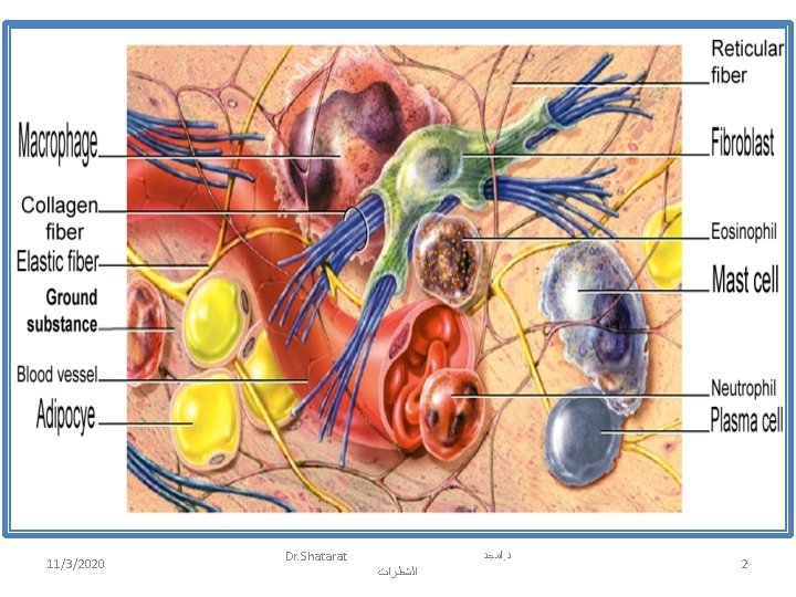
Connective Tissue Functions: Gives, maintains and supports shape of the body and its structures. Variations in the composition and amount of the 3 components are responsible for the typing of connective tissue. Has 3 components: 1. Cells. 2. Fibers. 3. Ground substance. 11/3/2020 Dr. Shatarat ﺍﻣﺠﺪ. ﺩ ﺍﻟﺸﻄﺮﺍﺕ 3
Cells of the Connective Tissue Cells are found at different stages: – Immature cells e. g. Fibro/blast (blast= to bud). • Maintain the capacity for cell division. • Secrete the extracellular matrix. • Can differentiate into mature cells Mature cells e. g. . Fibro/cyte (cyte= cell) Decrease capacity for cell division and matrix formation. maintain the extracellular matrix. 11/3/2020 Dr. Shatarat ﺍﻣﺠﺪ. ﺩ ﺍﻟﺸﻄﺮﺍﺕ 4
Connective Tissue Cells 1. Fibroblasts. Originate locally from mesenchymal cells (embryonic cells of CT). The most common and numerous cells in connective tissue. Functions: Synthesis of fibers. Synthesis ground substance. Scar formation in healing of wounds 11/3/2020 Dr. Shatarat 5
The macrophages, plasma cells and mast cells originate from stem cells in the bone marrow. Remember 2. Macrophages: (large eaters) – Defense, by phagocytosis of foreign substances and bacteria. 3. Plasma cells: – Production of antibodies ( important for immunity). 4. Mast cells: – Plenty alongside blood vessels. – Produce histamine as reaction to infection. • Histamine increase vascular permeability that is important in inflammation. • Promote the allergic reactions. e. g. . Flu, Hay fever. 11/3/2020 Dr. Shatarat ﺍﻣﺠﺪ. ﺩ ﺍﻟﺸﻄﺮﺍﺕ 6
5. Adipocytes: § Fat cells that store triglycerides. § Specialized for storing energy and heat production. 6. White blood cells: • They migrate from blood into CT in response to certain conditions. • They produce immunocompetent (skilled resistant) cells. As in infection and allergy. 11/3/2020 Dr. Shatarat ﺍﻣﺠﺪ. ﺩ ﺍﻟﺸﻄﺮﺍﺕ 7
Extracellular Matrix • • It fills the space between the cells. Has 2 major components: 1. Fibers: (collagen, reticular, and elastic) 2. Ground substance: • • • A mixture of macromolecules that fills the space between cells and fibers. May be fluid, semifluid or calcified. Extracellular matrix consist of different combinations of protein fibers + ground substance that instruct strength and rigidity to the matrix. 11/3/2020 Dr. Shatarat ﺍﻣﺠﺪ. ﺩ ﺍﻟﺸﻄﺮﺍﺕ 8
CT Fibers They function to strengthen and support connective tissues. • Collagen fibers: – The most abundant protein in the human body. – Organize in parallel bundles. – Very strong and resist tension, but allow flexibility. – Found in bone, cartilage, tendons to and ligaments. 11/3/2020 Dr. Shatarat ﺍﻣﺠﺪ. ﺩ ﺍﻟﺸﻄﺮﺍﺕ 9
CT Fibers • Elastic fibers: – Has small diameter. – Form a network within a connective tissue. – Possess elasticity, ability to return to its original shape after being stretched. – Found in skin, wall of blood vessels, and lung tissue. 11/3/2020 Dr. Shatarat ﺍﻣﺠﺪ. ﺩ ﺍﻟﺸﻄﺮﺍﺕ 10
CT Fibers • Reticular fibers: – Produced by fibroblasts. – Form of collagen arranged in fine bundles. – Form branching networks that support soft organs, such as the spleen, liver and lymph nodes. 11/3/2020 Dr. Shatarat ﺍﻣﺠﺪ. ﺩ ﺍﻟﺸﻄﺮﺍﺕ 11
Classification of CT A. Loose connective tissues 1. Areolar connective tissue 2. Adipose tissue 3. Reticular connective tissue B. Dense connective tissues 1. Dense regular connective tissue 2. Dense irregular connective tissue 3. Elastic connective tissue C. Cartilage 1. Hyaline cartilage 2. Fibrocartilage 3. Elastic cartilage D. Bone tissue E. Liquid connective tissue 1. Blood tissue 2. Lymph 11/3/2020 Dr. Shatarat ﺍﻣﺠﺪ. ﺩ ﺍﻟﺸﻄﺮﺍﺕ 12
1. Areolar connective tissue areol- = small space • Widely distributed CT • Contains numerous cells + 3 types of fibers embedded in semifluid ground substance. • Location: In and around of most body structures. Like, – Subcutaneous layer deep to skin. – Around body organs. 11/3/2020 Dr. Shatarat ﺍﻣﺠﺪ. ﺩ ﺍﻟﺸﻄﺮﺍﺕ 13
2. Adipose tissue Consists of adipocytes (fat cells) specialized to store fat. 2 types: – White fat in adults. – Brown fat in newborn. Functions: – Good insulators (reduce heat loss). – Major store of energy. 11/3/2020 Dr. Shatarat ﺍﻣﺠﺪ. ﺩ ﺍﻟﺸﻄﺮﺍﺕ 14
3. Reticular connective tissue Consists of thin network of reticular fibers and reticular cells. Location: Form the supporting framework of liver, spleen and lymph nodes. Functions: § Forms stroma of organs; § Connect smooth muscle tissue cells. § Filters and removes worn-out blood cells in spleen and microbes in lymph nodes. 11/3/2020 Dr. Shatarat ﺍﻣﺠﺪ. ﺩ ﺍﻟﺸﻄﺮﺍﺕ 15
Dense connective tissue Ø Contains more numerous, thicker and dense fibers but fewer cells than loose CT. Ø Has 3 types: 1. Dense regular CT. § Has collagen fibers regularly arranged in bundles with fibroblasts in rows between bundles. § Locations: Tendons and ligaments. § Function: § Give great strength. § Resist tension along axis of fibers. 11/3/2020 Dr. Shatarat ﺍﻣﺠﺪ. ﺩ ﺍﻟﺸﻄﺮﺍﺕ 16
2. Dense irregular CT. • Has irregularly arranged collagen fibers and few fibroblasts. • Found where pulling forces occurs in different directions. E. g. . – Dermis of skin – Pericardium – Periosteum • Function: Provides strength. 11/3/2020 Dr. Shatarat ﺍﻣﺠﺪ. ﺩ ﺍﻟﺸﻄﺮﺍﺕ 17
3. Elastic CT. • Elastic fibers predominate. With fibroblasts between fibers. • Location: Lung tissue, walls of elastic arteries, and trachea. • Function: Strong and can recoil to original shape after being stretched. 11/3/2020 Dr. Shatarat ﺍﻣﺠﺪ. ﺩ ﺍﻟﺸﻄﺮﺍﺕ 18
Cartilage • Consist of a dense net of collagen fibers and elastic fibers embedded firmly in a gel-like part of the ground substance. • Has few cells and large quantities of extracellular matrix. • Resilience, has the ability to return its original shape after pressure. • Has no nerves. • Has no blood supply poor healing after an injury. • Chondrocytes: Mature cartilage cells found singly or in groups within spaces called lacunae =small lakes. • Perichondrium (Peri= around): – A dense irregular connective tissue membrane covers the surface of most cartilage. – Contains blood vessels and nerves. – The source of new cartilage cells. • Has 3 types: Hyaline, Fibrocartilage and Elastic cartilage. 11/3/2020 Dr. Shatarat ﺍﻣﺠﺪ. ﺩ ﺍﻟﺸﻄﺮﺍﺕ 19
1. Hyaline cartilage (hyalinos = glassy) • contains a resilient gel as ground substance. • Most abundant cartilage in body. – Ends of long bones, nose, parts of larynx, trachea. • Function: – Provides smooth surfaces at joints. – Affords flexibility, and support. 11/3/2020 Dr. Shatarat ﺍﻣﺠﺪ. ﺩ ﺍﻟﺸﻄﺮﺍﺕ 20
2. Fibrocartilage • Chondrocytes spread within thick bundles of collagen fibers inside extracellular matrix. • No perichondrium. • Location: Intervertebral discs, Pubic symphysis and menisci cartilage of knee. • Function: • The strongest type of cartilage due to its strength and rigidity. 11/3/2020 Dr. Shatarat ﺍﻣﺠﺪ. ﺩ ﺍﻟﺸﻄﺮﺍﺕ 21
3. Elastic cartilage • Chondrocytes located within fine network of elastic fibers in the matrix. • Has perichondrium. • Location: Epiglottis, auricle of ear. • Function: – Provides strength and elasticity; maintains shape of some structures as the external ear. 11/3/2020 Dr. Shatarat ﺍﻣﺠﺪ. ﺩ ﺍﻟﺸﻄﺮﺍﺕ 22