Connective tisse By Dr PARDEEP KUMAR Connective tissue

Connective tisse By Dr. PARDEEP KUMAR

Connective tissue Groups of elements derived largely from embryonic mesoderm & composed of cells, fibres & amorphous ground substance Originate from embryonic tissue called mesenchyme Most diverse and abundant type of tissue Function: to protect, support and bind together other tissues • Bones, ligaments, tendons • Areolar cushions; adipose insulates and is food source • Blood cells replenished; body tissues repaired Cells separated from one another by large amount of nonliving extracellular matrix

Connective Tissues Components • Specialized cells • Extracellular matrix • Protein fibers • Fluid phase (the ground substance)

Components of Connective Tissue Cells Extra Cellular Material/Matrix A Cells • Fibroblast & fibrocytes • Macrophages & histocytes • Plasma cells • Mast Cells • Fat Cells • Reticular cells • Lymphocytes • Chondroblast & Chondrocytes • Neuroglial Cells • Osteoblasts & Osteoclasts • RBC, WBC & Platelets 4

B Matrix/ Extracellular substances Nonliving material between cells Produced by the cells and then extruded Responsible for the strength Two components 1. Ground substance ¨ Of fluid, adhesion proteins, proteoglycans ¨ Liquid, semisolid, gel-like or very hard 2. Fibers: collagen, elastic or reticular 5

Ground Substance Amorphous, colloidal, gel like material of various viscosity which the fibres & cells of connective tissue are embedded Water, salts and other low molecular substances are contained within the ground substance, but its main structural constituent are • Glycosaminoglycans [95%] (hyaluronic acid) • Glycoprotein [5%] 6

Connective Tissues Functions • • Structural framework Fluid and solute transport Physical protection Tissue interconnection Fat storage Microorganism defense Repair and healing

Connective Tissues Classifying Connective Tissues • Connective tissue proper • Fluid connective tissues • Supporting connective tissues

Connective Tissues Major Types of Connective Tissue Figure 4 -7

Connective Tissues Connective tissue proper Contains varied cell populations and fiber types surrounded by a syrupy ground substance

Connective Tissues Connective Tissue Proper • Resident and migrating cells • Fibroblasts • Macrophages • Fat cells • Mast cells • Other white cells

Connective Tissues Connective Tissue Proper • Protein fibers • Collagen fibers • Provides toughness • Reticular fibers • Supports cells • Elastic fibers • Provides resilience

Connective Tissues Cells and Fibers of Connective Tissue Proper Figure 4 -8

Connective Tissues Connective Tissue Proper • Three types • Loose connective tissue • Example: beneath dermis of skin • Adipose tissue • Dense connective tissue • Examples: dermis, tendons, ligaments Copyright © 2007 Pearson Education, Inc. , publishing as Benjamin Cummings

LOOSE AREOLAR CONNECTIVE TISSUE Consists of loosely arranged fibres and cells in ground substance Areolar, because there are spaces in between the loosely arranged fibres. The spaces are filled with ground substance. The loosely arranged fibres are- collagen fibres, elastic fibres and reticular fibres. The cells are fibroblasts and fibrocytes, histocytes, macrophages, mast cells, reticular cells, plasma cells and fat cells etc. 15
")
Connective Tissues Loose Connective Tissue Figure 4 -9(a)
")
Connective Tissues Adipose Tissue Figure 4 -9(b)

ADIPOSE TISSUE 1. consists mainly of fat cells 2. Intercellular substance is scanty. 3. The cells may remain singly or in groups. 4. Irregularly arranged collagen, elastic or reticular fibres surround the cells. 18

Functions of adipose tissue: Fat is a source of good energy and may be transformed to glycogen in times of need. Acts as a shock absorber Fat conserves body heat, because it is non conductor of heat. 19
")
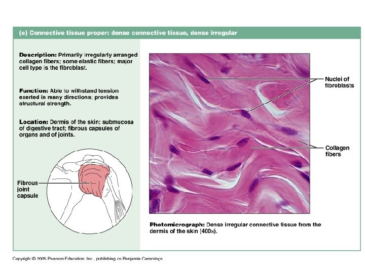
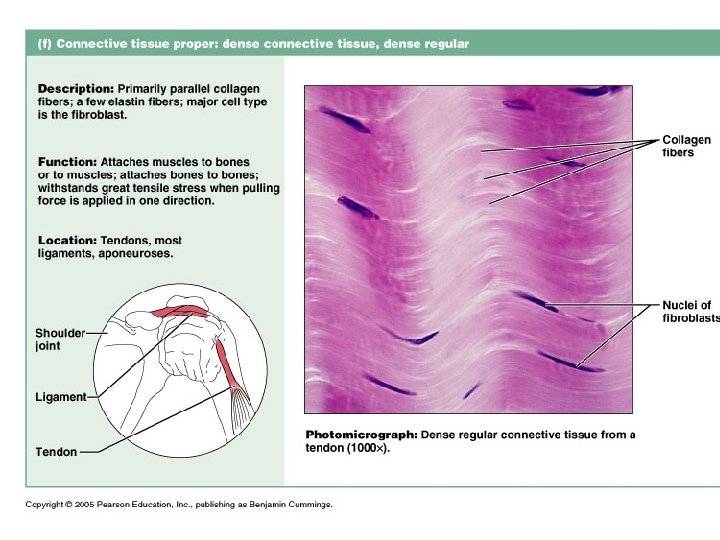
Connective Tissues Dense Connective Tissues Figure 4 -9(c)

Dense Connective Tissue It contains mainly fibres with some cells & a little ground substance. 1. White Fibrous Tissue It is dense connective tissue. It appears white in Colour where collagen fibres predominate. Cells are fewer in relation to loose areolar tissue 21

Two Types Regular: Collagen Bundles arranged in Parallel sequence Distribution • Tendon, Ligament, Aponeurosis Irregular: Collagen bundles arranged in an intertwining network Distribution • Dermis of Skin • Fibrous capsule of Liver, lymph Node, Testes • Sub mucous of Digestive Tract • Perichondrium, Periosteum 22

Connective Tissues Fluid Connective Tissues • Cells + a liquid ground substance • Blood • RBCs, WBCs, platelets + plasma • Lymphocytes + lymph fluid
 •")
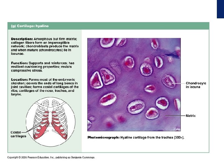
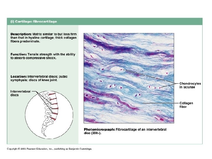
Connective Tissues Supporting Connective Tissue • Support the body • Bone (osseous tissue) • Osteocytes + collagen + calcium salts • Cartilage • Chondrocytes + firm gel

Connective Tissues Supporting Connective Tissue • Cartilage • Avascular • Covered by a fibrous perichondrium • Three types of cartilage • Hyaline cartilage • Elastic cartilage • Fibrocartilage

Connective Tissues Bone Figure 4 -11
 • Matrix provides unique properties • Collagen fibers resist")
Connective Tissues Bone (Osseous Tissue) • Matrix provides unique properties • Collagen fibers resist bending • Calcium salts resist compression • Diffusion through canaliculi nourishes osteocytes • Covered by periosteum Copyright © 2007 Pearson Education, Inc. , publishing as Benjamin Cummings

Membranes • A thin, flexible layer of tissue that covers, lines, separates, or connects cells or parts of an organism. Membranes are usually made up of • Lipids (cholestrol, phospholipid) • Protein • Carbohydrate (glycoprotein) • membranes are permeable to water and fat-soluble substances.

Membranes • Membranes that combine epithelial sheets plus underlying connective tissue proper

Membranes Types of Membranes • Mucous • Lines cavities that connect to exterior • Mucus moistens surface • Examples: oral cavity, airways Serous • Line internal cavities • Watery fluid moistens surface • Example: peritoneal membrane

Membranes • Cutaneous • Covers body surface • Dry surface waterproofs the body • Example: the skin • Synovial • Lines joints • Secretes slippery synovial fluid • Lubricates joints • Examples: knee, elbow

Membranes Figure 4 -12

Muscle Tissue Properties of Muscle Tissue • Capable of contraction • Actin filaments • Myosin filaments

Overview of Muscular Tissue • Functions of Muscular Tissue • Producing Body Movements • Walking and running • Stabilizing Body Positions • Posture • Moving Substances Within the Body • Heart muscle pumping blood • Moving substances in the digestive tract • Generating heat • Contracting muscle produces heat • Shivering increases heat production

Overview of Muscular Tissue • Types of Muscular Tissue • The three types of muscular tissue • Skeletal • Cardiac • Smooth • Skeletal Muscle Tissue • So named because most skeletal muscles move bones • Skeletal muscle tissue is striated: • Alternating light and dark bands (striations) as seen when examined with a microscope • Multinucleated cell • Skeletal muscle tissue works mainly in a voluntary manner • Its activity can be consciously controlled • Most skeletal muscles also are controlled subconsciously to some extent • Ex: the diaphragm alternately contracts and relaxes without conscious control

Overview of Muscular Tissue • Cardiac Muscle Tissue • Found only in the walls of the heart • Striated like skeletal muscle • Action is involuntary • Contraction and relaxation of the heart is not consciously controlled • Contraction of the heart is initiated by a node of tissue called the “pacemaker” • Smooth Muscle Tissue • Located in the walls of hollow internal structures • Blood vessels, airways, and many organs • Lacks the striations of skeletal and cardiac muscle tissue • Usually involuntary
 • Fibers tied together")
Muscle Tissue Skeletal Muscle Tissue • Contains elongated cells (fibers) • Fibers tied together by loose connective tissue • Possesses microscopic striations • Contains many nuclei • Controlled by voluntary nervous system • Moves and stabilizes the skeleton
 Skeletal muscle Description: Long, cylindrical, multinucleate cells; obvious striations. Striations Function: Voluntary movement;")
(a) Skeletal muscle Description: Long, cylindrical, multinucleate cells; obvious striations. Striations Function: Voluntary movement; locomotion; facial expression; voluntary control. Location: In skeletal muscles attached to bones or occasionally to skin. Nuclei Part of muscle fiber (cell) Photomicrograph: Skeletal muscle (approx. 460 x). Notice the obvious banding pattern and the fact that these large cells are multinucleate. Copyright © 2010 Pearson Education, Inc.
")
Muscle Tissue Skeletal Muscle Tissue Figure 4 -13(a)

Muscle Tissue Cardiac Muscle Tissue • • Only in heart Short, branched fibers Single nucleus Striated Involuntary contraction Blood circulation Blood pressure
 Cardiac muscle Description: Branching, striated, generally uninucleate cells that interdigitate at specialized junctions")
(b) Cardiac muscle Description: Branching, striated, generally uninucleate cells that interdigitate at specialized junctions (intercalated discs). Striations Intercalated discs Function: As it contracts, it propels blood into the circulation; involuntary control. Location: The walls of the heart. Nucleus Photomicrograph: Cardiac muscle (500 X); notice the striations, branching of cells, and the intercalated discs. Copyright © 2010 Pearson Education, Inc.

Cardiac Muscle Tissue • Principal tissue in the heart wall • Intercalated discs connect the ends of cardiac muscle fibers to one another • Allow muscle action potentials to spread from one cardiac muscle fiber to another • Cardiac muscle tissue contracts when stimulated by its own autorhythmic muscle fibers • Continuous, rhythmic activity is a major physiological difference between cardiac and skeletal muscle tissue • Contractions lasts longer than a skeletal muscle twitch • Have the same arrangement of actin and myosin as skeletal muscle fibers • Mitochondria are large and numerous • Depends on aerobic respiration to generate ATP • Requires a constant supply of oxygen • Able to use lactic acid produced by skeletal muscle fibers to make ATP
")
Muscle Tissue Cardiac Muscle Tissue Figure 4 -13(b)

Smooth Muscle Tissue • Usually activated involuntarily • Action potentials are spread through the fibers by gap junctions • Fibers are stimulated by certain neurotransmitter, hormone, or autorhythmic signals • Found in the • • • Walls of arteries and veins Walls of hollow organs Walls of airways to the lungs Muscles that attach to hair follicles Muscles that adjust pupil diameter Muscles that adjust focus of the lens in the eye

Muscle Tissue Smooth Muscle Tissue • Short, tapering cells • No striations • Involuntary contraction • Blood vessels • Urinary bladder • Digestive organs • Uterus
 Smooth muscle Description: Spindle-shaped cells with central nuclei; no striations; cells arranged closely")
(c) Smooth muscle Description: Spindle-shaped cells with central nuclei; no striations; cells arranged closely to form sheets. Function: Propels substances or objects (foodstuffs, urine, a baby) along internal passageways; involuntary control. Smooth muscle cell Location: Mostly in the walls of hollow organs. Nuclei Photomicrograph: Sheet of smooth muscle (200 x). Copyright © 2010 Pearson Education, Inc.
")
Muscle Tissue Smooth Muscle Tissue Figure 4 -13(c)

Neural tissue

Neural Tissue Properties of Neural Tissue • Conduct electrical impulses • Transfer, process, and store information • Comprises neurons and neuroglia

Neural Tissue Neurons • Dendrites • Information entry • Cell body • Information integration • Axon (nerve fibers) • Information transmission • Synaptic terminals • Information transfer

Neural Tissue Neuroglia • • • Several types of neuroglia Provide physical support Maintain extracellular chemistry Supply nutrients Defend against infection

Neural Tissue Figure 4 -14

Tissue Injuries and Repair • An injury harms multiple tissues simultaneously • Tissues make coordinated response • Responses restore homeostasis(the state of balance in the internal environment of the body achieved by various control mechanisms • Two response types • Inflammation • Restoration . )

Tissue Injuries and Repair Inflammation • Isolation of injured area • Cleanup of damaged tissue • cardinal signs of inflammation Calor: Heat (Temperature) Rubor: Redness Tumor: Swelling Dolar: Pain Laesa Function: Loss of Function

Tissue Injuries and Repair Regeneration • • • Repair/replacement of damaged tissue Restoration of tissue function Scar tissue from fibroblast activity Complete, partial, or no regeneration Tissue fibrosis common outcome

Tissues and Aging Tissues Change with Age • • • Healing slows Epithelia become thinner Connective tissues become more fragile Bones weaken, become brittle Neuron and muscle fiber losses accumulate • Lifestyle interventions slow decline

Tissues and Aging and Cancer Incidence • 25% develop cancer • Cancer risk rises with age • After heart disease, cancer second leading cause of death • Smoking linked to 40% of cancers • 75% caused by environment
- Slides: 61