Connective and Supportive tissues Type of body tissues

• Connective and Supportive tissues: Type of body tissues specialized to physically support and connect other tissues and maintain water required for metabolite diffusion to and from cells. • Function of con. T. : • Mechanical: ex- cartilage, bone and tendon. • Nutrition: ex – blood. • Protection: ex – WBCs, Macrophages. • Repair: ex – fibroblast, osteocytes.

General Con. T. composed of: First: Fibers of con. T. : Fiber: - Elongated structures formed from proteins. Types of fiber are: • Collagen (white) fiber: Fibers sheets and networks extremely strong and resistance to normal tearing forces. • Elastic (yellow) fibers: thin single fibers form a network with ability of elasticity. • Reticular fibers: small fibers appeared black in color.

Second : cells of con. T. : • Mesenchyme cells ------- common cells, produce fibers and interstitial material. • Adiposities ----- store fat. • Plasma cells -----antibody producing cells. • Macrophages ------- phagocytosis. • Mast cells ----- release histamine and cytokines. • White blood cells (WBC). • Pigment cells ---- in skin.

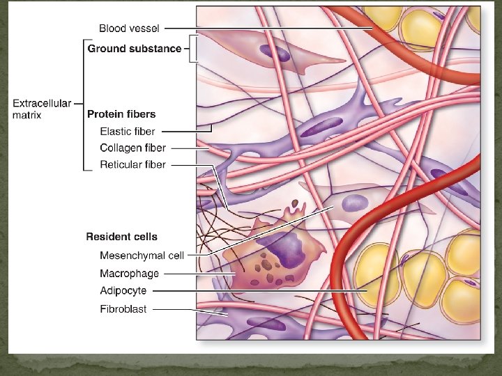
Cell types Macrophage Extracellular matrix Ground substance Fibers • Collagen fiber • Elastic fiber • Reticular fiber Fibroblast Lymphocyte Fat cell Mast cell Neutrophil Copyright © 2010 Pearson Education, Inc. Capillary

Third: ground substance. Watery unstained extracellular material contains water, polysaccharide mucouprotein and protein such as albumin, glycoprotein, and globulin. • Classification of con. T. : First type of con. T. : General con. T. : • Loose (areolar) con. T. : - many cells and little collagen, randomly distributed, thick layer beneath the epithelial in the digestive system.
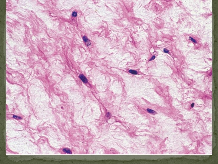
 Connective tissue proper: loose connective tissue, areolar")
Figure 4. 8 a Connective tissues. (a) Connective tissue proper: loose connective tissue, areolar Description: Gel-like matrix with all three fiber types; cells: fibroblasts, macrophages, mast cells, and some white blood cells. Elastic fibers Function: Wraps and cushions organs; its macrophages phagocytize bacteria; plays important role in inflammation; holds and conveys tissue fluid. Collagen fibers Location: Widely distributed under epithelia of body, e. g. , forms lamina propria of mucous membranes; packages organs; surrounds capillaries. Fibroblast nuclei Epithelium Lamina propria Copyright © 2010 Pearson Education, Inc. Photomicrograph: Areolar connective tissue, a soft packaging tissue of the body (300 x).

• Reticular tissue : specialized con. T. consist of collagen fiber produced by reticular cells. this tissue form a delicate network supports various type of cells and lymphocytes in most lymphoid organs (spleen). ●Dense con. T. : • less flexible. • more resistance than loose con. T. , • adopted to offer stress resistance and protection. .
 Connective tissue proper: loose connective tissue, reticular")
Figure 4. 8 c Connective tissues. (c) Connective tissue proper: loose connective tissue, reticular Description: Network of reticular fibers in a typical loose ground substance; reticular cells lie on the network. Function: Fibers form a soft internal skeleton (stroma) that supports other cell types including white blood cells, mast cells, and macrophages. Location: Lymphoid organs (lymph nodes, bone marrow, and spleen). White blood cell (lymphocyte) Reticular fibers Spleen Photomicrograph: Dark-staining network of reticular connective tissue fibers forming the internal skeleton of the spleen (350 x). Copyright © 2010 Pearson Education, Inc.

Classified according to fiber arrangement to Dense regular con. T : q consists of bundles of collagen fibers and fibroblasts qforms tendons, ligaments and aponeuroses ( flat sheet of dense fibrous). § Function : provide strong attachment between various structures
 4. 8 d Connective tissues. tissue proper: dense connective tissue, dense regular Description:")
Figure(d) 4. 8 d Connective tissues. tissue proper: dense connective tissue, dense regular Description: Primarily parallel collagen fibers; a few elastic fibers; major cell type is the fibroblast. Collagen fibers Function: Attaches muscles to bones or to muscles; attaches bones to bones; withstands great tensile stress when pulling force is applied in one direction. Location: Tendons, most ligaments, aponeuroses. Nuclei of fibroblasts Shoulder joint Ligament Tendon Copyright © 2010 Pearson Education, Inc. Photomicrograph: Dense regular connective tissue from a tendon (500 x).

§ Dense irregular con. T. : § Few cells much collagen fibers randomly arranged. § Providing resistance to stress from all direction. § ex – Dermis of the skin and sub mucosa of digestive system.
 4. 8 e Connective tissues. tissue proper: dense connective tissue, dense irregular Connective")
Figure(e) 4. 8 e Connective tissues. tissue proper: dense connective tissue, dense irregular Connective Description: Primarily irregularly arranged collagen fibers; some elastic fibers; major cell type is the fibroblast. Nuclei of fibroblasts Function: Able to withstand tension exerted in many directions; provides structural strength. Location: Fibrous capsules of organs and of joints; dermis of the skin; submucosa of digestive tract. Collagen fibers Fibrous joint capsule Photomicrograph: Dense irregular connective tissue from the dermis of the skin (400 x). Copyright © 2010 Pearson Education, Inc.

Second type of con. T. : - Embryonic con. T. : • Mesenchyme : - Con. Tissue developing mainly from mesoderm in embryo. • Composition: • Ground substance. • Sparse collagen fibers, reticular fibers. • Mesenchyme cells Function: q secretion of ground substance, fibers q proliferation and differentiation into different connective tissue cell types, smooth muscle cells, blood cells.
 con. T. Location: umbilical cord Composition: • Ground substance rich in hialuronic")
Mucoid (mucous) con. T. Location: umbilical cord Composition: • Ground substance rich in hialuronic acid • Collagen fibers • Mucocytes (fibroblasts) Function: secretion of ground substance, fibers.
- Slides: 16