Congestive heart failure Dr Jamal Dabbas Interventional cardiologist

Congestive heart failure Dr. Jamal Dabbas Interventional cardiologist & internist
: a clinical syndrome in which the heart is unable")
Definition Congestive heart failure (CHF): a clinical syndrome in which the heart is unable to pump enough blood to meet the metabolic needs of the body; characterized by ventricular dysfunction that results in low cardiac output
 Diastolic dysfunction: CHF")
Systolic dysfunction: CHF with reduced stroke volume and ejection fraction (EF) Diastolic dysfunction: CHF with reduced stroke volume and preserved ejection fraction
: CHF due to right ventricular dysfunction; characterized by backward heart")
Right heart failure (RHF): CHF due to right ventricular dysfunction; characterized by backward heart failure Left heart failure (LHF): CHF due to left ventricular dysfunction; characterized by forward heart failure Biventricular (global) CHF: CHF in which both the left and right ventricle are affected, resulting in simultaneous backward and forward CHF

Chronic compensated CHF: clinically compensated CHF; the patient has signs of CHF on echocardiography but is asymptomatic or symptomatic and stable (see “Diagnostics” below) Acute decompensated CHF: sudden deterioration of CHF or new onset of severe CHF due to an acute cardiac condition (e. g. , myocardial infarction)

Epidemiology Systolic heart disease is the most common form of CHF overall.

Etiology
 due to: Reduced contractility: Damage and loss of myocytes")
Systolic ventricular dysfunction (most common) due to: Reduced contractility: Damage and loss of myocytes reduce ventricular contractility and stroke volume.

Diastolic ventricular dysfunction due to: Decreased ventricular compliance: increased stiffness or impaired relaxation of the ventricle

Consequences of systolic and diastolic dysfunction Forward failure: reduced cardiac output → poor organ perfusion → organ dysfunction (e. g. , hypotension, renal dysfunction) Backward failure Increased left-ventricular volume and pressure → backup of blood into lungs → increased pulmonary capillary pressure → cardioge nic pulmonary edema Reduced cardiac output → systemic venous congestion → edema and progressive congestion of internal organs

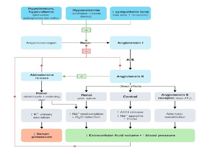
Compensation mechanisms Aim: maintain cardiac output if stroke volume is reduced ↑ Adrenergic activity → increase in heart rate, blood pressure, and ventricular contractility
: activated following decrease in renal perfusion")
• Increase of renin-angiotensin-aldosterone system activity (RAAS): activated following decrease in renal perfusion secondary to reduction of stroke volume and cardiac output • ↑ Angiotensin II secretion → vasoconstriction → ↑ systemic blood pressure → ↑ afterload • Kidney: vasoconstriction of the efferent arterioles and, to a lesser degree, the afferent arterioles → ↓ net renal blood flow and ↑ intraglomerular pressure to maintain GFR • ↑ Aldosterone secretion → ↑ renal Na+ and H 2 O resorption → ↑ preload
: ventricular myocyte hormone released in response to increased")
• Brain natriuretic peptide (BNP): ventricular myocyte hormone released in response to increased ventricular filling and stretching • ↑ Intracellular smooth muscle c. GMP → vasodilation → hypotension and decreased pulmonary capillary wedge pressure • CHF is characterized by reduced cardiac output that results in venous congestion and poor systemic perfusion!

General features of heart failure • Nocturia • Fatigue • Tachycardia, various arrhythmias • Heart sounds: S 3/S 4 gallop • Pulsus alternans

High-output heart failure Definition: heart failure secondary to conditions associated with a high-output state, in which cardiac output is elevated to meet the demands of peripheral tissue oxygenation
 • Anemia • Systemic")
Etiology: conditions that lead to increased cardiac demand (highoutput state) • Anemia • Systemic arteriovenous fistulas • Sepsis • Hyperthyroidism • Multiple myeloma • Glomerulonephritis • Polycythemia vera • Wet beri (vitamin B 1 deficiency) • Physiological causes: pregnancy, fever, exercise

Clinical features Symptoms of lowoutput CHF; particularly tachycardia, tachypnea, low blood pressure, and jugular distention with an audible hum over the internal jugular vein Pulsatile tinnitus Bounding peripheral pulses Laterally displaced apical heart beat Midsystolic murmur, S 3 gallop (indicates rapid ventricular filling)

Diagnostics Primarily a clinical diagnosis X-ray and echocardiography: cardiomegaly

Therapy Manage heart failure: symptom relief, hemodynamic stabilization Treat underlying condition
 functional classification system is")
Stages NYHA functional classification The NYHA (New York Heart Association) functional classification system is used to assess the patient's functional capacities (i. e. , limitations of physical activity and symptoms) and has prognostic value.

NYHA class Characteristics Class I No limitations of physical activity; no symptoms of CHF Class II Slight limitations of moderate or prolonged physical activity (e. g. , symptoms after climbing 2 flights of stairs or heavy lifting); comfortable at rest Class III Marked limitations of physical activity (symptoms during daily activities like dressing, walking across rooms); comfortable only at rest Class IV Confined to bed, discomfort during any form of physical activity; symptoms present at rest
 Classification (2013) The AHA classification system classifies patients according to")
American Heart Association (AHA) Classification (2013) The AHA classification system classifies patients according to their stage of disease. It takes objective findings (patient history, diagnostic findings) as well as symptoms of CHF into account.

Stages Stage A Objective assessment Corresponding NYHA functional class High risk of developing heart failure No (e. g. , pre-existing arterial corresponding NYHA hypertension, CAD, diabetes class mellitus); no structural cardiac changes Stage B Structural damage to the heart (e. g. , infarct scars, dilatation, hypertrophy), without signs or symptoms of heart failure NYHA I Stage C Structural damage to the heart + signs or symptoms of heart failure NYHA I, III, IV Stage D Heart failure at its terminal stage NYHA IV

Diagnostics The presence of CHF means severe pathology and advanced etiology. Heart failure is primarily a clinical diagnosis. Laboratory tests and imaging tests, including a chest x-ray and echocardiogram, are useful for evaluating the severity and cause of the condition.

Diagnostic approach 1. Medical history, including preexisting conditions and history of alcohol and recreational or prescribed drug use 2. Initial evaluation involves a range of routine laboratory tests and a test for BNP level, ECG, and chest x-ray. 3. Echocardiography is the gold standard tool for assessing cardiac morphology and function, as well as investigating the underlying cause of CHF. 4. Other procedures (exercise testing, angiography) may be required for further investigation.

Laboratory analysis • Elevated BNP and NT-pro BNP • High levels of BNP in patients with classic symptoms of CHF confirm the diagnosis (high predictive index). BNP (pg/m. L) NT-pro BNP (pg/m. L) CHF unlikely < 100 < 300 CHF likely > 500 > 450
: • Complete blood count: may show anemia")
• Elevated atrial natriuretic peptide (ANP): • Complete blood count: may show anemia • Serum electrolyte levels: hyponatremia → indicates a poor prognosis • Kidney function tests: ↑ creatinine, ↓ sodium • Urine analysis: rule out concurrent renal impairment • Fasting glucose: to screen for diabetes mellitus, which is a common comorbidity • Fasting lipid profile: to detect dyslipidemia associated with a higher cardiovascular risk
 • ECG abnormalities in CHF are common, but are mostly nonspecific and")
Electrocardiogram (ECG) • ECG abnormalities in CHF are common, but are mostly nonspecific and nondiagnostic. • Signs of left ventricular hypertrophy • ↑ QRS voltage (in the left chest leads and limb leads I and a. VL) → positive Sokolow-Lyon index • ↑ QRS duration (incomplete or complete left bundle branch block) • Left axis deviation • Assessment of prior or concurrent heart conditions • Previous or acute MI: see ECG changes in STEMI • Arrhythmias (e. g. , atrial fibrillation, ventricular arrhythmias, sinus tachycardia or bradycardia, AV block) • Signs of pericardial effusion and tamponade: low voltage ECG

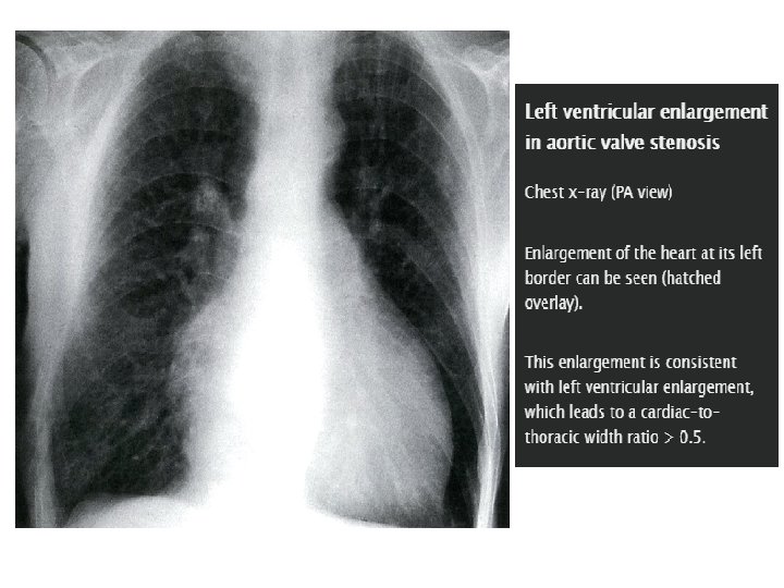
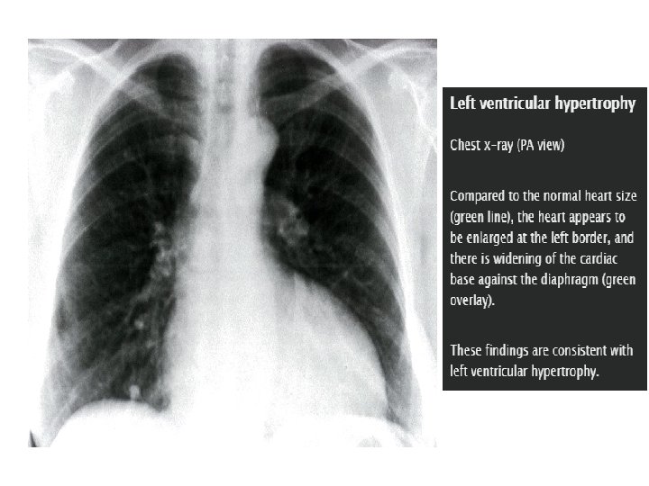
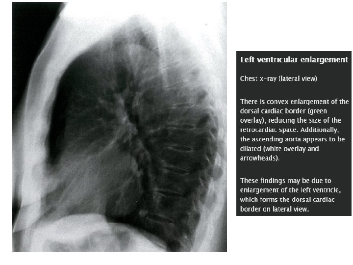
Chest x-ray Useful diagnostic tool to evaluate a patient with dyspnea and differentiate CHF from pulmonary disease Signs of cardiomegaly Cardiac-to-thoracic width ratio > 0. 5 Boot-shaped heart on PA view (due to left ventricular enlargement) Assess pulmonary congestion (see x-ray findings in pulmonary congestion)
 Right")
Investigate etiology Valvular heart disease Wall motion abnormalities (indicate prior or acute MI) Right ventricular strain
: to assess the functional impairment due")
Further tests Cardiac stress test (exercise tolerance test): to assess the functional impairment due to CHF or other conditions (particularly CHD!) Radionuclide ventriculography : indicated to assess left ventricular volume and ejection fraction (LVEF) Cardiac MRI: particularly useful for assessing cardiac morphology and function Cardiac size and volumes, wall thickness, valvular defects, wall motion abnormalities Coronary angiography (left heart catheterization): indicated to detect/confirm CHD and possible percutaneous coronary intervention Right heart catheterization: if pulmonary hypertension is suspected, to assess the severity of systolic dysfunction, and/or to differentiate between types of shock Sv. O 2: will be low in decompensated heart failure Endomyocardial biopsy: may be performed if a specific diagnosis is suspected in patients with rapidly progressive clinical CHF or in case the results would alter the management of the patient, e. g. , in amyloidosis
 Fluid restriction in patients")
Treatment General measures Lifestyle modifications Salt restriction (< 3 g/day) Fluid restriction in patients with edema and/or hyponatremia Weight loss and exercise Cessation of smoking and alcohol consumption Immunization: pneumococcal vaccine and seasonal influenza vaccine

Patient education Self-monitoring and symptom recognition Daily weight check Weight gain > 2 kg within 3 days: consult the doctor Monitoring of potential side effects (e. g. , hypotension caused by ACE inhibitors, hyperkalemia caused by aldosteroneantagonists, sensitivity to sunlight caused by amiodarone) Treat any underlying conditions and contributing comorbidities.
 (loop diuretics and thiaz ide diuretics)")
Drug I NYHA stages II IV Diuretics (✓) (loop diuretics and thiaz ide diuretics) ✓ ✓ Indications First-line drugs • Begin treatment with loop diuretics (furosemi de) to treat volume overload • Thiazides may be added for a synergistic effect Contraindica tions and important side effects Benefits • Monitor • Improve for hypokale symptoms mia and hyp onatremia, weight gain, and volume status

Drug NYHA stages I II IV Indications Contraindica tions and important side effects Benefits ACE ✓ ✓ • Initiate treatment with ACE • Monitor • Improve inhibitors to for hyperkalem symptoms reduce preload, afterload, and ia, hypotension, and improve cardiac output ↑ creatinine (r prognosis • If the patient does not enal tolerate drug (e. g. , impairment) dry cough develops) → substitute with AT 2 receptor blocker Beta (✓) ✓ ✓ ✓ • Add a beta blocker once the • Contraindicate blockers patient is stable on ACE d in acute inhibitor decompensate • Particularly beneficial for d heart failure! patients with hypertension and postmyocardial infarction Aldostero (✓) ✓ ✓ • In select patients, • Monitor ne an aldosterone antagonist may for hyperkalem antagonist be beneficial. ia s • If EF < 35%, and after myocardial infarction • Spironolactone; eplerenone a s an alternative
 (✓) Indications Second-line drugs • If")
Drug Ivabradine NYHA stages I II IV (✓) (✓) Indications Second-line drugs • If the highest tolerable dose of beta blocker is reached and the patient is still symptomatic or if the patient has a contraindication to beta-blocker use • If EF < 35% and the patient has a sinus rhythm with a resting heart rate > 70/min Contraindica tions and important side effects Benefits • Contraindica ted in severe brady cardia • Improves symptoms • Reduces hospitalizati on rate
 (✓ )")
Drug Hydralazin e plus nitr ate NYHA stages I II IV (✓) (✓ ) Indications Contraindica tions and important side effects Benefits • If EF < 40%; particularly beneficial for African. American patients • Alternative if ACE inhibitors and AT 1 blockers are not tolerated • Monitor for volume depletion and hypotensi on • Improves symptoms; may improve prognosis
 (✓) Indications Contraindica tions and important")
Drug Digoxin NYHA stages I II IV (✓) (✓) Indications Contraindica tions and important side effects • In heart failure with reduced • Contraindicat ejection fraction ed in severe AV • If symptoms persist despite block treatment with beta blocker, ACE inhibitor, diuretics, and aldosterone antagonists • May be given to control ventricular rate in atrial fibrillation (if beta blockers are contraindicated) Benefits • Improves symptoms • Reduces hospitalizatio n rate
 (✓)")
Drug NYHA stages I II IV Indications ARNI (angi otensin re ceptorneprilysini nhibitor) (✓) (✓) • Persistent or worsening symptoms despite adequate treatment regimen with firstline drugs • Administered as combination valsartansacubitril Contraindica tions and important side effects • Angioedema, hypotension, h yperkalemia, and progression of chronic renal disease(↑ cre atinine) Benefits • Improves prognosis • Reduces hospitalization rate
 NYHA stages I II IV Indications Contraindica tions and important")
Drug Nesiritide (BNPderiv ative) NYHA stages I II IV Indications Contraindica tions and important side effects • Acute decompensated heart failure • Rarely used today due to side effects and longer halflife compared to other vasodilators (e. g. , nitrogly cerin) • Contraindicat ed in patients with hypotens ion and/or car diogenic shock • Adverse effects include hypot ension and decrease in pulmonary capillary wedge pressure (✓): see “Indications” column for detailed information Benefits

Drugs that improve prognosis: beta blockers, ACE inhibitors, and aldosterone antagonists!
!")
Drugs that improve symptoms: diuretics and digoxin (significantly reduce the number of hospitalizations)!
 is mandatory if")
Conducting regular blood tests to assess electrolyte levels (potassium and sodium) is mandatory if the patient is on diuretics!
 •")
Contraindicated drugs • NSAIDs • Worsen renal perfusion (see “Side effects” of NSAIDs) • Reduce the effect of diuretics • May trigger acute cardiac decompensation • Calcium channel blockers (verapamil and diltiazem): negative inotropic effect; worsen symptoms and prognosis • Thiazolidinediones: promote the progression of CHF (↑ fluid retention and edema) and increase the hospitalization rate • Moxonidine: increases mortality in CHF with reduced ejection fraction (systolic dysfunction)
: prevents sudden cardiac death • Primary prophylaxis")
Invasive procedures • Implantable cardiac defibrillator (ICD): prevents sudden cardiac death • Primary prophylaxis indications • CHF with EF < 35% and prior myocardial infarction/CHD • Increased risk of life-threatening cardiac arrhythmias • Secondary prophylaxis indications: history of sudden cardiac arrest, ventricular flutter, or ventricular fibrillation
: improves cardiac function • Indications: CHF with")
• Cardiac resynchronization therapy (biventricular pacemaker): improves cardiac function • Indications: CHF with EF < 35%, dilated cardiomyopathy, and left bundle branch block • Can be combined with an ICD • Coronary revascularization with PCTA or bypass surgery may be indicated if CAD is present. • Valvular surgery if valvular heart defects are present • Ventricular assist devices: may be implanted to support ventricular function; may be indicated for temporary or long-termsupport (e. g. , to bridge time until transplantation) of decompensated CHF • Cardiac transplantation: for patients with endstage CHF (NYHA class IV), ejection fraction < 20%, and no other viable treatment options
 • Cardiorenal syndrome • Cardiac")
Complications • Acute decompensated heart failure (see section below) • Cardiorenal syndrome • Cardiac arrhythmias • Central sleep apnea syndrome • Cardiogenic shock
 •")
• Stroke; increased risk of arterial thromboembolisms (especially with concurrent atrial fibrillation) • Chronic kidney disease • Cardiac cirrhosis (congestive hepatopathy): Cirrhosis due to chronic hepatic vein congestion in patients with right‑sided heart failure. • Venous stasis, leg ulcers

Acute decompensated heart failure Cardiac decompensation is the most common reason for hospital admissions and is the most important complication of congestive heart failure.

Etiology ADHF typically occurs in patients who have a history of CHF or other cardiac conditions in which an acute cause precipitates the deterioration of cardiac function. • Exacerbation of congestive heart failure (e. g. , through pneumonia, anemia, volume overload, medication noncompliance) • Acute myocardial infarction

• Atrial fibrillation, severe bradycardia, and other arrhythmias • Myocarditis • Hypertensive crisis • Pulmonary embolism • Pericardial tamponade • Aortic dissection • Cardiotoxic substances • Renal failure • Cardiodepressant medication (e. g. , beta blockers, CCBs)

Clinical features • Rapid exacerbation of symptoms of CHF (see symptoms of left heart failure and symptoms of right heart failure)

• Pulmonary congestion with: • Acute, severe dyspnea and orthopnea; worse when supine • Cough (occasionally with frothing, blood-tinged sputum) • Cyanosis • Auscultation of the lungs: rales accompanied by wheezing • Flash pulmonary edema: rapid, lifethreatening accumulation of fluid associated with the risk of acute respiratory distress • Weakness, fatigue, and cold, clammy skin

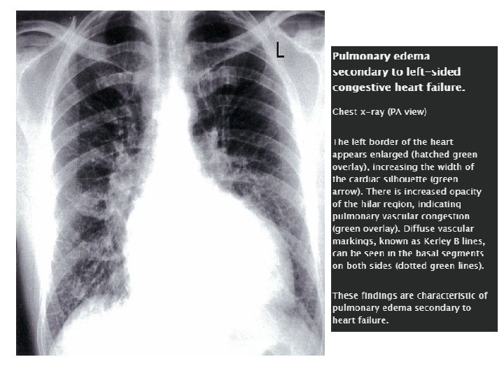
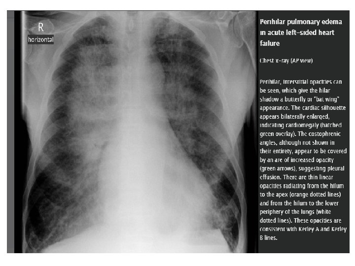
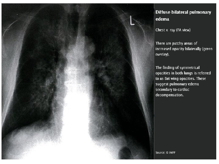
Diagnostics • X-ray findings in pulmonary congestion • Cardiomegaly • Prominent pulmonary vessels and perihilar pulmonary edema (butterfly or “bat's wings” appearance of the hilar shadow) • Kerley B lines: visible horizontal interlobular septa caused by pulmonary edema • Basilar edema • Bilateral pleural effusions
")
• Sputum analysis: heart failure cells (hemosiderin-containing cells)

• Thoracentesis • Indicated if the etiology of the pleural effusion is unclear • Pleural fluid analysis: Transudate effusions are typical of cardiogenic causes.

The radiologic signs of pulmonary congestion can be remembered with “ABCDE”: A = Alveolar edema (bat's wings), B = Kerley B lines (interstitial edema), C = Cardiomegaly, D = Dilated prominent pulmonary vessels, and E= Effusions!

Differential diagnosis of pulmonary edema and respiratory distress Noncardiogenic pulmonary edema due to ARDS, pulmonary embolism, transfusionrelated acute lung injury, high altitude Asthma Pneumonia

Treatment • Sufficient oxygenation and ventilation ; assisted ventilation as needed (e. g. , CPAP). • Fluid management: • Aggressive diuresis (e. g. , IV furosemide) to reduce volume overload • Vasodilators: (e. g. , IV nitroglycerine) can be considered as adjunct treatment in patients without hypotension.
 in case of systolic dysfunction")
• Hemodynamic stabilization: inotropes (e. g. , dobutamine) in case of systolic dysfunction • Treat the cause of decompensation. • Hemodialysis if volume overload is symptomatic (pulmonary edema, pleural effusion, ascites) and resistant to treatment • ECLS may temporarily substitute pulmonary function. • Ventricular assist devices (see “Treatment of heart failure” above)

Beta blockers must be used cautiously in decompensated heart failure!
, M =")
Management of ADHF can be remembered with “LMNOP”: L = Lasix (furosemide), M = Morphine, N = Nitrates, O= Oxygen, P = Position (with elevated upper body).

Cardiorenal syndrome is a complication of acute heart failure and CHF. Definition: a complex syndrome in which renal function progressively declines as a result of severe cardiac dysfunction; occurs in ∼ 20– 30% of cases of ADHF

Pathophysiology Cardiac forward failure → renal hypoperfusion → prerenal kidney failure Cardiac backward failure → systemic venous congestion → renal venous congestion → decreased transglomerular pressure gradient → ↓ GFR→ worsening kidney function RAAS activation → salt and fluid retention, hypertension → hypertensive nephropathy

Diagnosis: ↓ GFR, ↑ creatinine that cannot be explained by underlying kidney disease Treatment: treat heart failure; manage renal failure (see treatment of acute renal injury) Prognosis: CHF with reduced GFR is associated with a poor prognosis.

Prognosis • The prognosis depends on the patient, type and severity of heart disease, medication regimens, and lifestyle changes. • The prognosis for patients with preserved EF is similar to or better than for patients with decreased EF • Risk stratification scales may be used to evaluate the prognosis (e. g. , CHARM and CORONA risk scores).

• Factors associated with worse prognosis. Elevated BNP • Hyponatremia • Systolic BP < 120 mm Hg • Diabetes • Anemia • Weight loss or underweight • S 3 heart sound • Implantable cardioverter-defibrillator use • Frequent hospitalizations due to CHF

1 -year survival according to NYHA stage • Stage I: ∼ 95% • Stage II: ∼ 85% • Stage IV: ∼ 35%
- Slides: 77