Congenital Coronary artery anomalies DR SANMATH SHETTY K


 Serious prognostic implications in young")


. Anomalous path")


 Usually involves anterior/left surface of")
 Bland White Garland syndrome Described in")


 Stenosis due to membrane")

 Defined as a segment of variable length covered by")


 myocardial ischemia is seen in all except A. Myocardial bridge")
- Slides: 72
Congenital Coronary artery anomalies DR SANMATH SHETTY K SENIOR RESIDENT DEPT OF CARDIOLOGY MCH, CALICUT
Overview Embryology Incidence and classification. Anomalies of origin and course. Anomalies of intrinsic coronary anatomy Anomalies of termination Coronary anomalies associated with CHD
Normal coronary anatomy FEATURE RANGE No. of ostia 2 to 4 Location Right and left anterior sinuses Proximal orientation 45° to 90° off the aortic wall Proximal common stem Only left (LAD and Cx) Proximal course Direct from ostium to destination Mid-course Extramural (subepicardial) Branches Adequate for the dependent myocardium Essential territories RCA (RV free wall), LAD (anteroseptal), OM (LV free wall) Termination Capillary bed
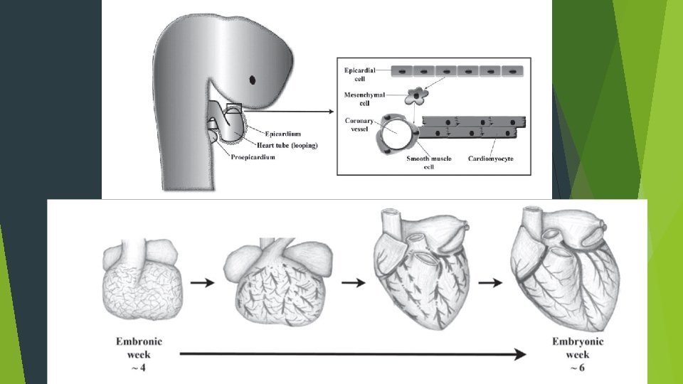
Coronary arteries- development Theories Outgrowth development hypothesis: fusion of subepicardial vascular network with endothelial buds that grew from the base of the truncus arteriosus. 1989, Bogers identified coronary arteries in aortic wall prior to emergence of coronary ostia ---- suggests ingrowth rather than outgrowth of these vessels.
Coronary arteries- development First directed migration- formation of the epicardium. Continuous simple squamous epithelium originating from septum transversum. Visible as a bulge in the sinoatrial pole of the heart - proepicardial organ(PEO). Epithelial to mesenchymal transition Epicardium thickens and some cells lose contact with epicardial epithelium Form freely migratory mesenchyme. Proepicardially derived mesenchymal cells coalesce to form channels. Channels fuse to form epicardial blood vessels. Second directed migration- movement of mesenchyme through myocardium Mesenchymal cells migrate into space in the developing myocardium. Migration is pervasive; i. e; each myocyte is in contact with PEO derived endothelial cell of coronary vasculature.
Coronary arteries development Sequence of development 1 st stage: Blood islands 2 nd stage: Development of venous connection between vascular network and coronary sinus. 3 rd stage: Coronary arteries join the aorta through an arterial connection to the vascular plexus. Occurs after aortopulmonary septation and formation of semilunar valves. 2 coronaries originate from the right and left sinus that face the RVOT. The sprouts or anlagen in the 3 rd aortic sinus and all 3 pulmonary sinuses undergo rapid involution or do not develop.
Basic principle of coronary classification Name of a coronary artery assigned not according to the site of origin or proximal course But according to the dependent territory. A common proximal trunk, which joins 2 or 3 coronary arteries is named a mixed trunk. RCA is the vessel that crosses the right AV groove and provides blood flow to the right ventricular free wall. LAD is the vessel that courses the anterior IV groove and provides blood flow to the anterior interventricular septum. LCX is the vessel that courses the left AV groove and provides blood flow to the free wall of the left ventricle, on the obtuse margin of the heart.
Anomalies of coronaries
Anomalies of origination and course
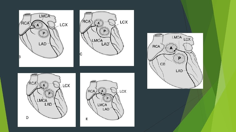
ACAOS (anomalous coronary artery from opposite sinus of valsalva) Serious prognostic implications in young individuals. Incidence Among, 9, 907 patients who underwent electron-beam CT or 64 -detector MDCT, incidence of ACAOS was 0. 7%. Prevalence RCA from left sinus 56%, LCX from right sinus 16. 7%, LM from right sinus 8. 3%, LAD from right sinus 2. 4%. Nezarat N, Luo Y, Darabian S, et al. PREVALENCE AND LONG TERM FOLLOW-UP OF ANOMALOUS CORONARY ARTERIES ORIGINATING FROM OPPOSITE SINUS (ACAOS). J Am Coll Cardiol. 2015; 65. Possible paths of connection to opposite cusps: Retrocardiac Retroaortic Preaortic, between aorta and PA Intraseptal(supracristal). Precardiac
Left circumflex artery from right coronary artery/sinus: ? Most common anomaly LCX passes behind aorta to reach its normal supply territory. No general clinical significance; may be compressed if both aortic and mitral prosthetic fixation rings are implanted. Unusually higher incidence of atherosclerosis. LAD from right coronary artery/sinus: Rare in absence of congenital heart disease. Common in TOF. Passes in front of RVOT or through IVS. Rarely passes in between aorta and RVOT.
Crossing pathways Retrocardiac Located behind the tricuspid and mitral valves in the posteriot AV groove. Retroaortic Most common Next to the posterior wall of the aorta, in the sulcus between the atria and the aorta (the transverse sinus), and finally reaches the left AV groove. 0. 1 -0. 9%; common in LCX from RCA. Less frequent in RCA from left sinus and Left main from right sinus. Preaortic Between aorta and PA Anomalous RCA, left main or LAD( never isolated LCX) crosses subepicardially across the aortopulmonary septum.
Crossing pathways Intraseptal Located inside upper anterior IV septum (embryologic conal septum). Anomalous path mostly intramural (systolic phasic narrowing on angio) and origin of 1 -2 septal perforators. Precardiac May be taken by ectopic RCA, LM or LAD, never isolated LCX. Common in patients with TOF (ectopic LAD from right sinus). Frequently gives infundibular branches; never septal braches.
The traditional concept-the aberrant artery was liable to a scissors-like mechanism especially during exertion. Unlikely because at the site of closest aortopulmonary proximity, the anomalous artery lies inside the aortic wall. IVUS imaging detected this anomaly to consist of intramural proximal intussusception of the ectopic artery at the aortic-root wall. Mechanisms of ischemia: Coronary hypoplasia: Intramural intussuscepted segment of the ectopic artery is smaller in circumference than the more distal extramural vessel. Lateral compression: Cross section of the intramural segment is characteristically not circular but ovoid. Smaller diameter is further compressed during each systole.
Manifestations of ACAOS SCD: associated with extreme exercise in young adults Low cardiac output and bradycardia or asystole typically occur early after extreme exercise. Terminal ventricular fibrillation may also occur. Dyspnea, palpitations, angina pectoris, dizziness and syncope: More frequently seen in older adults (specifically women) Related to the onset of hypertension.
Treatment Medical treatment/observation Beta blockers Restriction of activity Coronary angioplasty with stent deployment of the intramural segment Technically feasible for right ACAOS. Justifiable in the presence of (1) disabling symptoms and/or a high risk of sudden death (2) area stenosis more severe than 50% on IVUS (3) a large dependent myocardial territory (> 1/3 rd of total) (4) reversible ischemia, as documented by a nuclear stress test. Surgical repair Recommended for left-ACAOS that involves a large territory at risk. Direct reimplantation, unroofing of intramural coronary segment, osteoplasty
Single coronary A single aortic ostium or origination provides for all of the coronary blood flow. Incidence 0. 024%. 40% associated with cardiac malformations; TOF, TGA, truncus, BAV. Primary classification based on the location of single ostium.
Can arise from right or left sinus. May follow its usual course or separate branches may originate from a main coronary artery.
Single coronary Coronary blood flow is not affected unless congenital or acquired obstructive disease is present in the proximal trunk. Incidence of atherosclerotic disease not increased in the mixed trunk. An ectopic single coronary ostium more susceptible to congenital or acquired obstructive disease. Coronary angioplasty of the common trunk contraindicated. CABG: arterial grafts preferred Provide long lasting conduits. Proximal mixed trunk obstruction progress to total occlusion soon after successful bypass Graft occlusion fatal in such a scenario.
Ectopic Coronary Ostium in NCS Benign anomaly Most commonly involves LCA ostium Difficult cannulation-unexpected location, tangential or slitlike nature Nonselective angiography-longer than usual LMCA RAO and straight lateral projections are most useful For selective catheterization Amplatz or Multipurpose curved catheters offer best chance of success
Ectopic Coronary Ostium Arising Outside the Aortic Root(Ascending Aorta) Usually involves anterior/left surface of aorta Frequently have slit like orifices and tangential proximal course along aortic wall RCA is most frequent ectopic artery. Predisposed to more atherosclerosis. Association with Congenital aortic valve anomalies
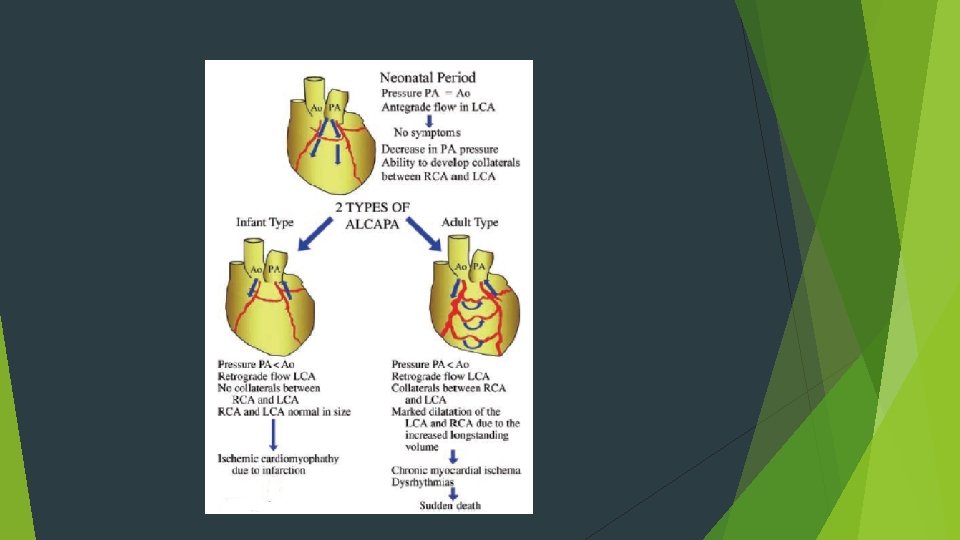
ALCAPA (anomalous left coronary artery from pulmonary artery) Bland White Garland syndrome Described in 1933. Incidence 1: 300, 000 live births. One of the most common causes for myocardial ischemia in infants and children. May occur in isolation; associated with VSD, TOF and coarctation. Usually originates from right pulmonary sinus (close proximity to left aortic sinus) Less commonly originates from other pulmonary sinuses or more distally in main or proximal branch pulmonary arteries.
Pathophysiology Early neonatal period: High PVR Antegrade perfusion of left coronary myocardial territory Perfusion by desaturated blood from PA. Later neonatal period: PVR falls ----- coronary perfusion pressure falls. Myocardial perfusion depends on collaterals from RCA. Further fall in PVR, flow in LAD becomes retrograde. “Steal” from right coronary circulation aggravating ischemia. Stenosis of LAD origin at this point- reduces the degree of steal and confers a degree of protection.
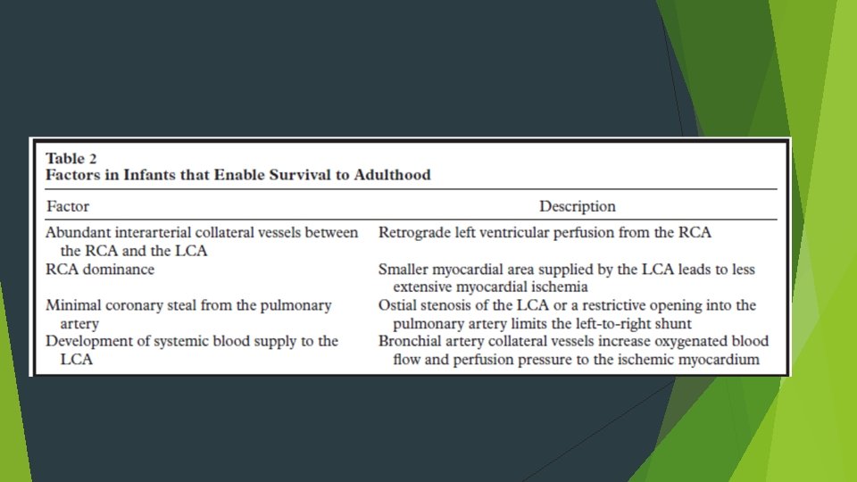
Clinical features Symptoms of congestive heart failure in early infancy Breathlessness, failure to thrive, sweating and wheezing. Acute episodes of irritability with associated pallor typically during feeding. Displaced apex beat, 3 rd or 4 th heart sounds, systolic murmur at apex (due to MR secondary to LV dilation or papillary muscle ischemia/rupture) 65% infants die within 1 st year without treatment. Mortality uncommon in first 2 months. Others present later in childhood or adulthood with continuous murmur, angina, ventricular arrhythmia or sudden death. Average life expectancy in this group 30 yrs. Patients surviving beyond 50 yrs, risk of sudden death decreases.
Investigations Chest X ray: Non specific Cardiomegaly LV enlargement Pulmonary venous congestion ECG: Pathological q waves inferolateral leads Poor R wave progression and T wave inversion. Q wave width > 30 ms in lead I, q wave depth >3 mm in a. VL and QR pattern in a. VL significantly associated with ALCAPA.
ECHO: LV dilated Impaired systolic and diastolic function Papillary muscles bright and echogenic ( due to infarction and fibrosis) Mitral regurgitation. 2 D imaging of coronaries: LCA takes a course very close to left aortic sinus (connection may be misinterpretated as normal in upto 70%). Doppler assessment with colour flow mapping of coronary flow Retrograde flow in LAD and retrograde jet of flow in PA RCA dilated – RCA: aorta ratio ≥ 0. 14.
CT/ MRI: useful in adult patients with poor acoustic window. CT demonstrates better spatial resolution. MRI allows functional assessment of LV function and myocardial viability. Cardiac Catheterisation: Gold standard for diagnosis. Elevated LVEDP, subsequently elevated pulmonary and RV pressures. Aortic root angiography or selective RCA : delineates anomalous LCA and extent of collateralization.
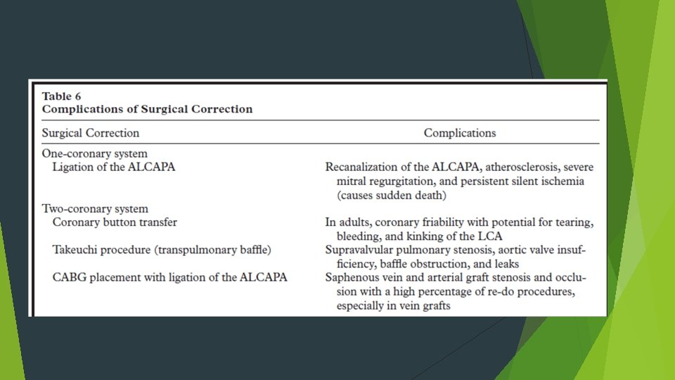
Management Medical supportive treatment: ventilation, inotropes, diuretics and afterload reduction. Surgical techniques:
Surgical outcomes Mortality rates reduced from 75– 80% in 80 s to 0 -23% in current era. Risk factors associated with increased 30 day mortality Young age at operation Reduced preoperative LV function Degree of MR not consistently found to influence early mortality. MR improves with time; consensus that mitral valve surgery at the time of ALCAPA repair is not necessary (unless there is papillary muscle rupture).
ARCAPA Rarer than ALCAPA. ? Lesion occurs more frequently and is underdiagnosed because of the relatively benign nature of the lesion. Diagnosis is usually made at autopsy or incidentally in asymptomatic adults. Less commonly present with myocardial ischemia and SCD, particularly in right dominant coronary pattern. Treatment : surgical reimplantation. With evidence of ischemia. All patients, to prevent SCD.
Isolated connection of LCX or LAD from pulmonary trunk: Less lethal than ALCAPA. Rarer than ARCAPA. Indication for surgery similar to ARCAPA. Total Anomalous Connection of Coronary Arteries to Pulmonary Trunk: Single ostium and trunk from which all branches emerge or from 2 ostia close together giving rise to left and right coronary systems. Symptoms appear within a few days of birth, and death follows within 2 weeks.
ANOMALIES OF INTRINSIC CORONARY ARTERIAL ANATOMY
Congenital Ostial Stenosis or Atresia (dimple seen from aortic side) Stenosis due to membrane or fibrotic ridge. Frequently occurs in pulmonary valve atresia with intact ventricular septum. Collateral circulation established prenatally. During myocardial stress testing, dependent myocardial territory may show reversible ischemia– collateral circulation inadequate for maximal myocardial demand.
Coronary ectasia or aneurysm Defined as localized dilations in an otherwise normal sized coronary artery. Morphologically defined as an increase in diameter of a coronary segment by >50% with respect to normal. Congenital or acquired(atherosclerosis, Kawasaki disease). Primary and secondary ectasia Primary: Localized segment has a disproportionately larger diameter in comparison to neighbouring segments Localized decrease in flow velocity. Secondary: Global coronary dilation secondary to increased or fistulous flow. Coronary diameters appropriate to increased flow. Increase in flow velocity.
Intramyocardial coronary artery (muscular bridge) Defined as a segment of variable length covered by myocardial fibres but that otherwise lies subepicardially. Reported prevalence 1. 7% by angiographic studies (only in LAD). 25% mean prevalence by pathologic studies, LAD most common. Length of bridges varies from 4 mm to 40 mm The myocardial fibres constitute the “bridge” whereas the underlying coronary artery segment represents the “bridged artery”. Angiographically, systolic narrowing of a coronary artery segment. Facilitated by intracoronary administration of NTG. Unlikely to cause absolute flow reduction- 85% of human coronary flow occurs in diastole. Usually clinically silent; rarely the longer and deeper bridges manifest with myocardial ischemia, ACS or SCD.
Atherosclerosis in myocardial bridge IVUS in 69 patients with myocardial bridge found increased incidence of atherosclerosis at segments proximal to the bridge, but no plague within or distal to the bridges. Ge J, Jeremias A, Rupp A, et al. New signs characteristic of myocardial bridging demonstrated by intracoronary ultrasound and Doppler. Eur Heart J 1999; 20: 1707 -16.
ANOMALIES OF TERMINATION
Coronary Arteriovenous Fistula A direct communication between a coronary artery and the lumen of any one of the four cardiac chambers, the coronary sinus or its tributary veins, or the superior vena cava, pulmonary artery or pulmonary veins close to the heart. First described by Krause in 1865. Coronary artery site- RCA or its branches- 50 -55% Left coronary artery 35% Both coronary arteries 5% Fistula occurs either in the main vessel that continues beyond the fistula (a side-to-side pattern) or at the termination of the main vessel itsel or at a branch (an end artery).
Coronary artery proximal to the fistula is always dilated, elongated and serpiginous, Artery beyond the fistula abruptly reduces in diameter. Site of Fistulous Connection > 90% of fistulae open into right heart chambers or their connecting vessels. True AV fistulae to the veins themselves (coronary sinus or its major branches or venae cavae) are uncommon 40% connect to RV, 25% to RA, 15%-20% to pulmonary artery, 7% to coronary sinus and only 1% to SVC. 8% of fistulae drain into left heart chambers or their tributaries, usually LA, less often LV(about 3%), and rarely the proximal pulmonary veins.
Fistulae entering the right side of the circulation rapid systolic and diastolic runoff from the aorta. L- R shunt, Q p/Q s is seldom larger than 1. 8. Left heart fistulae runoff from the aorta during both systole and diastole when fistulae enter LA or only during diastole when they enter LV. No L-R shunt.
Clinical features Most patients present late in life, occasionally in childhood and rarely in infancy. Most patients asymptomatic; present incidentally with continuous murmur. 80% of patients < 20 years are asymptomatic, only 40% of those older are asymptomatic. Most common symptoms are effort dyspnea and fatigue. Angina is uncommon (about 7%) and myocardial infarction rare (about 3%).
Clinical features Heart failure occurs in 12% to 15% of patients presenting for surgery. Heart failure more common in older patients. Review by Liberthson and colleagues, only 6% of patients <20 years and 19% of those >20 years had heart failure. Heart failure is more common in patients with fistulous connection to the coronary sinus (50% vs. 14% in the overall group as reported by Ogden and Stansel). More common with onset of atrial fibrillation; occurs more often when the connection is to the RA.
Natural history If not present at birth, develops early in life. Small fistulae remain small, and moderate fistulae slowly increase in size. Onset of dyspnea, heart failure and angina can occur in young patients with large fistulae. As shunt is usually moderate, symptoms often do not appear until later in life consequent to long-standing moderate LV volume overload. Maximum prevalence of heart failure occurs in the fifth and sixth decades. IE may precipitate symptoms 5% patients; may occur at any age. Aneurysm formation- 9% of children and 14 -29% adults. Spontaneous rupture not been reported in children. Liberthson and colleagues found that among 173 reported patients(mean age 24 yrs), fistula related death in 6%. Spontaneous closure recorded; very rare.
Diagnosis Clinical Examination: Diagnosis is often strongly suspected from physical signs Continuous murmur that is maximal to the right of the sternum when the fistula enters RA Lower left sternal edge when it enters RV/LV. Murmur may be only diastolic when it enters LV. Left infraclavicular area when it enters PA. Systolic thrill is occasionally palpable when the fistula lies anteriorly (entry into RA/RV). Large shunts- wide pulse pressure and jerky pulse.
Diagnosis ECG: Ø Normal > 50% cases; RV/LV overload in remainder. CXR: Ø May be normal or show mild CM with plethora. ECHO: Ø Can detect importantly enlarged coronary arteries and may also confirm specific chamber enlargement. Cardiac Catheterization: Aortography and selective coronary angiography Ø Gold standard Ø Calculate L-R shunts and Right heart pressures. CTA: Ø Diagnostic procedure of choice in cases that will not require hemodynamic measurements to make management decisions.
Management Indications for surgery: Recommended that diagnosis of a coronary AV fistula is an indication for operation unless the shunt is small (Q p/Q s < 1. 3). At least some of these fistulae will increase in size and eventually produce symptoms and heart failure Tendency for development of IE Low probability of spontaneous closure The safety and efficacy of operation.
Management Surgical options: external ligation or intracardiac closure on CPB. Percutaneous occlusion: detachable balloons, coils, vascular plugs, PDA/ASD/VSD devices. Ensure no coronary supply to myocardium distal to point of occlusion. If uncertain, observe ECG during temporary balloon occlusion of fistula. Results comparable to surgical results; expected mortality <1%. Proximal coronary artery persistently dilated- prone to thrombotic occlusion- long term antiplatelet therapy advisable and anticoagulation proposed in patients with moderate persistent aneurysmal dilation.
Anomalous collateral coronary arteries
Congenital anomalous collaterals- > 1 mm anastamotic communications between adjacent unobstructed coronary arteries or branches. Prognostic implications: protective in the event of occlusion of one of the connected vessel.
Functional classification
Coronary Artery Patterns with Congenital Heart Defects
TOF 40%- long, large conus artery may arise from a separate ostium. 4 -5%- LAD from RCA Passes across RVOT Makes transannular incision difficult at surgery. Occasionally, single coronary from either right or left sinus May pass across RVOT or behind the aorta.
D-TGA Important to identify anomalies prior to arterial switch Some patterns difficult to transfer with arterial switch. Sinuses facing RVOT – Facing sinuses. Nomenclature of facing sinuses depends on relation of great vessels. Side by side relationship- anterior or posterior. Oblique relationship- left anterior or right posterior. Anterior-posterior relationship- right or left.
Almost all patients, coronaries arise from facing sinuses. Increased incidence of coronary anomalies Presence of VSD Side by side great vessels. 60% cases – coronaries arise from appropriate sinuses and branch normally. More common with oblique orientation of great vessels. LM and LCX pass in anterior and leftward of RVOT. 20% cases- LCX from RCA Common in side by side orientation. LCX runs posterior to pulmonary artery. 4% cases- inverted arteries from inappropriate sinuses. 4% cases- partially inverted arteries.
L- TGA Aorta is anterior and to left of PA Coronary arteries come from facing sinuses Anterior sinus is usually noncoronary sinus Morphologic right and left coronaries are concordant with morphologic right and left ventricle. LCA Supplies LV, arises from right facing sinus. Passes in front of pulmonary annulus and divides into LAD and LCX. RCA Supplies RV, arises from left facing sinus. Runs in the AV groove in front of left atrial appendage to terminate as PDA. Most common variant- single coronary from right facing sinus.
Truncus arteriosus Depends on the no of cusps in truncal valve. Tricuspid- RCA from right anterior sinus, LCA from posterior sinus Bicuspid Anterior/ posterior- LCA from posterior sinus and RCA from anterior sinus. Right/ left- LCA from left and RCA from right sinus. Quadricuspid 2 anterior and 2 posterior cusps LCA from left posterior cusp and RCA from left or right anterior cusp High origins of right and left coronaries frequent.
Bicuspid Aortic Valve Associated with left coronary dominance High incidence of immediate bifurcation of LCA into LAD and LCX.
Coronary artery disease as a result of congenital heart disease
Coarctation of aorta Intimal proliferation and medial thickening due to hypertension. Risk of premature atherosclerosis. Supravalvular AS Ostial obstruction –aortic medial proliferation or adherence of aortic cusp. Thick walled, tortuous coronaries – prematurely atherosclerotic. Aortic regurgitation Coronary artery ectasia Large smooth walled coronaries Elongated, tortuous dilated coronaries in adults with cyanotic congenital heart disease. Pulmonary atresia with intact ventricular septum Development of ventriculocoronary connections due to suprasystemic pressure in RV.
THANK YOU
MCQ 1 Precardiac pathway is a crossing pathway taken by all except A. LM from right sinus. B. LAD from right sinus C. LCX from right sinus. D. RCA from left sinus.
MCQ 2 All the following are classified under coronary anomalies of origination except A. Single coronary B. ALCAPA C. Anomalous coronary from NCS. D. Coronary ostial stenosis.
MCQ 3 All the following regarding ACAOS is true except A. It is known to be associated with SCD especially in young adults. B. LAD from RCA common in TOF patients. C. Intraseptal pathway is diagnosed angiographically by presence of systolic narrowing and septal branches. D. Angioplasty is feasible for left ACAOS.
MCQ 4 Echo features suggestive of ALCAPA all except A. Dilated LV. B. RCA: Aorta ratio< 0. 14. C. Bright, echogenic papillary muscles. D. Retrograde LAD flow.
MCQ 5 All the following are associated with SCD except A. Ectopic coronary ostium in NCS B. ACAOS C. ALCAPA D. ARCAPA
MCQ 6 Secondary (episodic) myocardial ischemia is seen in all except A. Myocardial bridge B. Coronary ectasia C. Coronary ostial stenosis D. ALCAPA