Congenital Bone diseases Dysostosis localized abnormal bone formation

Congenital Bone diseases • Dysostosis ﺧﻠﻞ ﺍﻟﻌﻈﻢ : localized abnormal bone formation Craniofacial dysostosis • Bone aplasia ﻋﺪﻡ ﺗﻨﺴﺞ : absence of finger or limb • Supernumerary digits or ribs • Abnormal bone fusion (rib fusion, craniosynostosis ( ) ﺗﻌﻈﻢ ﺍﻟﺪﺭﻭﺯ ﺍﻟﺒﺎﻛﺮ • Phocomelia ﺗﻔ ﻡ ﺍﻷﻄﺮﺍﻑ : multiple absent or short, fused bones in limbs, Thalidomide, genetic

Osteogenesis Imperfecta ﺗﻜﻮﻥ ﺍﻟﻌﻈﻢ ﺍﻟﻨﺎﻗﺺ • Group of hereditary disorders caused by defective synthesis of type I collagen • Gene mutations in the coding sequences for α 1 or α 2 chains • Mutant type 1 collagen is defective and prematurely degraded • Bone matrix amount is too little, results in bone fragility, deformity • Different types of mutations, range in severity • Type 1: most common, normal life expectancy • Type 2: severest, death early in life or in utero (severe fractures) • Also affects skin, joints, and eyes (blue sclera), hearing loss (conduction defects in the middle and inner ear bones), and small misshapen teeth are a result of dentin deficiency

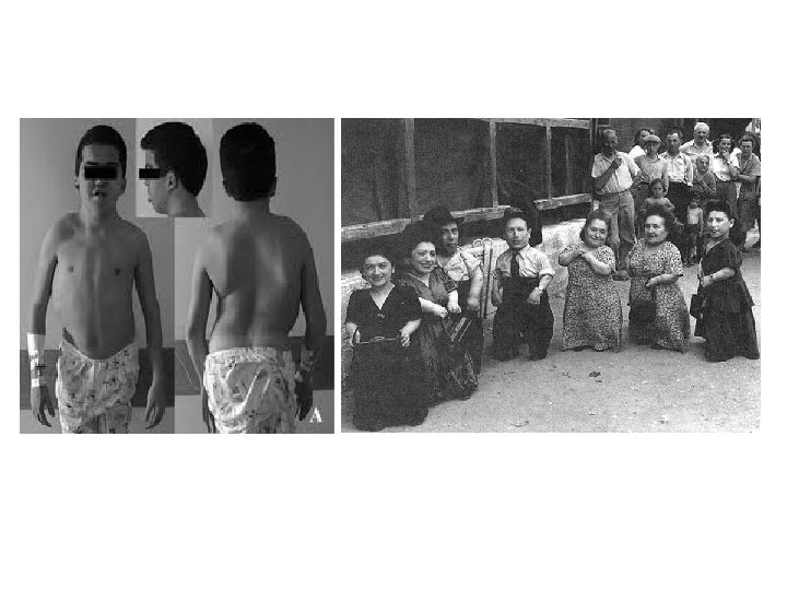
Achondroplasia ﻭﺩﺍﻧﺔ ﺍﻟﻌﻈﻢ • Major cause of dwarfism • Point mutation in the fibroblast growth factor receptor 3 (FGFR 3) that results in its constitutive activation • Activated FGFR 3 inhibits chondrocyte proliferation; as a result, the normal epiphyseal growth plate of the long bones is suppressed • Patients have normal head and trunk, but short bowing limbs • AD, most cases represent new acquired mutation • The affected individuals are typically heterozygotes, since homozygosity leads to abnormalities in chest development and death from respiratory failure soon after birth

Osteopetrosis ﺗﺼﺨﺮ ﺍﻟﻌﻈﻢ • Rare genetic disorders characterized by reduced osteoclastmediated bone resorption and therefore defective bone remodelling • Normally, bone matrix degradation requires first acidification of contact site • Multiple mechanisms, one is known, as deficiency in enzyme Carbonic Anhydrase II, required for the osteoclast hydrogen ion excretion important for acidification • The affected bone is grossly dense and stone-like. Paradoxically, the architecture is unsound and fractures are common • Cranial nerves compressions, deafness • Decreased hematopoiesis (infections) • Hepatosplenomegaly (extramedullary hematopoiesis)

• Osteopetrosis: markedly thickened bone trabeculae, marrow spaces are minimal

Acquired diseases of bone development • • Osteoporosis ﻫﺸﺎﺷﺔ ﺍﻟﻌﻈﻢ Paget disease Vitamin D deficiency Hyperparathyroidism

osteoporosis • • Decreased bone mass, prone to fracture Grossly: bone appears porous and spongy Localized: disuse of a limb Generalized: primary, secondary
 shortened by compression fractures, compared with a normal vertebral body.")
Osteoporotic vertebral body (right) shortened by compression fractures, compared with a normal vertebral body. Note that the osteoporotic vertebra has a characteristic loss of horizontal trabeculae and thickened vertical trabeculae

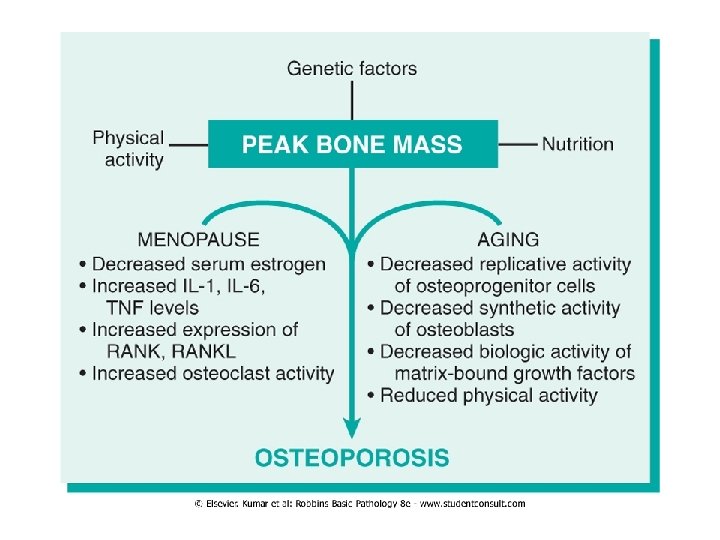
Primary Osteoporosis • The most common form of osteoporosis; senile, postmenopausal • Senile osteoporosis affects all aging individuals • Postmenopausal osteoporosis obviously affects only women after menopause • Peak bone mass is achieved during young adulthood, then in the third or fourth decade in both sexes, bone resorption begins to outpace bone deposition • Such losses generally occur in areas containing abundant cancelloues (trabecular) bone and are therefore more pronounced in the spine and femoral neck • The rate of loss can be accelerated by the postmenopausal state • Osteoporosis-related fractures cause significant morbidity and mortality
 Receptor Activator for Nuclear factor κB (RANK)- ligand")
Pathogenesis Osteoblasts and stroma synthesize 1) Receptor Activator for Nuclear factor κB (RANK)- ligand 2) Macrophage colony-stimulating factor (M-CSF) 3) Osteoprotegerin (OPG) Together, RANK ligand M-CSF conspire to convert macrophages into bone-crunching osteoclasts • OPG bind RANK ligand blocks osteoclast formation • Dysregulation of these interactions occur with aging and decreased estrogen, resulting in relative increased osteoclast activity compared to osteoblast function • Secondary causes: high cortisol, hyperthyroidism, drugs • • •

• Paracrine mechanisms regulating osteoclast formation and function. Osteoclasts are derived from the same stem cells that produce macrophages. RANK (receptor activator for nuclear factor-κB) receptors on osteoclast precursors bind RANK ligand (RANKL) expressed by osteoblasts and marrow stromal cells. Along with macrophage colony-stimulating factor (M -CSF), the RANK-RANKL interaction drives the differentiation of functional osteoclasts. Stromal cells also secrete osteoprotegerin (OPG) that acts as a decoy receptor for RANKL, preventing it from binding the RANK receptor on osteoclast precursors. Consequently OPG prevents bone resorption by inhibiting osteoclast differentiation

Paget disease • Repetitive episodes of severe, regional osteoclastic activity and bone resorption (osteolytic stage), followed by exuberant bone formation (mixed osteoclastic-osteoblastic stage), and finally by an apparent exhaustion of cellular activity (osteosclerotic stage) • The net effect of this process is a gain in bone mass; however, the newly formed bone is disordered and lacks strength • Does not occur until mid-adulthood but becomes progressively more common thereafter • White population
 • Paramyxovirus antigens can be demonstrated in osteoclasts")
Pathogenesis • Original name (osteitis deformans) • Paramyxovirus antigens can be demonstrated in osteoclasts (but the virus is not isolated from tissue) • IL-1 and M-CSF are secreted in large amounts from infected cells, activating other osteoclasts • Genetic background: osteoclasts in Paget disease appear to be intrinsically hyper-responsive to activating agents such as vitamin D and RANK ligand
, in 15% of")
Morphology • Paget disease may present as a solitary lesion (monostotic), in 15% of cases, or may occur at multiple sites in the skeleton (polyostotic), in the remainder, with marked variation at each location • Axial bones and proximal femur most commonly affected • In the initial lytic phase, osteoclasts (and their associated Howship lacunae) are numerous and abnormally large

Morphology • Mixed phase: persistent osteoclasts but the bone surfaces become lined by prominent osteoblasts. The marrow is replaced by loose connective tissue containing osteoprogenitor cells, as well as numerous blood vessels needed to meet the increased metabolic demands of the tissue. The newly formed bone may be woven or lamellar • The pathognomonic histologic feature is a mosaic pattern of lamellar bone (likened to a jigsaw puzzle) • As the osteoblastic activity burns out, the resulting cortex is softened and prone to deformation and fracture under stress • Sarcoma may complicate the disease in 1% of patients

• Mosaic pattern of lamellar bone pathognomonic of Paget disease

Vitamin D deficiency • The fundamental change is defective bone mineralization resulting in overabundant nonmineralized osteoid • This contrasts with osteoporosis, where the mineral content of the remaining bone is normal, but the total bone mass is decreased • Rickets ﺳﺎﺡ refers to a childhood disorder in which deranged bone growth produces distinctive skeletal deformities • Osteomalacia ﺗﻠﻴﻦ ﺍﻟﻌﻈﻢ is the adult counterpart; bone that forms during the remodeling process is undermineralized, resulting in osteopenia ﻗﻠﺔ ﺍﻟﻌﻈﻢ and predisposition to fractures

Hyperparathyroidism • PTH increases RANKL production by osteoblasts => activating osteoclasts • Increased urinary excretion of phosphate • Increased synthesis of active vitamin D, 1, 25(OH)2 -D, by the kidneys, which in turn enhances calcium absorption from the gut and mobilizes bone calcium (hypercalcemia)

Morphology • Increased osteoclasts, bone resorption, especially in periosteum, prominent in phalanges • Increased connective tissue (fibrosavcular core) • With persistent disease, repeated hemorrhage and increased osteoclasts appear as brown tumor, cysts follow (osteitis fibrosa cystica)

• Bony manifestations of hyperparathyroid ism. A, Osteoclasts gnawing into and disrupting lamellar bone. B, Resected rib, with expansile cystic mass ("brown tumor").

Pyogenic Osteomyelitits • Most commonly secondary to hematogenous spread (then direct extension and traumatic implantation) • Staph Aureus is the most common organism • Group B-strep and E. Coli are the most common in neonates • Sickle cell disease patients have increased proclivity to Salmonella • Mixed bacteria, including anaerobes, occur in traumatic osteomyelitis • In 50% of clinical practice cases, no bacteria is isolated
")
Morphology • Acute stage: sheets of neutophils, dead cells, entrapped necrotic bone (called sequestrum) • Bacteria and inflammation reach the periosteum through Haversian system • In children, the periosteum is loosely attached to the cortex; therefore, subperiosteal abscesses can form and extend along the bone surface • Lifting of the periosteum further impairs the blood supply to the affected region, and both suppurative and ischemic injury can cause segmental bone necrosis • Rupture of the periosteum can lead to an abscess in the surrounding soft tissue and formation of a draining sinus
, epiphyseal infection can spread into the adjoining")
• In infants (uncommonly in adults), epiphyseal infection can spread into the adjoining joint to produce suppurative arthritis, sometimes with extensive destruction of the articular cartilage and permanent disability • An analogous process can involve vertebrae, with an infection destroying intervertebral discs and spreading into adjacent vertebrae • After the first week of infection chronic inflammatory cells become more numerous. Leukocyte cytokine release stimulates osteoclastic bone resorption, fibrous tissue growth, and bone formation in the periphery (woven and lamellar) which forms a shell of living tissue around a segment of devitalized (involucrum) • Viable organisms can persist in the sequestrum for years after the original infection.
")
• Resected femur from a person with chronic osteomyelitis. Necrotic bone (the sequestrum) visible in the center of a draining sinus tract is surrounded by a rim of new bone (the involucrum)

Mycobacterial osteomyelitis • • Mycobacterial infection of bone complicates 1% to 3% of cases of pulmonary tuberculosis The organisms usually reach the bone through the bloodstream, although direct spread from a contiguous focus of infection (e. g. , from mediastinal nodes to the vertebrae) can also occur With hematogenous spread, long bones and vertebrae are favored sites. The lesions are often solitary but can be multicentric, particularly in patients with an underlying immunodeficiency Because the tubercle bacillus is microaerophilic, the synovium, with its higher oxygen pressures, is a common site of initial infection The infection then spreads to the adjacent epiphysis, where it causes a typical granulomatous inflammation with caseous necrosis and extensive bone destruction Tuberculosis of the vertebral bodies, or Pott disease, is an important form of osteomyelitis. Infection at this site causes vertebral deformity and collapse, with secondary neurologic deficits Extension of the infection to the adjacent soft tissues with the development of psoas muscle abscesses is fairly common in Pott disease.

Bone tumors • Metastatic tumors are more common • Primary tumor can be benign or malignant • Benign, matrix producing tumors are more common, especially before 40 • Tumors are classified according to cell of origin and pattern of differentiation • Osteochondroma is most common benign tumor • Osteosarcoma is most common cancer • Long bones are more commonly affected
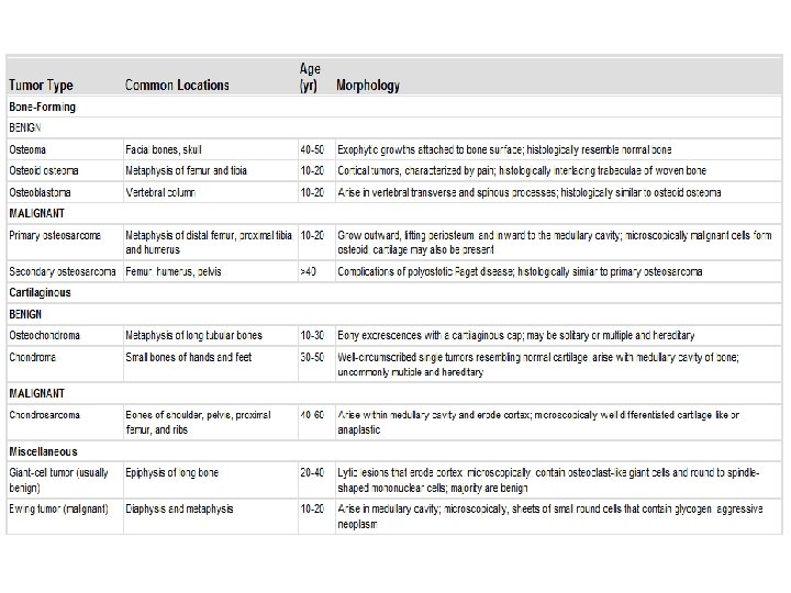

Osteoma • Developmental abnormality rather than true neoplasm • Middle age patients • Occurs in skull, face and sinuses • Bony hard protrusions • Solitary, multiple in Gardner syndrome • Mixture of woven and lamellar bones • Does not transform

Osteoid osteoma and osteoblastoma Benign tumors Teenage and middle age Arise in the cortex Similar histology and radiology (radiolucent nidus) • OO arises in proximal femur and tibia (<2 cm), painful (respond to aspirin) • Osteoblastoma arises in vertebra (large), painful, does not respond to aspirin • Treatment: excision, NO radiation • •

• Osteoid osteoma showing randomly oriented trabeculae of woven bone rimmed by prominent osteoblasts. The intertrabecular spaces are filled by vascular loose connective tissue
")
Osteosarcoma • Bone-producing tumor • Most common primary bone tumor (after excluding hematopoietic tumors) • Occur at all age groups, but >75% less than 20 • Occur at all sites, but Metaphysis of long bones (60% around knee) • Most osteosarcoma is primary, solitary, intramedullary, and poorly differentiated, producing a predominantly bony matrix

Pathogenesis • RB gene mutations occur in 60% to 70% of sporadic tumors, and individuals with hereditary retinoblastomas (due to germ-line mutations in the RB gene) • Spontaneous osteosarcomas also frequently exhibit mutations in genes that regulate the cell cycle including p 53, cyclins, cyclin-dependent kinases, and kinase inhibitors • Many osteosarcomas develop at sites of greatest bone growth

Morphology • Grossly, gray-white tumors exhibiting hemorrhage and cystic degeneration • Tumors frequently destroy the surrounding cortices and produce soft tissue masses • They spread extensively in the medullary canal, infiltrating and replacing the marrow, but only infrequently penetrating the epiphyseal plate or entering the joint space • Microscopically: cells vary in size and shape, and frequently have large hyperchromatic nuclei and mitoses • Production of mineralized or unmineralized bone (osteoid) by malignant cells • The neoplastic bone is typically coarse and ragged but can also be deposited in broad sheets • Cartilage and fibrous tissue can also be present in varying amounts. When malignant cartilage is abundant, the tumor is called a chondroblastic osteosarcoma

• Osteosarcoma. A, Mass involving the upper end of the tibia. The tan-white tumor fills most of the medullary cavity of the metaphysis and proximal diaphysis. It has infiltrated through the cortex, lifted the periosteum, and formed soft tissue masses on both sides of the bone. B, Histologic appearance, with coarse, lacelike pattern of neoplastic bone (arrow) produced by anaplastic tumor cells. Note the wildly aberrant mitotic figures (arrowheads).
")
Clinical • Painful enlarging mass • Spontaneous fracture • X-ray: triangular Growth (Codman triangle) between Cortex and periosteum Hematogenous spread

Osteochondroma • AKA exostoses, benign neoplasm • they are relatively common benign cartilage-capped outgrowths attached by a bony stalk to the underlying skeleton • Solitary osteochondromas are usually first diagnosed in late adolescence and early adulthood (male-to-female ratio of 3: 1) • multiple osteochondromas become apparent during childhood, occurring as multiple hereditary exostosis, an autosomal dominant disorder • Inactivation of both copies of the EXT gene in chondrocytes is implicated in both sporadic and hereditary osteochondromas • Osteochondromas develop at the metaphysis near the growth plate of long tubular bones, especially about the knee, also in pelvis, scapula, ribs, phalanges

Chondroma • Chondromas are benign tumors of hyaline cartilage • When they arise within the medulla, they are termed enchondromas; when on the bone surface they are called juxtacortical chondromas • Enchondromas are usually diagnosed in persons between ages 20 and 50; they are typically solitary and located in the metaphyseal region of tubular bones, the favored sites being the short tubular bones of the hands and feet • Ollier disease is characterized by multiple chondromas preferentially involving one side of the body • Maffucci syndrome is characterized by multiple chondromas associated with benign soft tissue angiomas • Enchondromas are translucent, central calcification, O-ring sign • Enchondromas are usually smaller than 3 cm. Microscopically, there is well -circumscribed hyaline matrix and cytologically benign chondrocytes

Chondrosarcoma • • • Cartilage-producing malignant tumors Can be intramedullary or juxtacortical Less common than osteosarcoma Usually >40 years Tumor grade is determined by cellularity, cytologic atypia, and mitotic activity Low-grade tumors resemble normal cartilage. Higher grade lesions contain pleomorphic chondrocytes with frequent mitotic figures Multinucleate cells are present with lacunae containing two or more chondrocytes Approximately 10% of patients with conventional low-grade chondrosarcomas have a second high-grade poorly differentiated component, might include osteosarcoma Most common sites: pelvis, shoulder, ribs (not long bones)

• Chondrosarcoma. A, Islands of hyaline and myxoid cartilage expand the medullary cavity and grow through the cortex to form a sessile paracortical mass. B, Anaplastic chondrocytes within a chondroid matrix.

Fibrous Cortical Defect and Nonossifying Fibroma • Fibrous cortical defects occur in 30% to 50% of all children >2 • Developmental defects rather than true neoplasms • The vast majority are smaller than 0. 5 cm and arise in the metaphysis of the distal femur or proximal tibia • 50% are bilateral or multiple. Larger lesions (5 -6 cm) develop into nonossifying fibromas • Fibrous cortical defects and nonossifying fibromas both present as sharply demarcated radiolucencies surrounded by a thin zone of sclerosis • Microscopically are cellular lesions composed of cytologically benign fibroblasts and activated macrophages, including multinucleated forms. The fibroblasts classically exhibit a storiform (pinwheel) pattern • Usually asymptomatic and resolve, if large, may cause pathologic fracture

• Fibrous cortical defect or nonossifying fibroma. Characteristic storiform pattern of spindle cells interspersed with scattered osteoclast-type giant cells

Fibrous Dysplasia • • FD is a benign tumor that is better regarded as developmental arrest All components of normal bone are present, but they fail to differentiate into mature structures FD occurs as one of three clinical patterns: (1) involvement of a single bone (monostotic), 70% of cases, early adolescents, In descending order of frequency: ribs, femur, tibia, jawbones, calvaria, and humerus are most commonly affected. Lesions are asymptomatic and usually discovered incidentally (2) involvement of multiple bones (polyostotic) 29%, earlier age, progress, femur is most common, causes marked deformity and fractures (3) polyostotic disease, associated with café au lait skin pigmentations and endocrine abnormalities, especially precocious puberty (Mc. Cune-Albright syndrome) result from a somatic (not hereditary) embryonic mutation yielding a Gprotein that constitutively activates adenyl cyclase with resultant cyclic adenosine monophosphate overproduction and cellular hyperfunctioning (precocious puberty) However, fibrous dysplasia can cause marked enlargement and distortion of bone, so that if the face or skull is involved, disfigurement can occur
 arising in a")
• Fibrous dysplasia. Curved trabeculae of woven bone (Chinese letters) arising in a fibrous tissue. Note the absence of osteoblasts rimming the bones

Morphology and clinical • Well-circumscribed, intramedullary lesions of varying sizes • By x-ray, lesions exhibit a characteristic groundglass appearance with well-defined margins • Symptomatic lesions are readily cured by conservative surgery • Polyostotic involvement is frequently associated with progressive disease, and more severe skeletal complications • Rarely, polyostotic disease can transform into osteosarcoma, especially following radiotherapy
 are primary malignant small")
Ewing sarcoma • Ewing sarcoma and primitive neuroectodermal tumors (PNETs) are primary malignant small round-cell tumors of bone and soft tissue • They share an identical chromosome translocation, they should be viewed as the same tumor, differing only in degree of differentiation PNETs demonstrate neural differentiation whereas Ewing sarcomas are undifferentiated by traditional marker analysis

Pathogenesis • Next to osteosarcomas, they are the second most common pediatric bone sarcomas • Most patients are 10 to 15 years old • 80% are younger than age 20 • Boys are affected slightly more frequently than girls • Racial predilection; blacks are rarely afflicted • Translocation that causes fusion of the EWS gene on 22 q 12 with a member of the ETS family of transcription factors, most commonly FL 1 gene on 11 q 24, and the ERG gene on 21 q 22 • The resulting chimeric protein functions as a constitutively active transcription factor to stimulate cell proliferation

• Ewing sarcoma. Sheets of small round cells with scant, cleared cytoplasm
, pelvis Aggressive, highly invasive, necrotic")
Arises in medullary cavity Most common: long bones (femur), pelvis Aggressive, highly invasive, necrotic Sheets of primitive cells, slightly larger than lymphocytes, with occasional presence of Homer. Wright rosettes (tumor cells circled about a central fibrillary space) indicates neural differentiation • X-ray: bone mass, lytic lesions. There is a characteristic periosteal reaction depositing bone in an onionskin fashion • •

Metastasis More common than primary tumors Rich vascularity, nutrients, slow perfusion Mostly in axial skeleton Cancer itself does not erode the bone Most cancer cells secret prostaglandins, interleukins, and PTHRp that stimulate osteoclastic bone resorption causing lytic lesions • Some cancers (prostate) stimulate osteobasts (sclerotic lesions) • Mixed lytic/blastic lesions are common • • •

Arthritis • • Osteoarthritis Gout Pseudogout Lyme disease

Osteoarthritis • • • Degenerative joint disease The most common joint disorder Important cause of physical disability in individuals over the age of 65 The fundamental feature of osteoarthritis is degeneration of the articular cartilage Bone changes are secondary Inflammation can be present as a minor component Primary OA: most common, insidious onset, no initiating factor, oligoarticular Secondary OA: 5% of cases, young age, history of trauma, DM, hemochromatosis, marked obesity, one or many joints Gender has some influence; knees and hands are more commonly affected in women, whereas hips are more commonly affected in men

Pathogenesis • Normal function of joint cartilage: prevents friction and spreads load across the joint to protect the underlying bone • Normal cartilage is elastic, regain normal architecture after compression (proteoglycans), and to have high tensile strength (collagen II), both produced by chondrocytes • Mechanical stresses and aging causes damage to chondrocytes • Genetic factors also seem to contribute to osteoarthritis susceptibility, particularly in the hands and hips, but the responsible genes are not known • Degenerating cartilage contains more water and less proteoglycan and collagen, its function is compromised • Chondrocytes in the deeper layers proliferate to compensate, but with time, they fail

Morphology • The earliest structural changes in osteoarthritis include enlargement, proliferation, and disorganization of the chondrocytes in the superficial part of the articular cartilage • Subsequently, vertical and horizontal cracking of the matrix occur as the superficial layers of the cartilage are degraded • With time, the subchondral bone plate is exposed • Friction smooths and burnishes the exposed bone, giving it the appearance of polished ivory (bone eburnation). The underlying cancellous bone becomes sclerotic and thickened • Small fractures can dislodge pieces of cartilage and subchondral bone into the joint, forming loose bodies (joint mice) • Mushroom-shaped osteophytes (bony outgrowths) develop at the margins of the articular surface • Synovium moves into subchondral bone and forms cysts. In severe disease, a fibrous synovial pannus covers the peripheral portions of the articular surface

• Osteoarthritis. A, Histologic demonstration of the characteristic fibrillation of the articular cartilage. B, Severe osteoarthritis with 1, Eburnated articular surface exposing subchondral bone. 2, Subchondral cyst. 3, Residual articular cartilage

Clinical Features • Osteoarthritis is an insidious • Patients, in their 50 s and 60 s • Characteristic symptoms include deep, aching pain exacerbated by use, morning stiffness, crepitus, and limited range of movement • Osteophytes cause nerve impingement (radicular pain, muscle spasms, muscle atrophy, and neurologic deficits) • Heberden nodes in the fingers, representing prominent osteophytes at the distal interphalangeal joints, are characteristic in women. • With time, significant joint deformity can occur, but unlike rheumatoid arthritis, fusion does not take place.

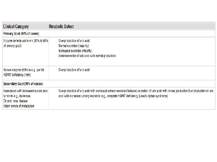
Gout ﺍﻟﻨﻘﺮﺱ • Accumulation of excessive amounts of uric acid (end product of purine metabolism) • Recurrent episodes of acute arthritis, sometimes accompanied by the formation of large crystalline aggregates called tophi, and chronic joint deformity • All of these result from precipitation of monosodium urate crystals • Although an elevated level of uric acid is an essential component of gout, not all such individuals develop gout, indicating that influences besides hyperuricemia contribute to the pathogenesis • Gout is traditionally divided into primary and secondary forms, accounting for about 90% and 10% of cases, respectively • Primary gout, basic cause is unknown or (less commonly) when it is due to an inborn metabolic defect that causes hyperuricemia • In secondary gout the cause is known, but gout is not necessarily the main or even dominant clinical disorder

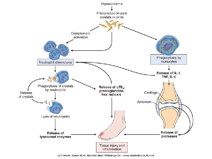
Types • Acute arthritis: dense neutrophilic infiltrate permeating the synovium and synovial fluid. Long, slender, needle-shaped monosodium urate crystals are frequently found in the cytoplasm of the neutrophils and in the synovium • Chronic tophaceous arthritis: repetitive acute attacks. Crust in the synovium (hyperplastia, fibrosis, inflammatory cells), then forms a pannus that destroys the underlying cartilage, and leading to bone erosions. In severe cases: ankylosis • Tophi: large aggregations of urate crystals surrounded by an intense inflammatory reaction of lymphocytes, macrophages, and foreign-body giant cells. Tophi appear in cartilage of joints, ligaments, tendons, and soft tissues, including the ear lobes, nasal cartilages, and skin of the fingertips • Gouty nephropathy: medullary tophi, intratubular precipitations, or free uric acid crystals and renal calculi. Secondary complications such as pyelonephritis can occur, especially when there is urinary obstruction

• Gout. A, Amputated great toe with white tophi involving the joint and soft tissues. B, Photomicrograph of a gouty tophus. An aggregate of dissolved urate crystals is surrounded by reactive fibroblasts, mononuclear inflammatory cells, and giant cells.

Pseudogout AKA chondrocalcinosis Calcium pyrophosphate crystal deposition disease Pseudogout typically first occurs in those age 50 or older Accumulation and crystallization with calcium Recruitment and activation of inflammatory cells Joint involvement can last from several days to weeks may be monoarticular or polyarticular; the knees, followed by the wrists, elbows, shoulders, and ankles, are most commonly affected • Ultimately, approximately 50% of patients experience significant joint damage • Therapy is supportive; no known treatment prevents or retards crystal formation • •

Infectious arthritis • Hematogenous, or complication of osteomyelitis • Rapid destruction of joint structures and permanent deformity (sudden pain, fever) • Haemophilus influenzae <2 years • S. aureus is the main causative agent in older children and adults • gonococcus is prevalent during late adolescence and young adulthood • Individuals with sickle cell disease are prone to infection with Salmonella at any age

Lyme arthritis • • • Borrelia burgdorferi, transmitted by deer ticks, common in the United States Involves multiple organ systems and is usually divided into three stages In stage 1 Borrelia spirochetes multiply at the site of the tick bite and cause an expanding area of redness (erythema chronicum migrans) + fever and lymphadenopathy but usually disappears in a few weeks' time In stage 2, the early disseminated stage, spirochetes spread hematogenously and cause secondary annular skin lesions, lymphadenopathy, migratory joint (large joints) and muscle pain, cardiac arrhythmias, and meningitis, often with cranial nerve involvement. If the disease is not treated, antibodies develop that are useful for serodiagnosis of Borrelia infection. Some spirochetes, however, escape host antibody and T-cell responses by sequestering themselves in the central nervous system or as intracellular forms within endothelial cells. In stage 3, the late disseminated stage, which occurs 2 or 3 years after the initial bite, Lyme Borrelia organisms cause a chronic arthritis, sometimes with severe damage to large joints, and an encephalitis that varies from mild to debilitating. Lyme arthritis develops in roughly 60% to 80% of untreated patients and is the dominant feature of late disease. The arthritis may be caused by immune responses against Borrelia antigens that cross-react with proteins in the joints. Histologically, there is a chronic papillary synovitis with synovial hyperplasia, fibrin deposition, mononuclear cell infiltrates In only 25% of cases do silver stains reveal a sprinkling of organisms Chronic arthritis with pannus formation and permanent deformities develops in roughly one of ten patients.
- Slides: 67