Congenital anomalies of CNS Abla sultan Felasten Abed

Congenital anomalies of CNS Abla sultan Felasten Abed

Abla sultan congenital anomalies of CNS Abla sultan Qudah Felasten Nasser Abed

Development of the neural tube

Development of the brain

Anencephaly

Anencephaly • Serious NTD caused by failure of closure of cranial neuropore in which : - The brain (cerebral hemispheres) hemispheres is absent - Cranial vault are grossly malformed. - The cerebellum are reduced or absent. - The hindbrain is present. -- leading to: early fetal loss, stillbirth, or neonatal death after few hours/ days. if a baby born with anencephaly is usually: blind, deaf, unaware of its surroundings and unable to feel pain Associated anomalies : - cleft lip/palate , omphalocele.

Etiology 1. Inadequate Folic acid or Antagonists such as : Valproic acid , carbamazepine , phenobarbital , methotrexate , trimethoprim 2 - Maternal type 1 diabetes mellitus 3 -Maternal Hyperthermia 4 -Genetics 5 - Amniotic band syndrome.

Screening Lab studies : mainly during second trimester : - maternal serum AFP , AF AFP ↑. Imaging studies : - US (identified from 13 week gestation) • absence of brain and calvaria superior to the orbits • reduced crown-rump length • Later on : polyhydramnios in 50%

Cephalocele • refers to the outward herniation of CNS contents through a defect in the cranium. ( failure of rostral / cranial end closure ) • Most commonly occipital then Parietal. • Comment on : 1 -Location 2 -Contents 3 -Associated anomalies 4 -Relation to vascular structure

1 -Location occipital ethmoidal parital frontal

2 -contents

3 -associated anomalies -Trisomy 13 , 18 -hydrocephalus / microceph -Meckle Gruber syndrome(occipital encephalocele with multiple renal cysts , postaxial polydactyly ). - and chiari 3 malformation

• Markers : MS AFP ↑. • Imaging : US , MRI. • Management : Surgery to remove the herniated sac ( tissue should be excised) and repair the opening in the skull.

Chiari Malformation • Caudal protrusion of “peg-shaped” cerebellar tonsils below foramen magnum • divided into 4 types

Type 1 • Herniation of cerebellar tonsils through foramen magnum • no herniation of brain stem • most common form • usually diagnosed in adolescence or adulthood • cause syringomyelia (in which a cyst or cavity forms within the spinal cord. This cyst, called a syrinx, can expand elongate over time)

Type 1 clinical presentation • Compression of upper cervical cord resulting in myelopathy. • Compression of cerebellum may result in ataxia, dysmetria, intentional tremor , nystagmus. • Disruption of CSF flow through foramen magnum : ↑ ICP , suboccipital headache. Neck pain, vomiting , visual defects , hydroceph < 10% of cases • Syringomyelia related symptoms: cape-like loss of pain and temp sense , weakness and wasting of the small muscle of the hand , and progressive motor deficit of the lower and upper limbs

Type 2 • caudal displacement of the cerebellar vermis , 4 th ventricle and medulla oblongata below foramen magnum , present in infancy , almost always association with myelomeningocele. -- signs of brainstem dysfunction predominate: swallowing/feeding difficulties, stridor, apnea, respiratory depression. nystagmus. Hydroceph. And syringomyelia are more common than type 1.

• Type 3 : similar to Chiari II but with an occipital and/or high cervical encephalocoele. Type 4 : cerebellar hypoplasia or agenesis. Management : posterior fossa decompression: your surgeon removes a small section of bone in the back of your skull, relieving pressure by giving your brain more room. If you have a syrinx or hydrocephalus, you may need a tube (shunt) to drain the excess fluid.

Arachnoid Cysts • Arachnoid cysts are the most common type of brain cyst. They are often benign congenital, or present at birth (primary arachnoid cysts). Head injury or trauma can also result in a secondary arachnoid cyst. • An arachnoid cyst forms when the layers of the arachnoid membrane split apart and become filled with cerebrospinal fluid. • The cysts are fluid-filled sacs, not tumors mostly asymptomatic Locations : • Sylvian fissure 50% • Cerebellopontine angle 10% • Quadrigeminal 10% • Suprasellar 10% • Vermian 8% • Cerebral convexity 5% • Other 7%

Sylvian fissure • Most common • Clinical features : 1 - ↑ICP : headache , Nausea , Vomiting. 2 - Seizures 3 - with minor head trauma > hrg > acute presentation.

Cerebellopontine angle • Clinical features as in acoustic neuroma : - SN hearing loss - Tinnitus - Vertigo • May cause compression on 5 th CN.
 • In children and adolescents. • Clinical features :")
Suprasellar (the only extradural one) • In children and adolescents. • Clinical features : 1 - hydrocephalus. 2 - visual impairment. 3 - Endoc. Dysfunction.

• Management : - asymptomatic , no ventricular distortion , enlargment : follow up at regular intervals. - otherwise : 1 - Craniotomy, excision of the cyst wall and opening of the membranes to allow drainage into the basal cisterns. 2 - Shunt : cyst – peritoneum

Dandy-Walker Malformation • Characterised by : 1 - Complete or partial agenesis of the cerebellar vermis(the part joining the two hemispheres of the cerebellum) 2 - Cystic dilatation of the 4 th ventricle( failure of foraminal outlets to open ) 3 - An enlarged posterior fossa with upward displacement of tentorium

• symptoms: symptoms 1 - 70 -90% of patients have hydrocephalus (present as increasing head size, vomiting, excessive sleepiness, irritability) 2 - If not diagnosed postnatally : in childhood the major presenting features are ataxia , and delayed motor development. 3 -Associated anomalies : agenesis of the corpus callosum, occipital encephalocele, spina bifida, syringomyelia , cleft palate , cardiac and renal anomalies. managment cyst-peritoneal shunt / VP shunt. .

Congenital spinal deformities Felasten Naser Abed

Spinal dysraphism : Is a Medical term used to describe group of congenital anomaly of the spine , characterized by midline defect affecting the nervous tissue and its bony and soft tissue covering. Spina Bifida : refer to defective fusion of posterior spinal bony elements but was and still widely used to refer to spinal dysraphism in general

Important terms : Placode: neural tissue herniating within spinal dysraphism Tethered cord syndrome : it is a clinical syndrome that involves progressive neurological deterioration whose picture includes motor and sensory dysfunction may be caused by low lying conus or normally positioned conus Tethered cord : imaging data refers to low lying conus being its end below l 2 level

Emryology The anterior neuropore closes at gestation day 25. The caudal neuropore closes at day 28 ( failure of spontanous closure of it. ) There is several risk factors have been identified: 1)History of neural tube defect in sibling 2)Folate deficiency in early pregnancy 3)Triosomy 13 and 18 4) Increase in risk associated with some anti-epilepsy drugs in pregnancy , particularly sodium valproate
 Open spinal dysraphism /Spina Bifida Aperta 2) Closed")
Major forms of spinal dysraphism 1) Open spinal dysraphism /Spina Bifida Aperta 2) Closed spinal dycraphism with subcutanous mass / Spina Bifida Cystica 3) Closed spinal syraphism without subcutanous mass / Spina Bifida Occulta
 spina bifida Aperta")
Back mass : spina bifida cystica ( covered by skin ) spina bifida Aperta (not covered by skin) No back mass : spina bifida occulta
 Myelomeningocele 2) Myelocele (Myeloschisis) 3) Hemimyelocele 4) Hemimyelomeningocele")
Spina bifida Aperta 1) Myelomeningocele 2) Myelocele (Myeloschisis) 3) Hemimyelocele 4) Hemimyelomeningocele

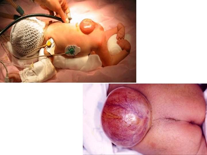
Myelomeningocele : -Congenital defect in vertebral arches with cystic dilatation of meninges and structural or functional abnormality of spinal cord or cauda equina -most common ocuurs 1 in 1000 birth and important form of dysraphism -May occur at any level but its most common in lumbar and lumbosacral ON MRI : extension of the neural placode to the skin surface caused by expansion of the underlying subarachnoid space which is charactarestic of myelomeningocele

Clinical features : ◦ Sensory and motor changes distal to anatomic level producing varying degrees of weakness ◦ Urinary and fecal incontinence ◦ wisted or abnormal legs and feet; for example, clubfoot ◦ 65 -85% of patients have hydrocephalus ◦ Most have Type II Chiari malformation Investigations : - Prenatal Measuring alpha-fetoprotein in amniotic fluid at 14 -16 weeks -Acetylcholinesterase (ACh. E) in amniotic fluid is a helpful confirmatory test -Prenatal ultrasound examination detect the myelomeningioceal as well as condition that can lead to it like hydrocephalis the chiari malphormation -Ultrasound examination can be confirmed by MRI and we perform fetal echocardiogram to rule out any problem in the heart

Management �Assessment of the sac and the coverings �Neurological evaluation �Examination of other associated conditions ◦ Within CNS e. g. hydrocephalus ◦ Extracranial e. g. gastrointestinal , urinary �Surgical procedure : �Closure of the defect over the spinal cord �Preserve all neural tissue and reduce it into the intervertebral canal �Obtain a watertight clocure of dura lining sac �Cover defect with muscle fscia and skin �Lower-extremity deformity correction to preserve neurological status prevent CNS infecion Most common cause of mortality are complications from Chiari malfomation ( respiratory arrest and aspiration ) , whereas late mortality is due to shunt mal function

Selective criteria were developed and the children were exclude from treatment where those with : 1) Paralysis at l 2 to l 3 or above 2)Marked hydrocephalus 3)Kyphosis 3)Other major congenital abnormalities or birth injury
: Exposed, unfused neural tissue without skin/meningeal covering.")
Myeloschisis (myelocele): Exposed, unfused neural tissue without skin/meningeal covering.

Q: why chiari 2 associated with spina bifida aperta ?

Spina bifida cystica:

Meningocele : -Congenital defect in vertebral arches with cystic distension of meninges, but no abnormality of neural tissue. One third have some neurologic deficit -Less common than myelomeningocele -Most commonly in lumbosacral area

• Etiology : 1. Primary failure of neural tube closure 2. Rupture of previously closed neural tube due to overdistention • Clinical features – Usually no disability – low incidence of associated anomalies like chiari 2 and hydrocephalus • Investigations : – Plain film , CT , MRI , genitourinary investigations • Treatment : – Surgical excision and tissue repair – ( excellent results )

Lipomeningocele -Spina bifida cystica -Placode-lipoma interface , is located within the spinal canal covered by subcutanous fatty mass -Less common disorder -Neurological examination is usually normal at birth and progressive neurological deficits occur resulting from growth and tethering of the spinal cord

lipomyelomeningocele: -Spina bifida cystica -Placode-lipoma interface , is located outside the spinal canal because of expansion of the subarachnoid space covered by subcutanous fatty mass
 Simple dysraphic state :")
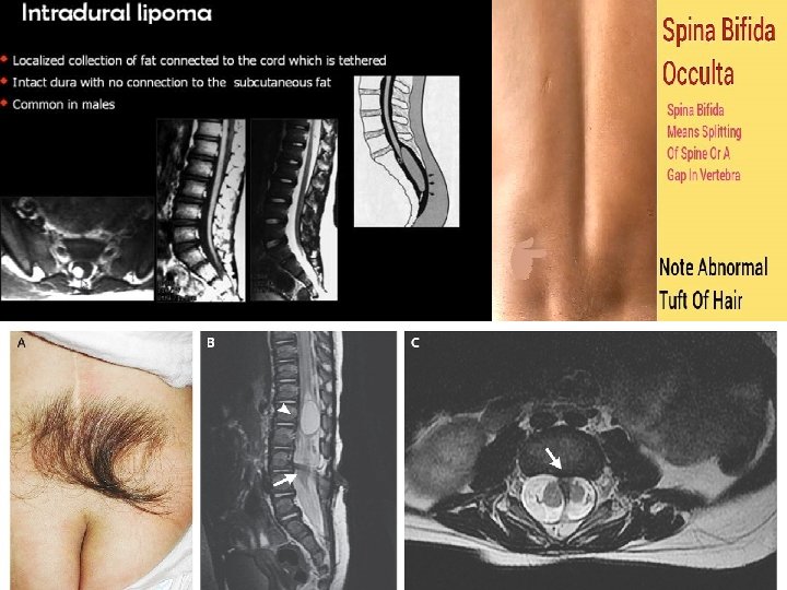
Spina bifida occulta Closed spinal dysraphism without sc mass 1) Simple dysraphic state : Usually baby come with tethered cord syndrome without cutanous (skin) symptoms -intradural lipoma -Filar lipoma - dermal sinus -tight filum terminale (fibrous thickening ) -persistance of the terminal ventricle
 Complex dysrapic state: There is cutanous manifestation ( hair , dimple , nevus")
2) Complex dysrapic state: There is cutanous manifestation ( hair , dimple , nevus , pigmentation) Disorder of the midline : -dorsal enteric fistula - neuroenteric cyst -diastematomyelia Disorder of notochordal formation -caudal regression - Segmental spinal dysgenesis

• A bony deficit usually found in the laminae of the lumbosacral spine to a medline fusion defect • it is an incidental radiological finding and is present in up to 20% of adults • Most common at L 5 or S 1 • In the vast majority there is NO neurological involvement and the lesion is asymptomatic throughout life • The underlying intraspinal lesion can often be suspected from an overlying skin lesion such as : – Dimples – Sinus tract – Fatty mass – Heamangioma – Abnormal tuft of hair

Slowly progressive neurological dysfunction involving : 1. Bowel and bladder disturbances 2. Progressive weakness of legs and foot deformities 3. Back pain 4. Sensory disability 5. Progressive scoliosis Depending on the level of abnormality neurological examination will show evidence of either upper/ lower motor neuron damage Less sever abnormalities will be associated with a low-lying conus with tethering of the cord by enlarged filum terminale or fatty tissue Treatment : If there is underlying lesion we remove it with preservation of neural tissue & untether the cord If NO underlying lesion no need for treatment

Investigations : X-ray : absence of the spinous process along with minor amounts of neural arch CT and MRI : to exclude spinal anomalies

Diastematomyelia - Focal Cleft /split cord -The spinal cord is bifid and the two hemi cords are separated by a bony spur or dural band -May or may not involve the dura -Progressive neurological dysfunction will occur due to traction on the cord that is transfixed during growth period

Tethered cord Spinal cord is abnormally attached to tissues around ( most commonly at the base of spine ) Abnormally low conus medullaris. Usually associated with a short, thickened filum terminale, or with an intradural lipoma (other lesions, e. g lipoma extending through dura, dermal sinuses tracts, dermoids. or diastematomyela Most common in myelomeningocele (MM). Diagnosis must be made clinically in MM, as almost all of these patients will have tethering radiographically. The spinal cord normally hangs loose in the canal free to move up and down with growth and bending and streching , a tethered cord is held taut at the end

Clinical features: In children : symptoms may include lesions , hairy patches , dimples , or fatty tumors on the lower back , Foot and spinal deformeties , weakness in the legs , Low back pain , Scoliosis , And incontenece In adults : onset of symptoms typically include severe pain ( in the lower back and radiating in to the legs , groin , and perineum ) , bilateral muscle weakness and numbness , bladder and bowel incontinence Treatment : • In children : early surgery is recommended to prevent further neurological deterioration • In adults : surgery to free (detether) the spinal cord can reduce the size and further development of cysts in the cord and may restore some function or alleviate other symptoms

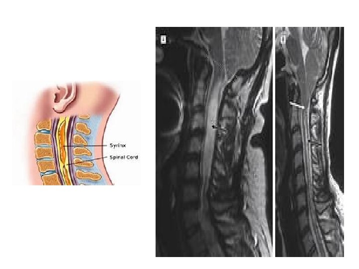
syringomyelia Is a genetic term referring to a disorder in which a cyst or cavity (fluid-filled )forms within the spinal cord, this cyst called a syrinx , can expand elongate over time destroying spinal cord Patients may present with sensory disturbances weakness of the hands loss of pain and temperature sensation or progressive kyphoscoliosis. Hindbrain herniation can lead to headache neck pain , ataxia , spasticity and lower cranial nerve palsies. Associated with Chiari I malformation , scoliosis and other congenital malformations; acquired causes include trauma and tumors. Most common location cervical > thoracic >> lumbar Syrinx = tube, as in “syringe. ”

Types of syringomyelia • Syringomyelia due to blockage of CSF circulation (without fourth ventricular communication) • Syringomyelia due to spinal cord injury • Syringomyelia and spinal dysraphism • Syringomyelia due to intramedullary tumors • idiopathic syringomyelia
 Treat underlying pathology : ◦ If associated with a type I Chiari")
Treatment 1) Treat underlying pathology : ◦ If associated with a type I Chiari , then a posterior cranial fossa decompression is indicated to restore cerebral spinal fluid outflow through formen of Magendie ◦ If the cause is an intramedullary spinal cord tumor , excision of tumor should restore the communication with central canal ◦ If post traumatic cause , the obstruction should be revealed by decompression 2) Not with standing this , fluid diversion to the peritoneum or plural space syingoperitonial or syringopleural ) has been used with moderate success

Thank you References : Essential neurosurgury
- Slides: 58