Conduction System of the Heart Faisal I Mohammed


")


 nerves, which release")


- Slides: 29
Conduction System of the Heart Faisal I. Mohammed, MD, Ph. D 1
The importance of calcium influx through the slow voltage gated calcium channels 2
Mechanism of Cardiac Muscle Excitation, Contraction & Relaxation
Intracellular Calcium Homeostasis… 2 4
Objectives l l l 5 List the parts that comprise the conduction system Explain the mechanism of slow response action potential (pacemaker potential) Point out the regulation of the conduction system potential by Autonomic Nerves
Structures of the conduction system 6
7
Conducting System of Heart 8
Heart Physiology: Sequence of Excitation 9
Autonomic Innervation of the Heart 10
Intrinsic Cardiac Conduction System Approximately 1% of cardiac muscle cells are autorhythmic rather than contractile 75/min 40 -60/min 30/min 11
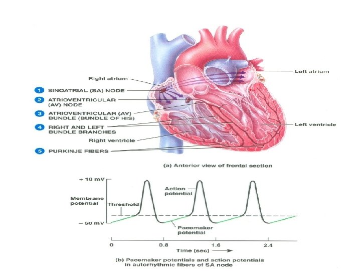
Intrinsic Conduction System Function: initiate & distribute impulses so heart depolarizes & contracts in orderly manner from atria to ventricles. SA node AV node Bundle of His Bundle Branches Purkinje fibers 12
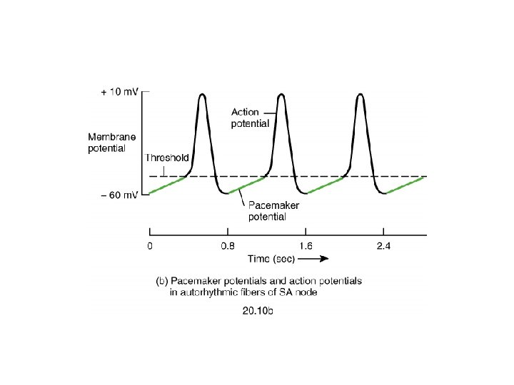
Sinus Node l l 13 Specialized cardiac muscle connected to atrial muscle. Acts as pacemaker because membrane leaks Na+ and membrane potential is -55 to -60 m. V When membrane potential reaches -40 m. V, slow Ca++ channels open causing action potential. After 100 -150 msec Ca++ channels close and K+channels open more thus returning membrane potential to -55 m. V.
Fast Response Action Potential of Contractile Cardiac Muscle Cell
Pacemaker and Action Potentials of the Heart 17
Slow Response Action Potential (Pacemaker Potential)
Intrinsic rate and speed of conduction of the components of the system SA node 60 -80 action potential /min (Pacemaker) l AV node 40 -60 action potential /min l Purkinje 15 -40 action potential /min Conduction Speed l SA node: slow speed of conduction l Ventricular and Atrial muscle: Moderate speed l AV node: slowest speed of conduction l Purkinje fibers: Fastest speed of conduction l Ectopic Pacemaker- Abnormal site of pacemaker l 19
Extrinsic Innervation of the Heart
Pacemaker Function
Autonomic neurotransmitters affect ion flow to change rate l l Sympathetic – increases heart rate by Ca+2 & If channel (net Na+) flow Parasympathetic – decreases rate by K+ efflux & Ca+2 influx What part of the graph is not changed by autonomic influences?
Effect of Sympathetic & Parasympathetic Stimulation Sympathetic 0 Parasympathetic 3 4
Regulation of the heart beat l l 24 Sympathetic from the cardiac plexus supplies all parts of the heart (atria, ventricle and all parts of the conduction system) Parasympathetic from Vagus nerves supply mainly the atria, SA and AV nodes, very little supply to ventricles Sympathetic: increase the permeability of the cardiac cells to Na+ and Ca++ i. e Positive Chronotropic and positive Inotropic action Parasympathetic: Increase the permeability of the cardiac cells to K+ and decrease its permeability to Na+ and Ca++
Sinus Node is Cardiac Pacemaker Normal rate of discharge in sinus node is 70 -80/min. ; A-V node - 40 -60/min. ; Purkinje fibers - 15 -40/min. l Sinus node is pacemaker because of its faster discharge rate l Intrinsic rate of subsequent parts is suppressed by “Overdrive suppression” l 25
Ectopic Pacemaker l This is a portion of the heart with a more rapid discharge than the sinus node. l Also occurs when transmission from sinus node to A-V node is blocked (A-V block). 26
Parasympathetic Effects on Heart Rate l l l 27 Parasympathetic (vagal) nerves, which release acetylcholine at their endings, innervate S-A node and A-V junctional fibers proximal to A-V node. Causes hyperpolarization because of increased K+ permeability in response to acetylcholine. This causes decreased transmission of impulses maybe temporarily stopping heart rate.
Sympathetic Effects on Heart Rate Releases norepinephrine at sympathetic ending l Causes increased sinus node discharge (Chronotropic effect) l Increases rate of conduction of impulse (Dromotropic effect) l Increases force of contraction in atria and ventricles (Inotropic effect) l 28
Thank You