Concentration techniques Flotation Method Concentration Methods 1 Sedimentation


- Slides: 28
Concentration techniques Flotation Method
Concentration Methods 1. Sedimentation method § Modified Formal- Ether sedimentation technique § Acid- Ether sedimentation technique 2. Flotation method § Saturated Salt Solution technique § Sheather’s Sugar Centrifugal Flotation technique § Zinc Sulphate Centrifugal Flotation technique
Flotation technique § These method use the high specific gravity of a solution to float the lighter ova and cyst. They can be improved by centrifugation. • Advantage: • Easy to perform. • Disadvantage: • Delay in examination can result in distortion. • Larvae and some fluke eggs do not concentrate. • Frequent checking of specific gravity.
Flotation Methods SATURATED SALT SOLUTION
Materials and Method 1. Boil granular sodium chloride in excess in water to produce a saturated solution which when cooled has a specific gravity of 1. 18 - 1. 2. 2. Half fill a wide- mounted flat bottomed container with the saturated salt solution. 3. Emulsify 1 gm of feces in the solution and strain it to remove the debris from the surface. 4. Pour the filtrate into meniscus and fill it to the top with saturated salt solution. 5. Lay a glass slide or large coverslip over the top, avoiding any bubbles being trapped. 6. Leave for 20 min before quickly inverting the slide. 7. Scan for ova using the 10 x objectives.
Saturated Salt solution technique § Advantages: – It is cheap preparation using simple apparatus. – It concentrates nematode ova well. § Disadvantages: – It doesn’t concentrate cysts.
Flotation Methods SHEATHER’S SUGAR CENTRIFUGAL FLOTATION TECHNIQUE
Materials and Method § Sheather’s sugar solution: – Table sugar -------------------500 gm – Distilled water ----------------- 320 ml – Phenol crystal ( melt in hot water bath) ----- 6. 5 gm
Procedures 1. Soften 1 gm of feces with water to a soft. 2. Strain the aqueous suspension through a wire sieve. 3. Mix 1 part aqueous suspension with 2 part of Sheather's sugar solution. 4. Pour into a centrifuge tube, centrifugation 1500 rpm for 10 minutes. 5. Pour the supernatant into a meniscus and add a sufficient solution to bring the meniscus to the top. 6. Place a coverslip and wait for 10 minutes. 7. Examine under microscope.
Sheather’s Sugar solution technique § Advantages: – Reveals most nematode eggs and protozoan cyst. § Disadvantages: – Flukes eggs and tape worm eggs are not demonstrate well. – Also most nematode larvae are not demonstrate well.
Intestinal Protozoa Balantidium coli
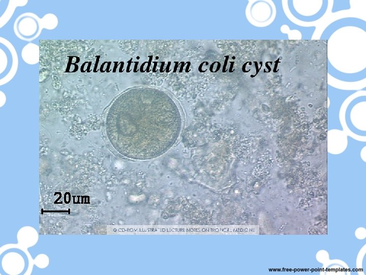
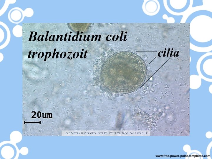
Balantidium coli § Is a parasitic species of ciliate protozoa that cause balantidiasis or Balantidium dysentery. § Balantidium coli has two developmental stages, a trophozoite stage and a cyst stage. § The cyst is the infective stage measures about 50 to 70 µm in diameter, characterized by the presence of a large kidney -shaped macronucleus. § The trophozoite is oval with 2 nuclei and measures approximately 50 to 100 µm long and 40 to 70 µm wide.
B. coli Cyst
B. coli Trophozoite
Intestinal Protozoa Cryptosporidium parvum
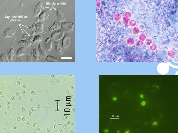
Cryptosporidium parvum § Infect human and most mammals. § The infective stage is oocyst containing sporozoites measuring 5 u in diameter. § The diagnostics stage is oocyst containing 4 sporozoites. § Diagnosis: – Detecting oocyst in stool. – Acid-fast stain.
Cryptosporidium parvum oocyst
Tissue Protozoa Toxoplasma gondii
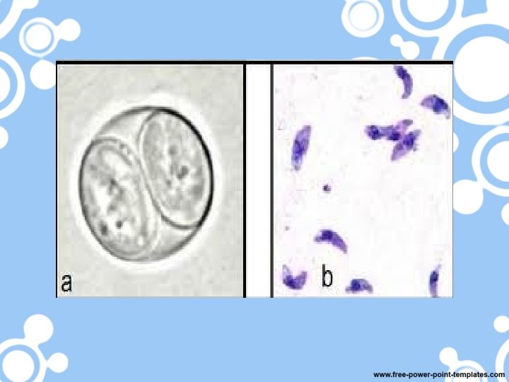
Toxoplasma gondii • Habitat: reticulo-endothelial system, monocyte, and muscle fiber and cause toxoplasmosis. • Humans can acquire Toxoplasma gondii infection by ingestion cyst or sporylated oocyst – Cyst: precipitated in flesh of cow. – Sporulated oocyst: cat feces. o Intermediate host: Human and cat. o Definitive host: Cat. • The tachyzoites are crescent-shaped and measure about 5 µm in length. • Diagnostic stages: – Diagnosis also can be done by detection antibody Ig. G, Ig. M, Ig. E and Ig. A.
Tachyzoites
Urogenital Protozoa Trichomonas vaginalis
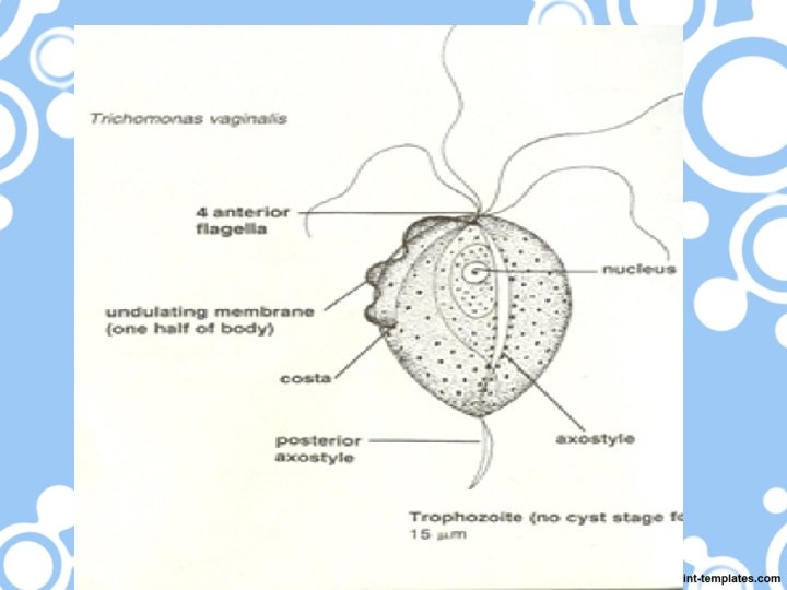
Trichomonas vaginalis § Is the most common cause of vaginitis, inhabit the urogenital system § There is no cyst stage but only have trophozoite stage. § The trophozoite measures about 15 x 10 µm. The trophozoite has a single nucleus and four flagella and undulating membrane. § Diagnosis: – Male: finding trophozoite in urethral prostatic discharge – Female: finding trophozoite in vaginal discharge.
T. vaginalis Trophozoite