composition Digestive tube Mouth Pharynx Esophagus Stomach Small

组成composition 消化管Digestive tube • 口腔 Mouth • 咽 Pharynx • 食管 Esophagus • 胃 Stomach • 小肠Small intestine Superior digestive tube 上消化道 十二指肠 Duodenum 空肠 Jejunum 回肠 Ileum Inferior digestive tube 下消化道 • 大肠Large intestine 消化腺Digestive glands • • • 大唾液腺Major salivary glands 肝Liver 胰Pancreas Function: ingestion, digestion, absorption, egesting

Mouth Major salivary glands Pharynx Esophagus Stomach Liver Pancreas Duodenum Large intestine Ileum Jejunum

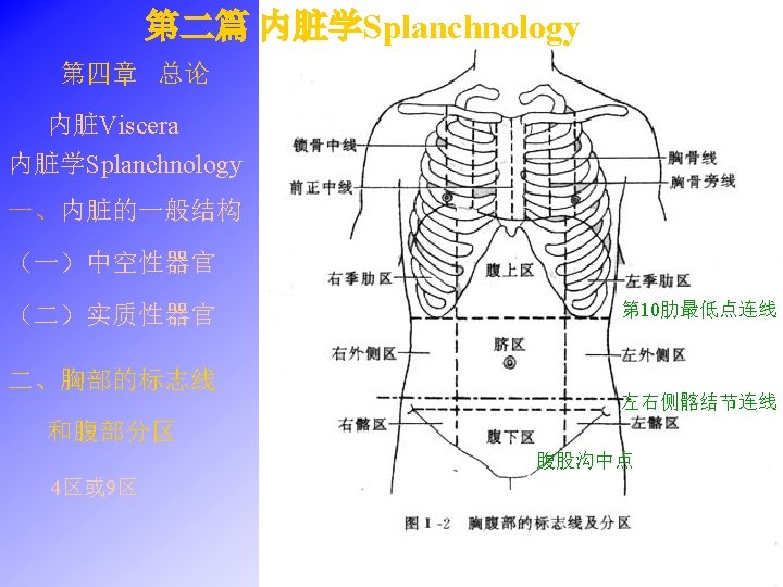
口腔 Oral Cavity Consists of two parts vestibule : between cheeks and lip and teeth • 固有口腔Oral cavity proper: within arch of teeth • 口腔前庭Oral Boundaries of oral cavity proper • • Anterior and lateral: gum and teeth Posterior: isthmus of fauces Roof: palate Floor: tongue, muscles and mucous membrane Oral vestibule leads, by the space behind the molar teeth, into the oral cavity proper

腭Palate Two parts • Hard palate: anterior 2/3, formed by the maxilla and palatine bone • Soft palate: posterior 1/3 – – 腭帆Velum palatinum 腭垂 Uvula 腭舌弓Palatoglossal arch 腭咽弓Palatopharyngeal arch 咽峡Isthmus of fauces formed by posterior border of velum palatinum, both side of palatoglossal arches, and root of tongue.

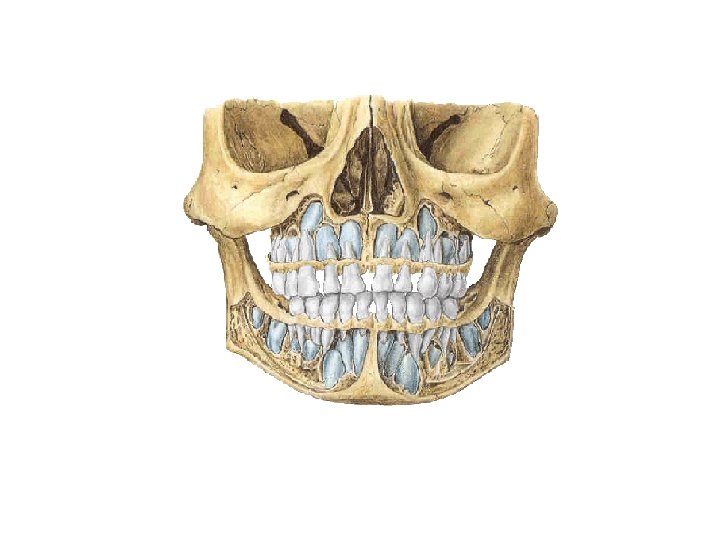
牙Teeth General features • Two sets: – 乳牙Deciduous – 恒牙Permanent • Classification: – – 切牙Incisors 尖牙Canine 前磨牙Premolars 磨牙Molars

Deciduous teeth: are 20 in number • Ten teeth in each mandibular and maxillary arch • Central incisor中切牙, lateral incisor侧切牙, canine尖牙, first molar第一磨牙 and second molar第二磨牙 in each quadrant Upper jaw Lower jaw Ⅰ in. Ⅱ Ⅲ Ⅳ Ⅴ total 20 in. can. mol. • Eruption: stars at about 6 mouth of age and continues to beginning of 3 rd year • Shedding: occurs between 6 th and 12 th years with replacement by permanent teeth
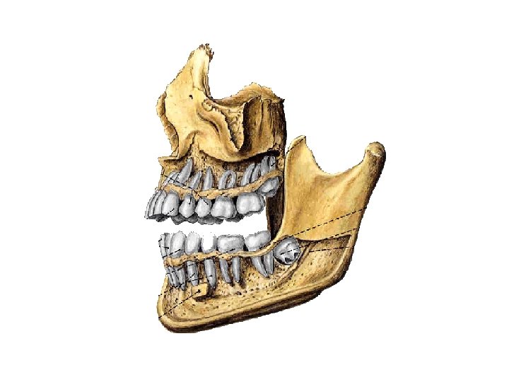
: are 32 in number • Sixteen in each mandibular and maxillary")
Permanent teeth (adult): are 32 in number • Sixteen in each mandibular and maxillary arch • Two incisors, one canine, two premolars, and three molars in each quadrant Upper jaw 1 2 3 4 5 6 7 8 total 32 Lower jaw • First permanent molar- appears at about 6 years • Third molars (wisdom teeth)-many erupt at any time after 12 years of age or not at all (impaction).

General description • Each tooth consists of 3 parts: – 牙冠Crown – 牙颈Neck – 牙根Root • 牙腔Dental cavity-contains connective tissue, blood vessels and nerves, and is continuous with the periodontal tissue through the root canal and apical foramen.

Calcified tissues 牙组织 • 牙质Dentine- is a yellowish white tissue, that forms the bulk of tooth. • 釉质Enamel -is a hard, brittle white tissue that covers the crown of the tooth • 牙骨质Cement -is an unusual form of bone that covers the root of the tooth • 牙髓 dental pulp 牙周组织Periodontal tissue • 牙周膜Periodontal membrane • 牙槽骨Alveolar bone • 牙龈Gum

舌Tongue -muscular organ Two parts: divided two parts by vshaped界沟terminal sulcus • Body of tongue 舌体-anterior 2/3, apex of tongue 舌尖-free rounded tip • Root of tongue 舌根- posterior 1/3 At the apex of terminal sulcus is a small median pit, the foramen cecum of tongue 舌盲孔

Lingual mucous membrane • Papillae of tongue 舌乳头 – – Filiform papillae 丝状乳头 fungiform papillae 菌状乳头 foliate papillae 叶状乳头 vallate papillae 轮廓乳头 • Lingual tonsil 舌扁桃体 -masses of submucosal tissue on the root of tongue contain taste buds lymphoid

Inferior surface of tongue • Frenulum of tongue 舌系带 -a midline fold of mucous membrane connecting tongue to floor of mouth • Sublingual caruncle 舌下阜 -small elevation • Sublingual fold 舌下襞

Muscles of tongue 舌肌 • Intrinsic muscles of tongue – Involved in changing shape of tongue – Include longitudinal, transverse and vertical muscles of tongue • Extrinsic muscles of tongue – Genioglossus 颏舌肌 • Arises from mental spine of mandible and inserts into either side of midline of tongue • Action: acting together draw tongue forward and downward (depresses and protrudes tongue ); acting along making apex of tongue to opposite side Hyoglossus 舌骨舌肌 Tyloglossus 茎突舌肌 – – – Involved in determining shape and position of tongue

大唾液腺Major salivary glands 腮腺Parotid gland • Superficial part: triangular in shape, lies below and in front of the external acoustic meatus, and partially covers the masseter. • Deep part: lies deep to medial pterygoid. • Parotid duct: arises front anterior border of gland, runs over the masseter a finger’s breadth below the zygomatic arch to pierce the buccinator and opens into the mouth cavity, opposite the upper second molar tooth

Submandibular gland 下颌下腺 • Position: lies in submandibular triangle, between anterior and posterior bellies of digastric • Duct opens on to sublingual caruncle Sublingual gland 舌下腺 • position: situated beneath the mucous membrane of the sublingual fold • Ducts – Major sublingual duct- opens onto the sublingual caruncle – Minor sublingual ducts- open onto the sublingual fold

The Pharynx 咽 General features • A –fibromuscular tube, part of digestive and respiratory systens • Extends from base of skull to the inferior border of cricoid cartilage (lower border of C 6 level) • Three segments

Nasopharynx 鼻咽 - posterior to nasal cavities • Extends from the base of skull to level of soft palate, below • Features – Pharyngeal opening of auditory tube 咽鼓管咽口Tubal torus咽鼓管圆枕 – Pharyngeal recess 咽隐窝 – Tubal tonsil 咽鼓管扁桃体 – Pharyngeal tonsil 咽扁桃体 Oropharynx 口咽 - posterior to oral cavity • Lies below soft palate, extends to upper border of epiglottis

Oropharynx 口咽 • Features – Median glossoepiglottic fold 舌会厌正中襞 – Epiglottic vallecula 会厌谷 – Palatine tonsil 腭扁桃体- lies within tonsillar fossa Lymphatic ring-consists of pharyngeal tonsil, tubal tonsil, palatine tonsil, and lingual tonsil, forming a circular band of lymphoid tissue at oropharyngeal isthmus

Laryngopharynx 喉咽-posterior to larynx • Extends from upper border of epiglottis to the level of lower border of C 6 • Piriform recess梨状隐窝-a deep depression on each side of aperture of larynx, common site for lodgement of foreign bodies (for example, fish bones)

The Esophagus 食管 General features -a muscular tuber about 25 cm long, connecting the pharynx at level of C 6 vertebra, passes through the diaphragm at level of T 10 vertebra and after 1~2 cm enters the stomach Division: • Cervical part • Thoracic part • Abdominal part

Three constrictions • At its beginning, 15 cm from incisors, lies at level of C 6, is the narrowest part of the esophagus • Where it is crossed by left main bronchus, 25 cm from incisors, lies at level of intervertebral disc between T 4 and T 5. • Where it passes through the esophageal hiatus of diaphragm, 40 cm from incisors, at level of T 10
- Slides: 28