COMPLICATIONS of Transfusion Therapy Dr Alizadeh LOW LEVELS


- Slides: 21
ﺑﺴﻢ ﺍﻟﻠﻪ ﺍﻟﺮﺣﻤﻦ ﺍﻟﺮﺣیﻢ COMPLICATIONS of Transfusion Therapy Dr. Alizadeh
LOW LEVELS OF FIBRINOGEN fibrinogen supplementation was primarily received from administration of FFP and cryoprecipitate. • Levy and colleagues : “fibrinogen is critical for effective clot formation, and its monitoring and guided supplementation as the treatment of major bleeding increasingly recognized. ” •
LOW LEVELS OF FIBRINOGEN • Another group concluded that current recommendations for fibrinogen replacement are too conservative. • Yet another group concluded that the clinical effectiveness of FFP was not apparent in trauma patients, but administration of fibrinogen concentrate generally improved outcome. • fibrinogen increased fibrin-based clot firmness after aortic surgery. • Furthermore, a dilutional coagulopathy from colloid administration was better treated with a combination of factor XIII concentrates and fibrinogen rather than fibrinogen alone—not a surprising result.
LOW LEVELS OF FACTORS V AND VIII • factors V and VIII gradually decrease to 15% and 50% of normal, respectively, in whole blood after 21 days of storage. • PRBCs even have fewer coagulation factors. Consequently, administration of FFP, which contains all the factors except platelets, has been recommended on a therapeutic or a prophylactic basis. • However, this practice is of questionable benefit because only 5% to 20% of factor V and 30% of factor VIII are needed for adequate hemostasis during surgery. • In other words, in spite of a patient receiving massive blood transfusion, factors V and VIII rarely decrease below those levels required for hemostasis.
LOW LEVELS OF FACTORS V AND VIII • Despite the PTTs (which measure all factors except VII and XIII) and platelets having returned to normal, bleeding persisted in every patient. Only when platelets in the form of fresh blood were administered did bleeding cease. • Although low factors I, V, and VIII are likely not the primary cause of bleeding during massive blood transfusion, such deficiencies may intensify bleeding from other causes, usually dilutional thrombocytopenia in the case of blood transfusion.
LOW LEVELS OF FIBRINOGEN AND FACTORS V AND VIII the following criteria are still relevant even though they did not have the advantage of ROTEM: 1. Generalized bleeding that cannot be controlled with surgical sutures or cautery. 2. PTT time at least 1. 5 times normal. 3. Platelet count greater than 70, 000/mm 3 (to ensure that thrombocytopenia is not the cause of bleeding).
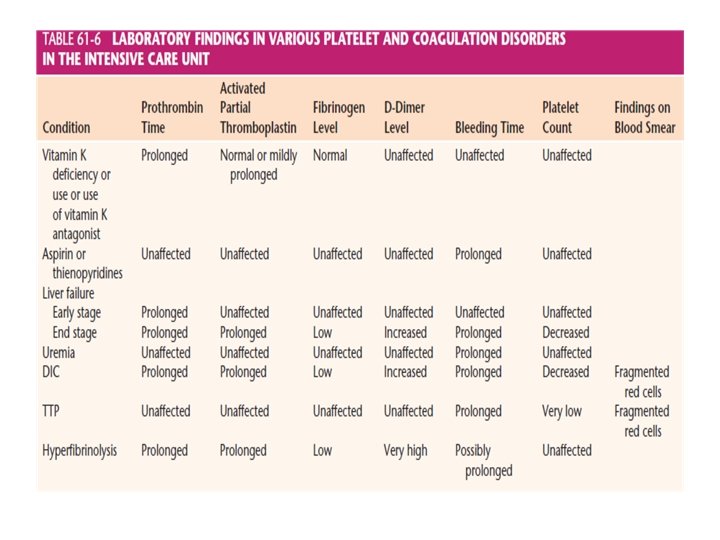
DISSEMINATED INTRAVASCULAR COAGULATION–LIKE SYNDROME • The coagulation system consists of clotting and fibrinolytic mechanisms. • The function of the former is to prevent excessive blood loss, and that of the latter is to ensure circulation within the vasculature. • With this DIClike syndrome, the clotting system is deranged and this leads to disseminated fibrin deposition, which renders the fluid blood unclottable. • The deposited fibrin may severely alter the microcirculation and lead to ischemic necrosis in various organs, particularly the kidney. The un clottable blood or circulating serum may induce a severe hemorrhagic diathesis.
DISSEMINATED INTRAVASCULAR COAGULATION–LIKE SYNDROME • The specific reasons for the development of DIC syndrome are usually not apparent. However, hypoxic acidotic tissues with stagnant blood flow probably release tissue thromboplastin directly or through liberation of some toxin as possibly modulated through the protein C pathway. • The release of tissue plasminogen activator from damaged tissue may cause fibrinolysis. • The extrinsic route of coagulation is activated by tumor necrosis factor and endotoxins. Presumably, tumor necrosis factor induces tissue factor expression on the surface of activated monocytes and possibly by exposure to sub endothelially localized tissue factor in blood.
DISSEMINATED INTRAVASCULAR COAGULATION–LIKE SYNDROME • Although the intrinsic system does not induce DIC, it may contribute to hypotension. • This triggers the coagulation process, resulting in consumption of factors I, II, V, and VIII and platelets. Supposedly, thrombi and fibrin are deposited in the microcirculation of vital organs, interrupting their blood flow. • In an attempt to counteract the hyper coagulable state, the fibrinolytic system is activated to lyse the excessive fibrin almost simultaneously; this is called secondary fibrinolysis.
DISSEMINATED INTRAVASCULAR COAGULATION–LIKE SYNDROME • DIC should not be considered a distinct disease entity but rather a sign of another disease. DIC has been associated with almost all life -threatening diseases. • Any condition in which tissue damage is sufficient to release tissue products or toxins into the circulation can be associated with DIC. • If enough thromboplastin lodges in the circulating blood, the result is massive focal necrosis or more generalized activation of the coagulation system. • Although treatment is more likely to be successful when the cause of the bleeding problem has been identified, precise diagnosis is often difficult.
DIAGNOSIS AND TREATMENT OF A HEMORRHAGIC DIATHESIS AFTER WHOLE BLOOD TRANSFUSIONS • In addition to clinical examination of the patient, various coagulation laboratory tests have been used for years. • One traditional approach has been to obtain a blood specimen on which the following tests can be performed: platelet count, PTT, and plasma fibrinogen level; observation of a clot for size, stability, and lysis; and observation of the plasma for evidence of hemolysis. • Provided the PTT is 1. 5 times normal or more and other tests are normal, the bleeding is probably a result of very low levels of factors V and VIII. • This can be treated with FFP, which contains all the coagulation factors except platelets, or with cryoprecipitate. • Although the preceding situation is a nice textbook description, I have never observed a clinical situation involving blood transfusions in which the PTT was increased without the presence of thrombocytopenia.
DIAGNOSIS AND TREATMENT OF A HEMORRHAGIC DIATHESIS AFTER WHOLE BLOOD TRANSFUSIONS • Point-of-care diagnosis of perioperative clinical coagulopathies is being increasingly facilitated by TEG and ROTEM. • Specifically, point-of-care ROTEM (viscoelastic and rotational) is being used to diagnose intraoperative coagulopathies. • All varieties of major surgeries and types of patients are being monitored with one of these devices, including liver transplantation and postpartum hemorrhage. • Data are available within 10 to 20 minutes and are increasingly used to guide hemostatic therapy in trauma patients and, more recently, in other clinical situations. • The theoretic advantages are assessment of clot formation in the context of whole blood, including contributions from platelets and RBCs and analysis of multiple stages of the clotting process from fibrin formation through fibrinolysis.
DIAGNOSIS AND TREATMENT OF A HEMORRHAGIC DIATHESIS AFTER WHOLE BLOOD TRANSFUSIONS • Dilutional thrombocytopenia in association with DIC, and hypo perfusion is the most likely cause of bleeding from blood transfusion. • When the platelet count is less than 100, 000/mm 3, a bleeding problem is likely to develop ; therefore, platelets are ordered. • The rule of thumb is based on the fact that a bleeding diathesis probably will develop after infusion of 20 units of stored blood in healthy patients and after lesser amounts in debilitated or small patients.
DIAGNOSIS AND TREATMENT OF A HEMORRHAGIC DIATHESIS AFTER WHOLE BLOOD TRANSFUSIONS • Whether platelets are administered in the form of fresh blood, plateletrich plasma, or platelet concentrates depends on intravascular volume replacement requirements, personal preference, and availability of laboratory personnel. • Fresh blood (<6 hours old) supplies the largest number of platelets per donation. More than 80% of the platelets can be given by platelet-rich plasma, which has half of the volume of a unit of blood. • However, because most blood banks advocate giving patients only components that are necessary, platelet concentrates are frequently recommended. • The remainder of the unit of blood, such as RBCs, plasma, and albumin, can be saved for other patients. Platelet concentrates are contained in a 50 -m. L unit and provide approximately 70% of the platelets in a unit of blood. • .
DIAGNOSIS AND TREATMENT OF A HEMORRHAGIC DIATHESIS AFTER WHOLE BLOOD TRANSFUSIONS • In a 70 -kg person, approximately 10 units of platelet concentrates are required to increase the platelet count by 100, 000/mm 3. • Although logistically difficult to obtain, fresh blood is extremely effective in treating transfusion-induced coagulopathies. • Lavee and associates found that 1 unit of fresh whole blood was as effective as, if not superior to, 8 to 10 platelet units. • I think that fresh blood also contains unidentified factors that make it far more effective than blood components.
DIAGNOSIS AND TREATMENT OF A HEMORRHAGIC DIATHESIS AFTER WHOLE BLOOD TRANSFUSIONS • Determining the plasma fibrinogen level is useful because this coagulation factor does not decrease in bank blood. • If the in vivo plasma fibrinogen level is low (<150 mg/d. L), it is not a result of a dilutional coagulopathy and strongly suggests DIC or a DIC-like syndrome. • DIC is likely with thrombocytopenia, hypofibrinogenemia, and lysis of a clot within 2 hours. • Unfortunately, fibrinogen levels in PRBCs decrease with increasing storage time. As a result, hypo fibrinogenemia occurs on a dilutional basis when multiple units of PRBCs are given.
DIAGNOSIS AND TREATMENT OF A HEMORRHAGIC DIATHESIS AFTER WHOLE BLOOD TRANSFUSIONS • ε-Aminocaproic acid (EACA) inhibits the formation of plasmin and attenuates fibrinolysis. EACA should not be used in the treatment of DIC. Blocking the fibrinolytic system and having the coagulation system activated have resulted in disseminated thrombosis. • Because primary fibrinolysis is rare other than in prostatectomy and liver transplantation, EACA should probably not be given unless the preceding diagnosis is clearly established after expert consultation. • Administration of recombinant activated coagulation factor VII (rf. VIIa, ) has produced successful treatment of such coagulopathies intraoperatively.
DIAGNOSIS AND TREATMENT OF A HEMORRHAGIC DIATHESIS AFTER WHOLE BLOOD TRANSFUSIONS • Most of these patients (coagulopathies intraoperatively) also had other conditions, such as necrotizing pancreatitis, cirrhosis, or severe trauma. • dilution of certain coagulation values may be more profound with the use of PRBCs rather than whole blood. • In general, the direction of coagulation changes was similar to that seen with whole blood, with one major exception. With use of PRBCs, fibrinogen levels decreased significantly in contrast to use of whole blood, in which fibrinogen levels remained unchanged unless DIC was present. Although all the coagulation factors decreased.
DIAGNOSIS AND TREATMENT OF HEMORRHAGIC DIATHESIS AFTER PACKED RED BLOOD CELL TRANSFUSIONS • When PRBCs are used to replace major blood loss, the clinician may be tempted to give FFP prophylactically. • However, Murray and co-workers specifically recommended not following the policy; they stated that FFP was needed only when prothrombin time (PT) and PTT were at least 1. 5 times normal and fibrinogen levels were less than 75 mg/d. L. • Leslie and Toy provided more specific guidelines when PRBCs are used for massive transfusions. • They believed that when 12 or more units of PRBCs or cell-saver blood had been given, coagulation factors (i. e. , FFP) were necessary. Patients who received 20 or more units often required platelet therapy, a finding identical to that of patients given whole blood.