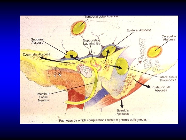
COMPLICATIONS OF SUPPURATIVE OTITIS MEDIA ROUTES OF SPREAD


 complications • Intracranial")

 COMPLICATIONS • Acute mastoiditis • Petrositis • Facial nerve paralysis • Labyrinthine")


 COMPLICATIONS • Acute mastoiditis • Petrositis (apical apicitis) • Facial nerve paralysis")
 An extension of infection from the middle ear into a pneumatized")
 – Retro-orbital pain –")

 COMPLICATIONS • Acute mastoiditis • Petrositis • Facial nerve paralysis • Labyrinthine")


 COMPLICATIONS • Acute mastoiditis • Petrositis (apical apicitis) • Facial nerve paralysis")


 • Suppuration of the subdural space • May be localized, multiple")


- Slides: 62
COMPLICATIONS OF SUPPURATIVE OTITIS MEDIA
ROUTES OF SPREAD • Direct extension • Thrombophlebitis • Normal anatomical pathways • Non anatomical bony defects
COMPLICATIONS OF SUPPURATIVE OTITIS MEDIA • Extracranial complications • Cranial (intra-temporal) complications • Intracranial complications
EXTRACRANIAL COMPLICATIONS • Otitis externa • Retropharyngeal abscess • Septicemia
CRANIAL (INTRATEMPORAL) COMPLICATIONS • Acute mastoiditis • Petrositis • Facial nerve paralysis • Labyrinthine fistula and labyrinthitis
ACUTE MASTOIDITIS
PATHOLOGY OF ACUTE MASTOIDITIS Involvement of the bone of the mastoid air cells by acute suppurative inflammation
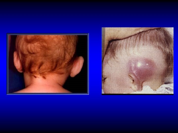
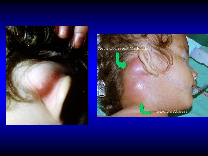
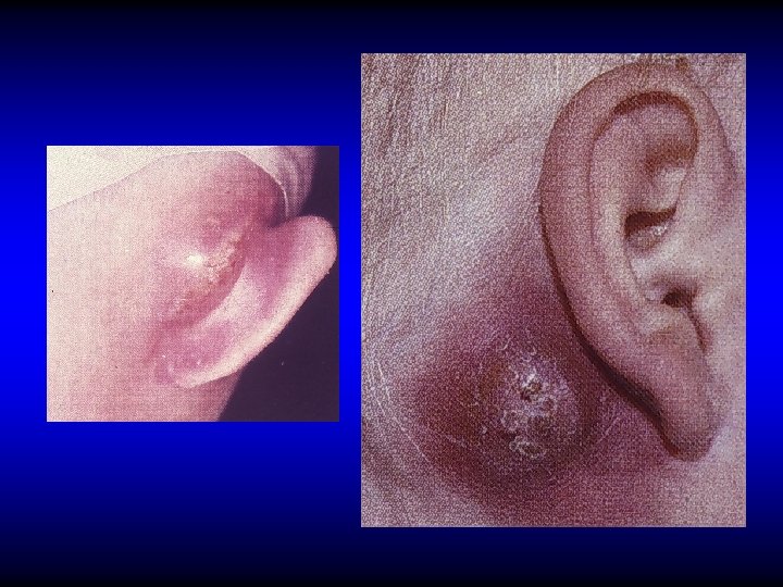
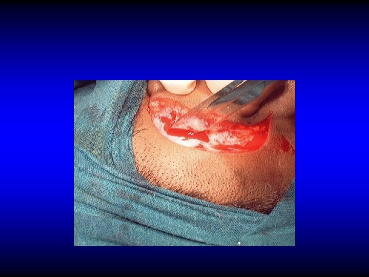
DIAGNOSIS OF ACUTE MASTOIDITIS • General constitutional manifestations • Tympanic membrane changes • Sagging of posterosuperior meatal wall • Otorrhea and reservoir sign • Retroauricular tender red swelling • Subperiosteal and Bezold’s abscess
DIAGNOSIS OF ACUTE MASTOIDITIS • General constitutional manifestations • Tympanic membrane changes • Sagging of posterosuperior meatal wall • Otorrhea and reservoir sign • Retroauricular tender red swelling • Subperiosteal and Bezold’s abscess • Imaging
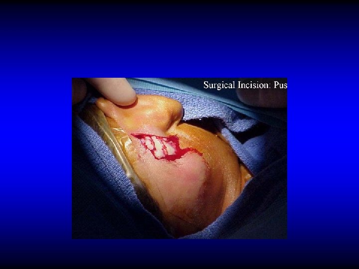
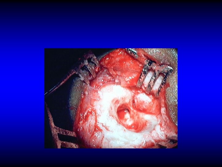
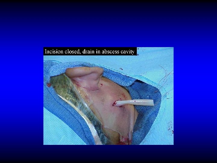
TREATMENT OF ACUTE MASTOIDITIS • IV antibiotics • Cortical mastoidectomy if medical treatment fails or if there are signs of abscess formation • Observe for other complication
CORTICAL “SIMPLE” MASTOIDECTOMY An operation in which the mastoid antrum and air cells are converted into one cavity without disturbing the middle or external ears. It may be combined with myringotomy.
CRANIAL (INTRATEMPORAL) COMPLICATIONS • Acute mastoiditis • Petrositis (apical apicitis) • Facial nerve paralysis • Labyrinthine fistula and labyrinthitis
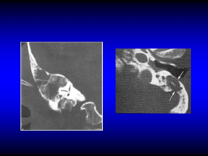
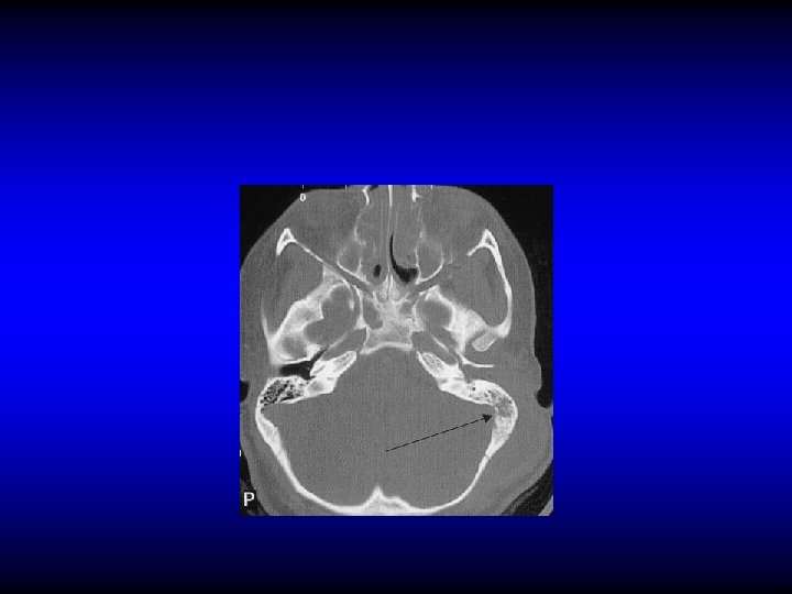
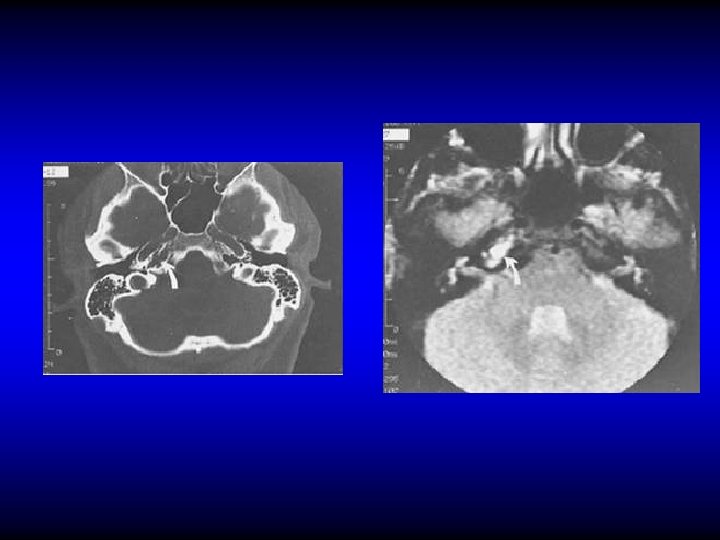
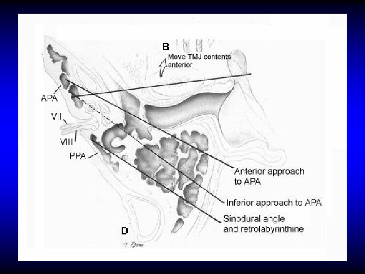
PETROSITIS (PETROUS APICITIS) An extension of infection from the middle ear into a pneumatized petrous apex.
DIAGNOSIS OF PETROSITIS • Gradenigo’s syndrome – Otitis media (otorrhea) – Retro-orbital pain – Squint (VI cranial nerve palsy) • Imaging
TREATMENT OF PETROSITIS • Antibiotics and myringotomy • Surgical drainage if medical treatment fails
CRANIAL (INTRATEMPORAL) COMPLICATIONS • Acute mastoiditis • Petrositis • Facial nerve paralysis • Labyrinthine fistula and labyrinthitis
FACIAL PARALYSIS IN AOM • Mostly due to pressure on a dehiscent nerve by inflammatory products • Usually is partial and sudden in onset • Treatment myringotomy is by antibiotics and
FACIAL PARALYSIS IN CSOM • Usually is due to pressure by cholesteatoma or granulation tissue • Insidious in onset • May be partial or complete • Treatment is by immediate surgical exploration and “proceed”
CRANIAL (INTRATEMPORAL) COMPLICATIONS • Acute mastoiditis • Petrositis (apical apicitis) • Facial nerve paralysis • Labyrinthine fistula and labyrinthitis
PATHOLOGY OF LABYRINTHITIS • Labyrinthine fistula • Circumscribed labyrinthitis • Acute diffuse serous labyrinthitis • Acute diffuse suppurative labyrinthitis • Chronic labyrinthitis
DEFINITION OF LABYRINTHINE FISTULA Loss of the bony labyrinthine wall exposing the endosteum
DIAGNOSIS OF LABYRITHINE FISTULA • No symptoms • Vertigo • SNHL • Fistula test • CT scan
INTRACRANIAL COMPLICATIONS • Extradural abscess • Lateral sinus thrombophlebitis • Subdural empyema • Meningitis • Brain abscess • Otitic hydrocephalus
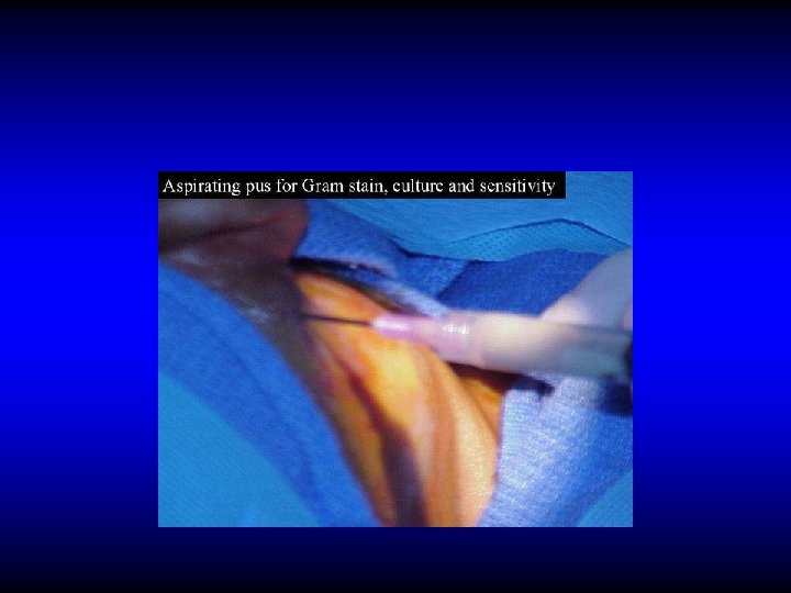
EXTRADURAL ABSCESS • Accumulation of pus between dura and bone • In the middle or posterior fossa (perisinus) • Causes headache but may be silent • Diagnosis is confirmed by CT or MRI • Treatment is by drainage
SUBDURAL ABSCESS (EMPYEMA) • Suppuration of the subdural space • May be localized, multiple or diffuse • Sever headache, fever, irritative and paralytic focal neurological symptoms • CT and MRI • Treatment is by neurosurgical drainage
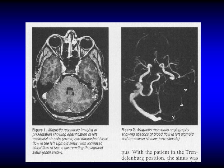
LATERAL SINUS THROMBOPHLEBITIS Pathology • Perisinusitis • Mural thrombus • Occluding thrombus • Suppuration • Embolization
LATERAL SINUS THROMBOPHLEBITIS • • Diagnosis Fever, rigor, and sweating Headache and neck pain Tenderness and edema in the neck Manifestation of increased IC pressure Propagation and embolic manifestations Blood culture, CSF manometry CT, MRI
CT Subtraction Angiogram MRI Angiogram
TREATMENT OF SINUS THROMBOPHLEBITIS • IV antibiotics • Surgery should follow within 48 hours unless there is dramatic clinical and radiological improvement
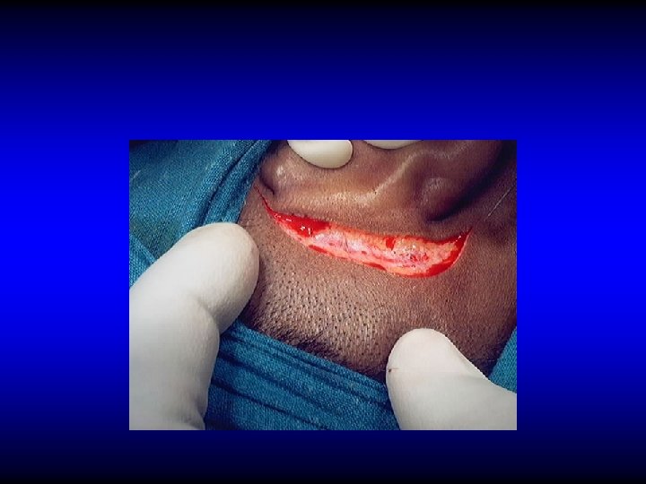
SURGICAL TREATMENT OF SINUS THROMBOPHLEBITIS • Exposure of healthy dura proximal and distal
SURGICAL TREATMENT OF SINUS THROMBOPHLEBITIS • Exposure of healthy dura proximal and distal • Verify the sinus content
SURGICAL TREATMENT OF SINUS THROMBOPHLEBITIS • Exposure of healthy dura proximal and distal • Verify the sinus content – Blood clot: leave alone – Pus: incise to drain • Ligate only if there is repeated embolisms or uncontrolled extension
INTRACRANIAL COMPLICATIONS • Extradural abscess • Lateral sinus thrombophlebitis • Subdural empyema • Meningitis • Brain abscess • Otitic hydrocephalus
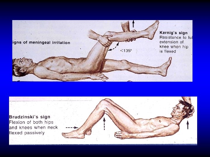
OTOGENIC MENINGITIS • Infection of the subarachnoid space • The most common intracranial complication • Fever, headache, neck stiffness, phonophobia, restlessness etc • Kernig’s & Brudziniski signs
OTOGENIC MENINGITIS • Infection of the subarachnoid space • The most common intracranial complication • Fever, headache, neck stiffness, phonophobia, restlessness etc • Kernig’s & Brudziniski signs • Lumber puncture
INTRACRANIAL COMPLICATIONS • Extradural abscess • Lateral sinus thrombophlebitis • Subdural empyema • Meningitis • Brain abscess • Otitic hydrocephalus
OTOGENIC BRAIN ABSCESS • 25% of children's and 50% of adult’s brain abscesses are otogenic • Mostly in temporal lobe or cerebellum (2: 1)
OTOGENIC BRAIN ABSCESS Clinical manifestations • General manifestations: fever, lethargy, headache. • Manifestation of raised IC pressure • Focal manifestations – Temporal: Aphasia, hemianopia, paralysis – Cerebellar: ataxia, vertigo, nystagmus, muscle incoordination
OTOGENIC BRAIN ABSCESS Diagnosis • • CT MRI LP Burr hole needling
CT
MRI
OTOGENIC BRAIN ABSCESS Treatment • Repeated aspiration • Excision
INTRACRANIAL COMPLICATIONS • Extradural abscess • Lateral sinus thrombophlebitis • Subdural empyema • Meningitis • Brain abscess • Otitic hydrocephalus
OTITIC HYDROCEPHALUS • Very rare • An idiopathic benign intracranial hypertension associated with ear disease. It most often follows lateral sinus thrombophlebitis • Clinically: Manifestations of increased IC pressure • Treatment: steroids, diuretics, hyperosmolar dehydrating agents, repeated LP
GENERAL PRINCIPLES OF TREATMENT OF THE COMPLICATIONS • Parental antibiotics • Surgery for the complication if applicable • Treatment of the ear lesion – Myringotomy in AOM – Mastoidectomy in CSOM
THANK YOU