Complications Of Bariatric Surgery Supervised by Dr Mohammed

Complications Of Bariatric Surgery Supervised by : Dr. Mohammed Nofal Presented by; qanetah shlouh saja AL faouri samar jaradat

Bariatric Surgeries Classification Restrictive Malabsorptive • Horizontal gastroplasty • Jejunoilial bypass • Vertical banded gastroplasty (VBG) • Biliopancreatic diversion (Scopinaro) • Adjustable gastric band • Biliopancreatic diversion w/ duodenal switch • Sleeve gastrectomy • Roux-en-Y gastric bypass

Possible Complications May Lead to Short or Longterm Hospitalization and/or Re-operation : � Cholelithiasis � VTE (DVT) � Infection, bleeding or leaking at suture/staple lines � Blockage of the intestines or pouch � Dehydration � Blood clots in legs or lungs � Vitamin and mineral deficiency � Protein malnutrition � Incisional hernia � Death

Cholelithiasis �Cholelithiasis occurs in about one-third of patients who undergo bariatric surgery. �The rapid post-surgical weight loss is a significant contributor to the development of gallstones as is the pre-surgical obesity.

Diagnosis and presenting symptoms : � Patient with cholelithiasis usually do not have any symptoms unless there is acute inflammation of the gallbladder or impaction of a stone within the biliary system. � Patients present with nausea, vomiting, and biliary colic localized to the epigastrium and right upper quadrant. �Diagnosis is confirmed with ultrasound or CT imaging
 �Venous thromboembolism (VTE), which includes deep vein thrombosis (DVT) and its")
Venous thromboembolism (VTE) �Venous thromboembolism (VTE), which includes deep vein thrombosis (DVT) and its complication, pulmonary embolism (PE), is a common cause of morbidity and mortality after bariatric surgery. �The reported postoperative incidence of VTE, however, varies widely, from 0. 2% to 1. 3% at 30 days to 0. 42% at 90 days.
 for VTE prevention after bariatric surgery")
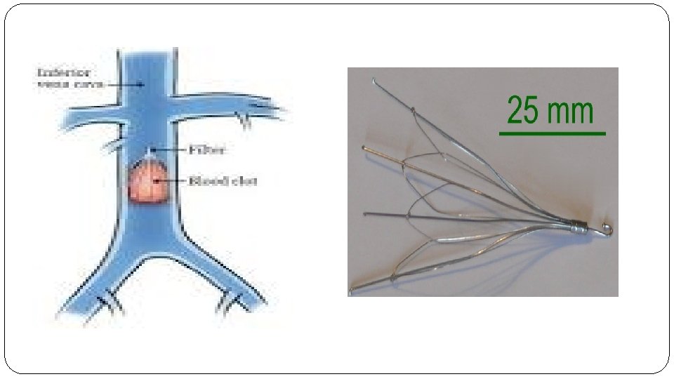
Treatment �The most commonly used pharmacological prophylaxis (91%) for VTE prevention after bariatric surgery was enoxaparin 40 mg subcutaneously twice daily for 10– 14 days after hospital discharge. �Consider an IVC FILTER for any patient with prior history of DVT/PE. �There were no reported cases of bleeding or VTE related mortality after 3 months.

LAP ADJUSTABLE GASTRIC BAND

LAGB complications

LAGB specific complications Prolapse Slippage Band erosion Port and tubing problems

1. Prolapse: The lower stomach is pushed upward and trapped within the lumen of the band.

Prolapse is perhaps the most common emergent complication that requires reoperation after LAGB. The incidence of reoperation is generally around 3%. Postoperative vomiting predisposes to this problem. Typical patient symptoms include immediate dysphagia, vomiting, and inability to take oral food or liquid. The initial evaluation for prolapse involves obtaining a plain film radiograph. If the band is in a horizontal position, prolapse must be strongly suspected. Initial treatment for a prolapse is to remove all the fluid from the system. This often allows reduction of the prolapse and resolution of symptoms. If symptoms resolve, the necessity of performing an upper gastrointestinal series is lessened. If they do not, an upper gastrointestinal series is indicated, and if prolapse persists, then reoperation laparoscopically to reduce the prolapse and resuture the band in place is indicated.

2. Slippage:

May present with vomiting due to pouch outlet distortion. Diagnosis confirmed using plain abdominal films, upper GI series, or abdominal CT scan. Treatment initially involves fluid removal from the band to relieve symptoms, followed by surgical revision of the band. Slippage has been greatly reduced by the pars flaccida technique. Operative need for repair now occurs in about 3% of cases in most series of short- term follow-up. Longerterm follow-up rates may show higher rates of prolapse.

3. Band erosion:

Band erosion is uncommon, reported in 1% to 2% of most series. The patient usually becomes ill but not floridly ill, developing either a port site infection or systemic fever and a low-grade abdominal inflammatory sepsis. Endoscopy can be diagnostic. The presence of otherwise unexplained free air on computed tomography (CT) scan should alert the surgeon to this diagnosis as well. Laparoscopic removal of the band is indicated, with repair of any gastric perforation. Often the perforation is already sealed by an inflammatory process, but if not, appropriate management of a gastric perforation must be followed.

4. Port and tubing problems :
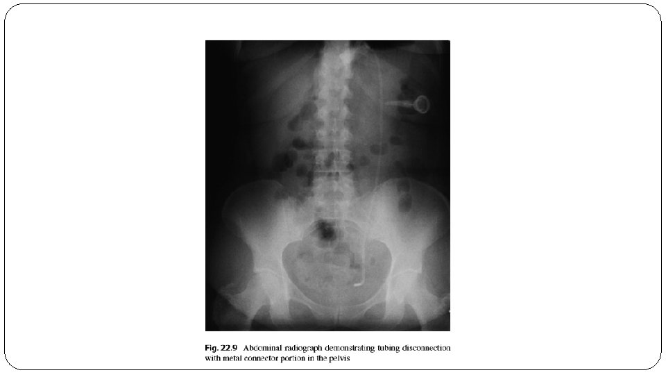

* Port and tubing problems : Port and tubing problems occur in approximately 5% of patients undergoing LAGB. These require revision of the port/ tubing system due to perforation, leaking, or kinking of the tubing or turning of the port such that access to the surface of the port for adding fluid is precluded. Usually a procedure under local anesthesia is all that is required to repair or realign the tubing or port.

Sleeve gastrectomy

Sleeve gastrectomy complications Early complications: 1. bleeding 2. staple line leak. Late complications: 1. Gastric sleeve stricture.

1. Bleeding: Usuallu occurs within the first 24 -48 hrs. can be defined by the presence of hematemesis or melena, and persistent large bloody output from a surgical drain, with or without the presence of tachycardia, hypotension, oliguria, and a decreasing hemoglobin and haematocrit. Postoperative bleeding can be classified based on bleeding site into intraluminal bleeding or intra-abdominal bleeding. In gastric sleeve patients, bleeding is mainly related to the long staple line closure where the stomach has been divided, short gastric vessel pedicles, or trocar sites. Incidence: less than 1%.

Treatment: Initial therapy: may include resuscitation with fluids and blood products and monitoring of urinary output. Definitive treatment: 1. Intraluminal bleeding: Controlled using endoscopic techniques; surgical exploration is reserved for failed endoscopic therapy. 2. Intraperitoneal bleeding: requires surgical exploration to control hemorrhage site.

2. Staple line leak: One of the most feared complications, which is a life threatening one. It occurs when a hole or gap develops somewhere along the staple line closure allowing stomach juice to escape into the abdominal cavity, Leakage usually appears as an acute complication (within 7 days), causing tachycardia, tachypnea, and fever very early on, indicating most often that the patient requires immediate intervention. Diagnosis: 1. Physical examination (abdominal): Unreliable; peritonitis is a late finding. 2. Abdominal CT: best diagnostic modality for stable patients. 3. Surgical exploration: definitive diagnostic tool and mandatory for diffuse peritonitis.

Treatment Fluid resuscitation, broad-spectrum antibiotics. 1. Wide peritoneal drainage of the leak site: surgically or percutaneously with image guidance; first-line treatment. 2. Direct repair of the leak site: occasionally successful and used in conjunction with wide drainage. 3. Endoscopic techniques: used to seal leaks, typically in conjunction with drainage.

3. stricture:

§ From postoperative scarring or a technical error in sleeve construction. § Incidence: 0. 5%. Clinical presentation: Most common symptoms are nausea and vomiting; The classic story is that the patient was initially able to tolerated solid foods, but after afew weeks he start to vomit solids but could handel liquids. Diagnosis: • 1. Physical examination: Usually unremarkable. Epigastric pain, if present, is mild. • 2. Radiologic evaluation: abdominal CT , upper GI series, and upper endoscopy. Treatment: • Endoscopic stenting and surgical stricturoplasty: successful in some cases.

Roux-en-Y gastric bypass surgery complications

• Predictors of complications �Age > 65 � BMI > 50 �medical co-morbidities ( obstructive sleep apnea, gastroesophageal reflux disease, history of a thrombotic event, diabetes, hyperlipidaemia, peripheral vascular disease hypertension, renal impairment, arthritis and asthma).

Classification : �Major complication as small bowel obstruction , major leak , abscess, anastomotic stricture , fistulae �Minor complication : marginal ulcer , pancreatitis , esophagitis, splenic abscess , cholithasis �Early complication less than one month �Late complication more than one month

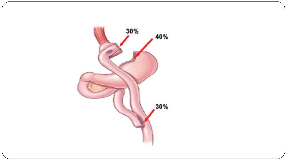
Early complications �Anastomotic or staple line leaks � There are five potential sites where a leak may form: gastrojejunostomy (most common site), gastric pouch staple line, gastric remnant staple line, Roux limb staple line and jejunojejunal anastomosis

�Diagnosis and presenting symptoms: � The diagnosis of ASL is based on a high clinical suspicion. Symptoms are nonspecific and variable including tachycardia, fever, abdominal pain, nausea, and vomiting and purulent drain output. In one study, sustained tachycardia > 120 beats per minute in the post-operative setting was highly specific for an ASL
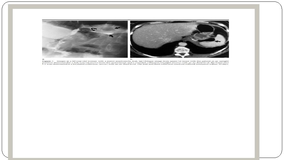

�Radiographic studies are occasionally helpful and may show fluid collection adjacent to the pouch, diffuse abdominal fluid or rarely, intraperitoneal air �Definitive investigation is made via ct-scan with contrast

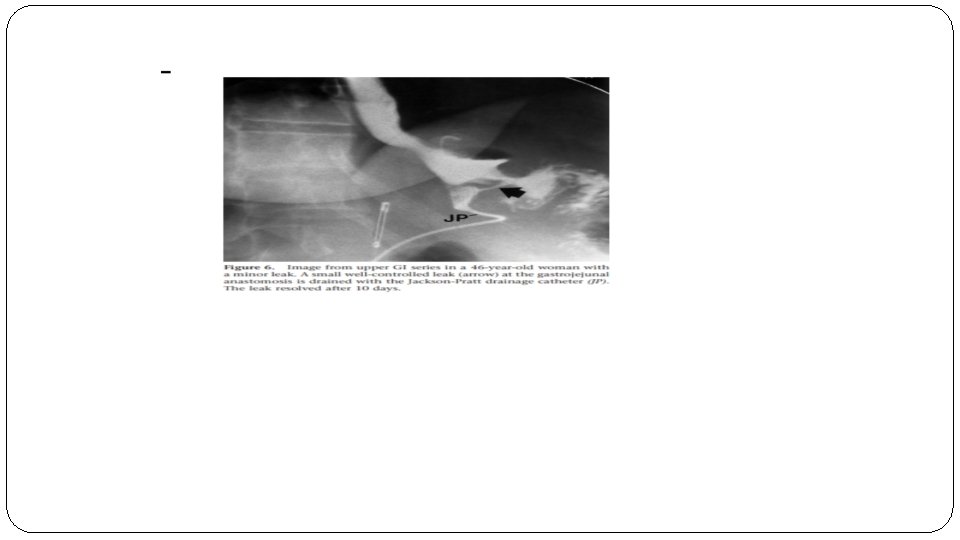
Management � Conservative management can be effective in non-septic, hemodynamically stable patients with contained leaks. The mainstay of this treatment are intravenous antibiotics, monitoring of secretions through drains, nasoenteral nutrition or total parenteral nutrition (depending on the case and the location of the leak), and if the leak is contained and accessible, a percutaneous treatment can be performed. This approach has been shown to be successful and lacks the morbidity associated with a reoperation. � But if the patient is hemodynamically unstable, has a complicated leak, or signs of sepsis, an operative treatment is mandatory. The operative goals are: to confirm and repair the leak, remove gastrointestinal contents from the abdominal cavity and place closed suction drains. � The repair of the leak would be the ideal situation, but often suturing the place of the leak can be challenging, as the acutely inflamed tissues might not be amenable to suture placement. In such cases, the removal of gastrointestinal contents and placement of drainage tubes may be the safest option. Depending on the surgical team skills the approach could be laparoscopic or open. � Maintain the nutrition is mandatory to allow healing the tissues in the place of the leak. In order to achieve this, the placement of a feeding gastrostomy into the gastric remnant or a feeding jejunostomy should be considered. This would allow for continued enteral nutrition while bowel rest is maintained at the site of the leak.
 after RYGB can be life threatening if not recognized")
Gastrointestinal bleeding �gastrointestinal bleeding (GIB) after RYGB can be life threatening if not recognized and treated early. �GIB after laparoscopic RYGB can be intraperitoneal ( bleeding into the abdominal cavity, possibly from staple lines ) or intraluminal (occurs into the lumen of the digestive tract ) and most commonly originates at one of the five potential staple lines : the gastric pouch, excluded stomach, Roux limb staple line, gastrojejunostomy, and jejunostom � the intraperitoneal bleeding occurs as an early bleeding.

�Diagnosis and presenting symptoms: � The presence of pallor, dizziness, confusion, tachycardia, hypotension, hematemesis, bright red blood per rectum, drop in the hemoglobin level, large quantity of bloody fluid from the abdominal drains and low urine output should alert the surgeon to ongoing postoperative bleeding. �The diagnosis is confirmed endoscopically and/or surgically

Management �The treatment depends on the timing of onset and the clinical presentation. In cases of late presentation (>48 hours) of gastrointestinal bleeding after surgery, it can be managed conservatively in most cases, especially when associated with no acute clinical symptoms, and melena, which might indicate the passage of old blood and inactive bleeding. In these cases discontinuation of DVT chemoprophylaxis and watchful waiting with supportive therapy can be successful. �In the other hand, early postoperative bleeding, occurring within a few hours after the surgery, manifested by hematemesis or bright red blood per rectum in the presence of clinical signs of bleeding is a clear indication for urgent surgical intervention. Abdominal reexploration using either a laparoscopic or open approach must be

�. �If is suspected that the bleeding source is proximal intraluminal the best treatment option is an endoscopic intervention, which is invaluable in controlling bleeding from the gastric pouch or gastrojejunostomy. Thermal coagulation, injection of vasoconstrictors, and clipping are all effective ways of controlling bleeding from these sites 4.

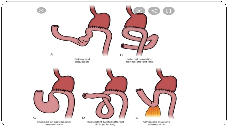
Gastrojejunal Stricture �Stricture of the proximal anastomosis, or gastrojejunostomy, is one of the most common Complications after gastric bypass. �The typical stricture patient presents 4– 6 weeks after surgery with solid food intolerance progressing to liquid intolerance as the stricture narrows.

Diagnosis and presenting symptoms �Symptoms of a gastrojejunal stenosis are usually acute onset nausea, vomiting , dysphagia to solid and liquid and abdominal pain, meanwhile in stenosis developing more distally, the symptoms may be more gradual in onset. �The diagnosis of stricture can usually be made based on history alone and confirmed with upper endoscopy. �Upper endoscopy is the primary diagnostic modality of choice, as it allows both rapid diagnosis and therapeutic
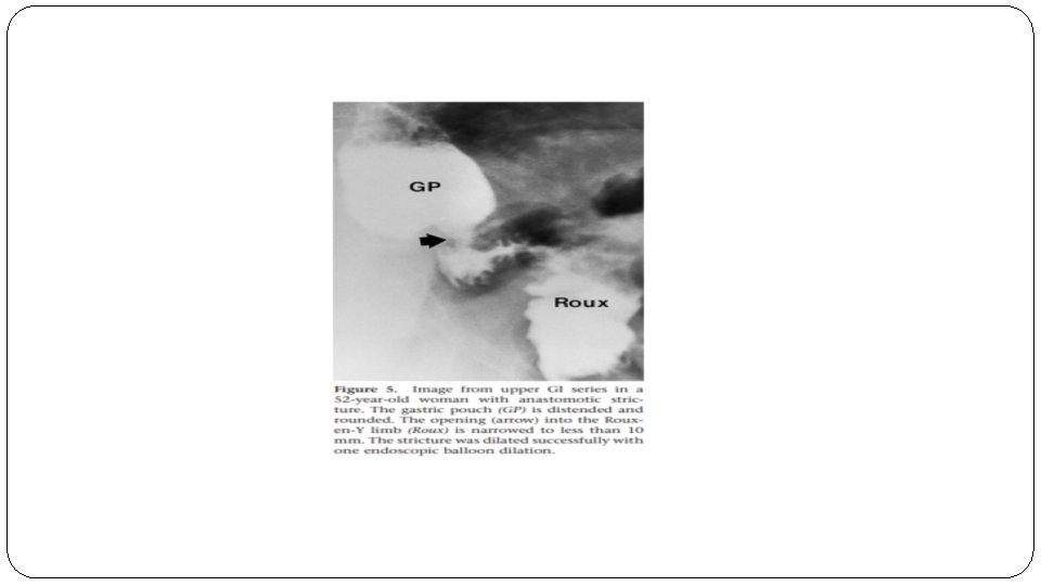

� Treatment : �When the stenosis is at the gastrojejunal anastomosis, balloon dilation with an EGD can be used. �In cases of a stenosis at the jejunojejunal anastomosis a single balloon enteroscope can be used to dilate the stenotic area.

Late Complications �Hernia �Fistula �Intussusception �Nutritional deficiencies �Dumping syndrome :

Hernia �Internal hernias are relatively common after gastric bypass and may result in bowel obstruction , intestinal ischemia, or both. � two different types of internal hernias may potentially occur: � Distal anastomosis mesenteric hernia—this space is bordered by the divided mesentery of the biliopancreatic limb and the mesentery of the Roux limb at the distal anastomosis. �Petersen hernia—the space between the mesentery of

�Typically, patients complain of intense pain in the midepigastrium, often radiating to the back. The pain may occasionally be relieved by leaning forward or getting “down on all fours, ” maneuvers that serve to reduce the compression on the entrapped bowel. �Computed tomography (CT) scan may demonstrate findings such as the “swirl sign, ” a spiraling of the mesentery. �Definitive diagnosis of internal hernia can only be achieved . through surgical exploration, either laparoscopic or open

Mesenteric swirl sign

�Treatment : �Cases of internal hernias and ventral hernias that are not incarcerated can be electively repaired, any case of incarcerated bowel requires emergent surgery due to risk of strangulation, ischemia, and perforation.

Intussusception �Small bowel intussusceptions may be transient or fixed and may result in an SBO after gastric bypass surgery. � They typically occur at or near the jejunojejunal anastomosis, with the staple line at the anastomosis presumably acting as the lead point for the intussusception.

�Diagnosis and presenting symptoms: �Intussusception may occur months or years after gastric bypass and is associated with nausea, persistent vomiting, abdominal pain, and bowel obstruction. �CT scan may reveal a “target sign” or dilated excluded stomach, but these findings are relatively insensitive �Treatment: A high level of clinical suspicion is usually the impetus for surgical exploration. Strangulation mandates resection of the affected area. In the absence of strangulation, resection or reduction and plication are accepted approaches with most surgeons favouring the former

�Explorative laparoscopy demonstrating a retrograde intussusception �
 is an abnormal communication between the gastric pouch and")
Fistulas �A gastrogastric fistula (GGF) is an abnormal communication between the gastric pouch and the excluded stomach. � The most common cause is secondary to incomplete gastric transection at the original surgery.
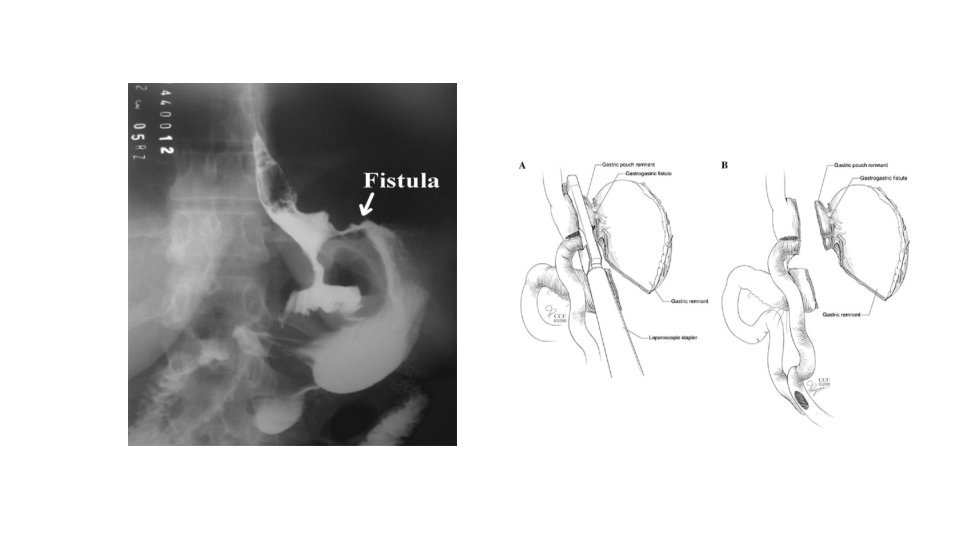

�Diagnosis and presenting symptoms: �The most common symptom is inadequate weight loss or weight gain. Patients may also present with epigastric pain and/or worsening acid reflux secondary to the new connection with the remnant stomach. � Diagnosis is made via endoscopic evaluation of the fistulous tract or via imaging with oral contrast flowing through the fistulous tract.

� Treatment: �Asymptomatic fistulous tracts are usually not a concern. However, symptomatic fistulous tracts can be closed endoscopically using specialized large endoclips to approximate the tissue surrounding the fistula.

Nutritional deficiencies �Alterations of gastrointestinal anatomy and physiology after gastric bypass elicit imbalances owing to reduced oral intake or excessive losses secondary to reconfiguration of GI motility, p. H and enzymatic profile �Anaemia is the most common complication.

�Diagnosis and presenting symptoms : � Patients may be asymptomatic or present with symptoms manifesting a particular nutritional deficiency. Patients with anaemia may present with dyspnoea, fatigue and inability to concentrate. Zinc deficiency can result in hair loss and low calcium levels can result in osteoporosis and fractures. � Blood work is recommended to detect and monitor vitamin and mineral deficiencies �Treatment : �Patients who undergo gastric bypass require a referral to a registered dietician for nutrition counselling, and lifelong vitamin and mineral supplementation is recommended.

-Dumping syndrome : �Effect of altered gastric reservoir function, abnormal postoperative gastric motor function, and pyloric emptying mechanism. �Dumping occurs when the patient eats refined high sugar concentration following gastric bypass, this causes symptoms of rapid heart beat, N&V, tremor and faint feeling, sometimes followed by diarrhea. � sweet lovers should avoid sweets after gastric bypass and this is a real help to them in their efforts to lose weight. �https: //youtu. be/m. Xh. NYj_oq. EU

A-Early Dumping Syndrome �Symptoms occurs 15 -30 minutes after meal. �Hypovolemia Pathophysiology: Loss of the reservoir function of the stomach. �it is an uncontrolled rapid emptying of hypertonic chyme into small intestine. �Fluid moves rapidly from intravascular space into intraluminal space producing acute intravascular volume depletion. S/S: �Anxiety, extreme weakness, tachycardia, diaphoresis, palpitations, and desire to lie down. Osmotic Diarrhea is common. �As simple sugars are absorbed and dilution of hypertonic

�Treatment: � Dietary modification should be the initial approach and is beneficial in most patients. Patients should be counseled to reduce portions, chew slowly and � thoroughly, and not drink fluids before an interval of 30 min after a solid meal. They should also be instructed to reduce the content of simple carbohydrates and select protein-rich foods. � somatostatin analogues such as octreotide and pasireotide may be an effective option. � Surgical interventions: Surgical options include gastric tube placement in the excluded stomach (if present), gastric outlet restriction, and gastric bypass reversal.

B-Late dumping syndrome: �Symptoms begin 3 hours after meals. �Hypoglycemia Pathophysiology: �Rapid changes in serum glucose and insulin levels. �Large glucose-bolus containing chyme presented to small intestine has a lot of glucose in it, glucose is absorbed faster than when the intact pylorus controls emptying of stomach. �this causes high levels of serum glucose shortly after meal and causes a big released of insulin. �Insulin response exceeds what is necessary to clear glucose from blood and hypoglycemia

S/S: �Sudden anxiety, diaphoresis, tachycardia, palpitations, weakness, fatigue, and desire to lie down, No diarrhea. � Treatment is dietary avoidance of Foods that are high in concentrated sugar content, Small frequent meals , Usually symptoms will resolve with time
 � Associated to bacteria overgrowth in the")
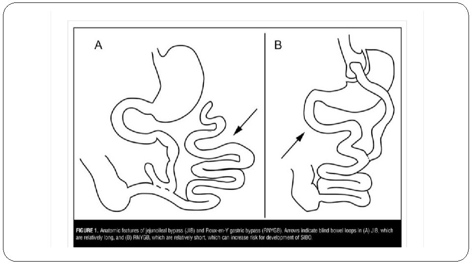
-Blind loop syndrome: (in afferent loop ) � Associated to bacteria overgrowth in the limb of the intestine excluded (not functioing ) from the flow of chyme. � This limb has bacteria that proliferate and interfere with folate and Vit B 12 metabolism, � Also bacterial overgrowth cause malabsorption of bile salts --steatorrhea , also as a result, No absorbtion the fat-soluble vitamins A, D, E and K and Ca. � B 12 deficiency leads to megaloblastic anemia S/S: � Abdominal pain, distention. � Diarrhea � Weight loss � Weakness , anemia.

�Treatment: �Empirical anti-biotic �Surgical mangment: �surgical management is reserved for fixing anatomical causes of bowel obstruction that interfere with normal function once they are amenable to such intervention. These conditions include: �Strictures �Fistulae �Diverticula

-Afferent loop syndrome : �Blockage of the afferent limb of loop causing the bile and pancreatic secretions to collect in the afferent limb with increasing pressure these enter the stomach and become dilated. �Causing projectile bilious vomiting not containing food and relieves symptoms. �Investigate with upper endoscopy and radio nucleotide scan.
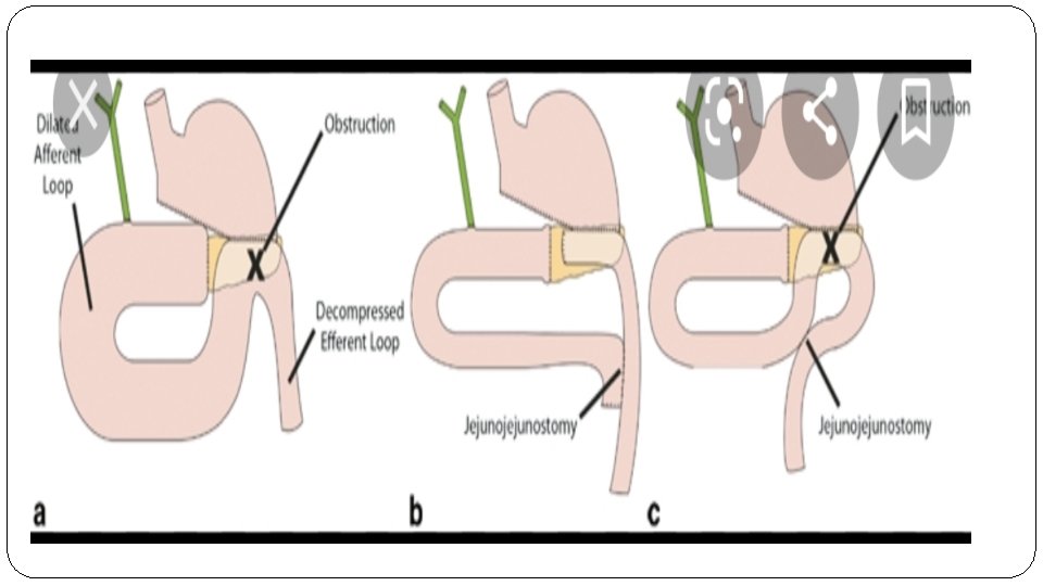

-Efferent loop syndrome : Ø Quite rare Ø Usually from internal herniation of limb behind anastomosis , When we create gastrojejunstomy anatomsis we make cut on jejunum and Lift it up to site of anastomsis (gastric pouch) so we need to cut the mesentery of Small Bowel to make it possible , Subsequently when we make this defect in mesentery (space ) , which make the intestines more susceptible to internal herniate through this defect. Ø Upper quadrant pain colicky in nature , bilious vomiting and abdominal distension. Ø Investigation – CT abdomen Ø new guideline to treatment is to make the anastomosis more high up( at level of esophagus ).

Biliopancreatic Diversion
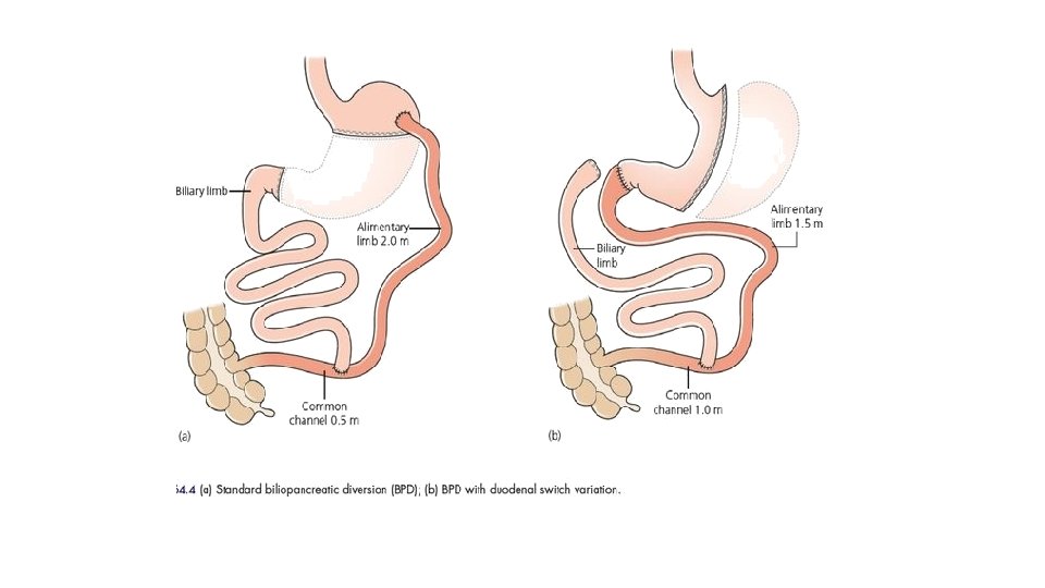

Biliopancreatic Diversion Involves resection of the distal half to twothirds of the stomach and creation of an alimentary tract of the most distal 200 cm of ileum, which is anastomosed to the stomach. The biliopancreatic limb is anastomosed to the alimentary tract either 75 or 100 cm proximal to the ileocecal valve.

Biliopancreatic Diversion This procedure which produces the most malabsorptive of all operations, is the most effective with 75 -85 % excess weight loss but at the expense of the highest mortality of 1 -2% Additionally as time goes by, if the patient does not adhere to their vitamins and micronutrients supplementation regimen, they are at severe risk of many deficiency syndromes.

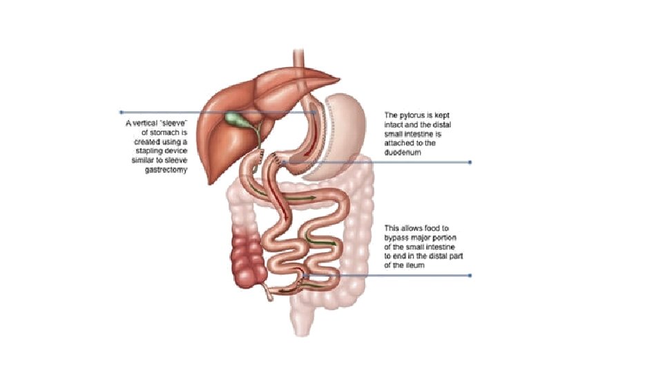
BPD ± Duodenal Switch The Duodenal switch variation of the BPD was designed to reduce the need for taking Vit B 12 and reduce the incidence of anastomotic stricture at the gastrojejunal anastomosis. In the standard BPD, approximately two-third of the distal stomach is removed while in the duodenal switch variation there is a vertical sleeve gastrectomy. In the duodenal switch variation, the anastomosis is made to the first part of the duodenum rather than the stomach as the standard BPD. https: //youtu. be/EX 68 RG 6 fa. Uo

Notes 1. 2. 3. 4. 5. 6. 7. Average weight loss after BPD and DS was over 72% Resolution of type 2 diabetes 98% Resolution of HTN 8% Improvement in dyslipidemia 100% Mortality rate of 1. 1% Complication rate of 27% to 33% Nutritional complication rate of 40% to 77%
 Immediate post operative: 1. Bleeding 2.")
Complications Intraoperative: Bleeding Injury (bowel, liver, spleen, esophagus) Immediate post operative: 1. Bleeding 2. DVT with or without pulmonary embolism 3. Infection 4. Abscess formation 5. Bowel obstruction 6. Anastomotic leaks � 1. 2.

Complications 5. Long term: Internal Herniation Bowel obstruction Osteopenia Diarrhea Malodorous bowel motions and flatus Marginal ulcer in 2. 8% of patients. Stomal ulcer. Gall stones. � 1. 2. 3. 4.

Complications � Nutritional : Iron deficiency anemia rate of less than 5%. � Alopecia from inadequate protein absorption - protein malnutrition rate of 7% � Night blindness from a lack of vitamin A �

Malnutrition The most severe and life-threatening. When it is diagnosed, the treatment is parenteral nutrition. Two episodes of required parenteral nutrition are usually considered adequate indication to lengthen the “common channel” of ileum—the ileum between the ileostomy of the biliopancreatic limb to the alimentary tract and the ileocecal valve.

�Thorough preoperative and postoperative counseling by a nutritionist versed in the operation and potential nutritional deficiencies is essential. � Vitamins and mineral supplements must be taken regularly on follow-up, including oral supplements for iron, calcium, and vitamin B 12 and a multivitamin. �Fat-soluble vitamins must be supplemented in parenteral form. Careful monitoring of protein intake.

References : § Schwartz principles of surgery 10 th edition. § The ASMBS Textbook of Bariatric Surgery § NMS surgery 6 th edition. § DR. mohammed nofal bariatric surgery lecture.

� Thank you
- Slides: 86