Complex malleolar fractures AO Trauma Advanced Principles Course


 forces • Plafond fractured • Prognostically significant")


 • Fibular fixation")


- Slides: 43
Complex malleolar fractures AO Trauma Advanced Principles Course
Learning objectives • Classify complex malleolar fractures and develop a treatment plan • Recognize typical concomitant soft-tissue injury • Apply reduction principles and choose appropriate implants for fixation • Explore key outcome publications
Malleolar fractures • Indirect forces • Shearing and tensile through talus • Plafond intact
Pilon fractures • Direct (impaction) forces • Plafond fractured • Prognostically significant
Malleolar fractures • Most common lower extremity fractures • Significant changes in management since 1970 s • Controversies remain • Complications still occur
6
44 A 2 Infrasyndesmotic fibula with medial malleolar fracture 44 A 3 Infrasyndesmotic fibula with posteromedial fracture 7
44 B Transsyndesmotic fracture 8
44 C Suprasyndesmotic fibula injury 9
Do all ankle fractures do well? • Minimum of 10 years of follow-ups • 36% moderate or severe arthrosis
What are we doing wrong?
Changing trends • Length of the fibula and rotation are important • Early range of movement • Anatomical reduction
Importance of the fibula • Length of the fibula is critical • Proximal migration (shortening) • Lateral talar displacement • Inaccurate reduction of distal tibia/fibula joint • Ankle incongruity—arthrosis
Importance of early range of movement • Is your fixation stable? • Do you trust your patient? • Why not move early?
Importance of anatomical reduction • Intraarticular fractures • No different to any other joint • Secondary arthrosis related to quality of reduction • 1 mm talar displacement—reduction of tibiotalar contact surface by 46%
Early management • Reduce dislocation immediately • Beware threatened skin, especially medially • Splint without delay • Immediate or delayed open reduction internal fixation (ORIF) • Always fix unstable injuries
Early management • Threatened skin
Weber A—fibula fixation “Hook” plate • Modified 1/3 tubular plate • Good distal fixation • Suitable for low fractures
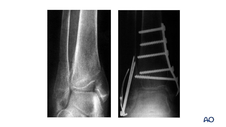
Weber A—medial malleolar fixation • Vertical shear injury • Always inspect joint • Restore and bone graft marginal impaction • Screws perpendicular to fracture plane • NOT from tip—malreduction • Buttress plate adds stability (antiglide) • 3. 5 screws better than “malleolar”
Marginal impaction
Marginal impaction
Marginal impaction
Vertical medial malleolar fracture
Management—Weber B • 60– 80% of ankle fractures • Be careful with superficial peroneal nerve (SPN) • Fibular fixation posterior or lateral? • Begin ORIF with fibula • Medial malleolar fixation—two 40 mm cancellous 4. 0 screws
Superficial peroneal nerve • • Injury to SPN common Variable course It may be lower than you think! Posterolateral incision
SPN over low Weber B fracture
Weber B—posterior fibular fixation • Posterolateral plate • Not subcutaneous • Biomechanically sound—antiglide • Lag screw(s) through the plate • No anterior stripping • Joint penetration less likely • Distal bicortical fixation
Weber B—when to fix? • Literature suggests: B-type fracture with no medial injury: ORIF = nonoperative • Surgery is NOT superior; maybe associated with more complications Mittal R, Harris IA, Adie S, Naylor JM; CROSSBAT Study Group. Surgery for Type B Ankle Fracture Treatment: a Combined Randomised and Observational Study (CROSSBAT). BMJ Open. 2017 Mar 27; 7(3)
Weber B—when to fix? • But many need ORIF • Significant instability • Significant medial or deltoid ligament injury and instability • Displaced medial malleolar fracture • Talar shift on x-ray Toth MJ, Yoon RS, Liporace FA, Koval KJ. : What's new in ankle fractures. Injury. 2017 Oct; 48(10): 2035 -2041.
Management—Weber C • Syndesmosis disrupted • Syndesmotic injury ligamentous or bony • Fibula—restoration of length critical • Fibular fracture may be high • Be careful with SPN • Accurate reduction and fixation of syndesmosis is paramount • Medial malleolar fixation—as for Weber B
Weber C—fibular fixation • Low fractures—anatomical reduction • High fractures (maisonneuve) • Fibular fixation not necessary if length is OK
Weber C—syndesmotic fixation • No lag screw—position • Reduce prior to insertion • 3 or 4 cortices • 3. 5 or 4. 0 cortical • Posterolateral to anteromedial • 2– 3 cm above ankle—parallel • Removal at 12 weeks or more
Syndesmotic reduction
Weber C—syndesmosis controversies • When is fixation necessary? • Best method—screw fixation or direct ligament repair? • Type of screw fixation? • Time to screw removal (syndesmosis healing)? • Time to weight bearing?
Screw removal?
Revision syndesmosis stabilization
Management—posterior tibial lip • Not true “malleolus” • Usually attached to posteroinferior tibiofibular ligament (PITFL) • Reduction with ORIF of fibula • Most authors—less than 20% ORIF • Approach PM or PL • AP or PA lag screws or posterior antiglide plate
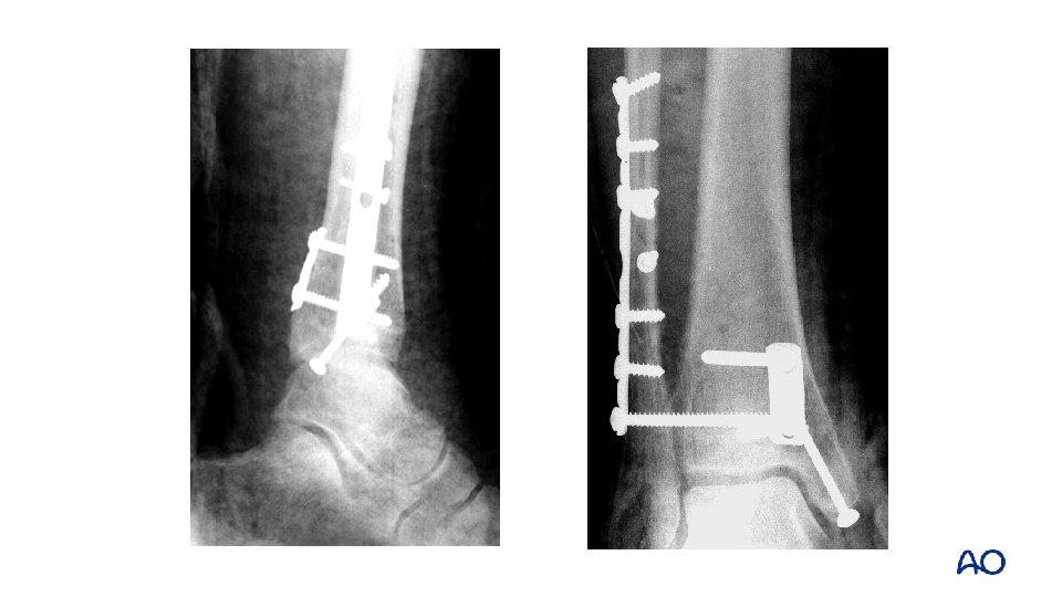
Osteoporotic fractures • Treat as if “normal” • Allow the implants to do the work • Aim: prevent progressive deformity and arthrosis • Better than arthrodesis • If fibula is osteoporotic and difficult to fix • Pass screws into tibia for added fixation and stability • Antiglide plate
Osteoporotic fractures—fibula pro-tibia technique
Neuropathic ankle fractures • Need more aggressive treatment • Double nonweight bearing • Double fixation • Double immobilization • Careful attention to soft tissues
Take-home messages • Not all ankle fractures are straightforward • Recognize more complex injuries • Apply sound fixation principles • Length of the fibula and rotation are critical • Restore anatomy • Recognize high-risk patients