
Completion of a Truncated Attenuation Image from the

- Slides: 15
Completion of a Truncated Attenuation Image from the Attenuated PET Emission Data Johan Nuyts, Christian Michel, Matthias Fenchel, Girish Bal, Charles Watson
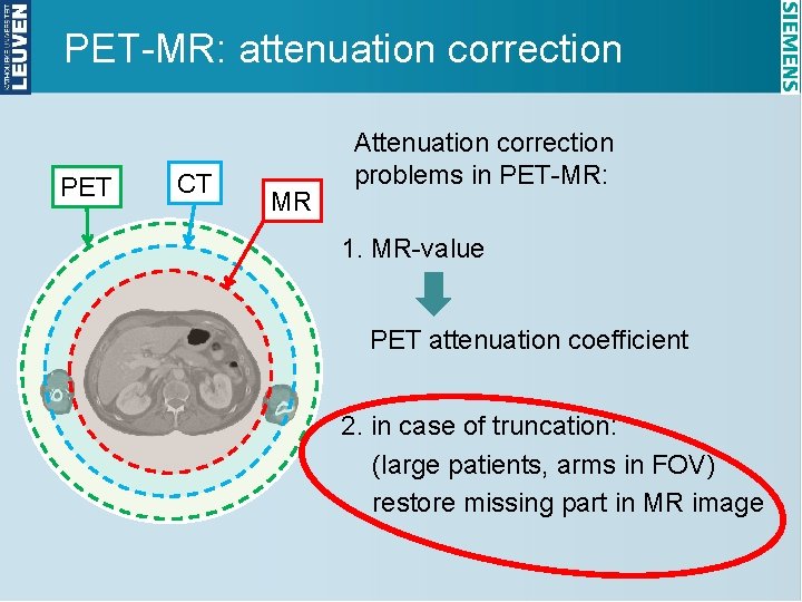
PET-MR: attenuation correction PET CT MR Attenuation correction problems in PET-MR: 1. MR-value PET attenuation coefficient 2. in case of truncation: (large patients, arms in FOV) restore missing part in MR image
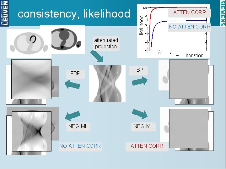
ATTEN CORR likelihood consistency, likelihood NO ATTEN CORR attenuated projection iteration FBP NEG-ML NO ATTEN CORR FBP NEG-ML ATTEN CORR
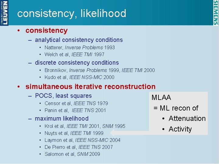
consistency, likelihood • consistency – analytical consistency conditions • Natterer, Inverse Problems 1993 • Welch et al, IEEE TMI 1997 – discrete consistency conditions • Bronnikov, Inverse Problems 1999, IEEE TMI 2000 • Kudo et al, IEEE NSS-MIC 2000 • simultaneous iterative reconstruction – POCS, least squares MLAA • Censor et al, IEEE TNS 1979 = ML recon of • Panin et al, IEEE TNS 2001 – maximum likelihood • Attenuation • Krol et al, IEEE TMI 2001, SNM 1995 • Activity • Nuyts et al, IEEE TMI 1999 • Laymon et al, IEEE NSS-MIC 2004 • De Pierro et al, IEEE TNS 2007 • Salomon et al, SNM 2009
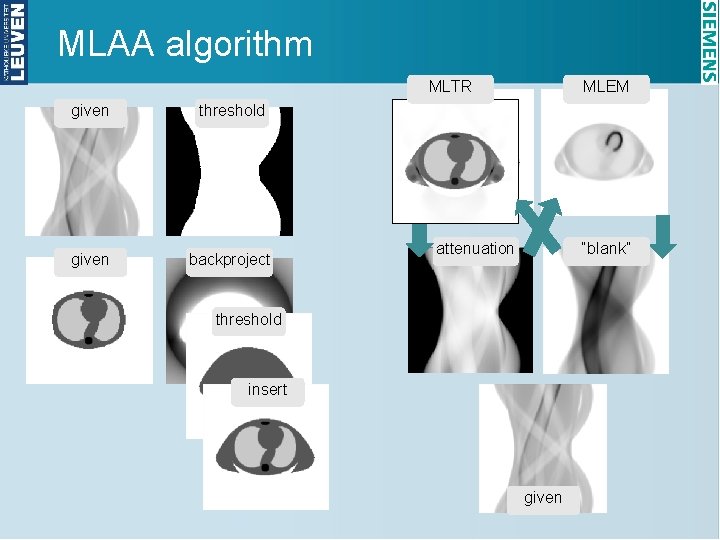
MLAA algorithm MLTR given threshold given backproject MLEM attenuation “blank” threshold insert given
MLAA: priors • emission update o smoothing Markov prior: relative difference prior • attenuation update o smoothing Markov prior: relative difference prior o intensity prior, favoring § air § tissue
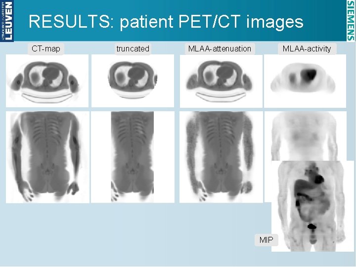
RESULTS: patient PET/CT images CT-map truncated MLAA-attenuation MLAA-activity MIP
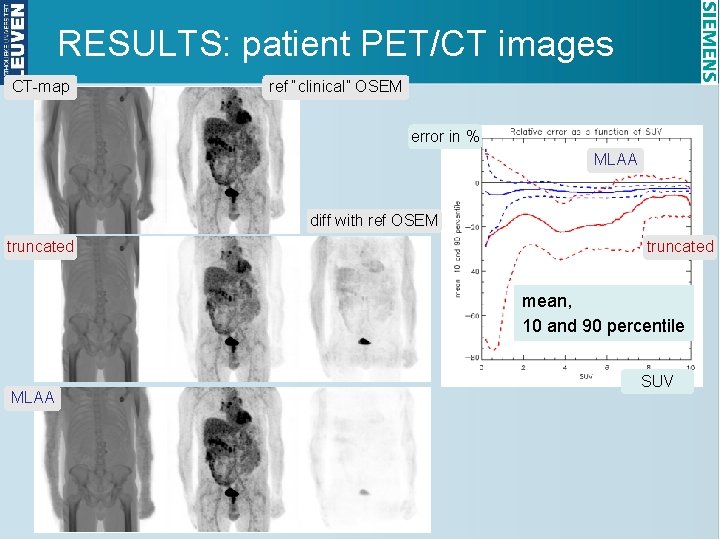
RESULTS: patient PET/CT images CT-map ref “clinical” OSEM error in % MLAA diff with ref OSEM truncated mean, 10 and 90 percentile MLAA SUV
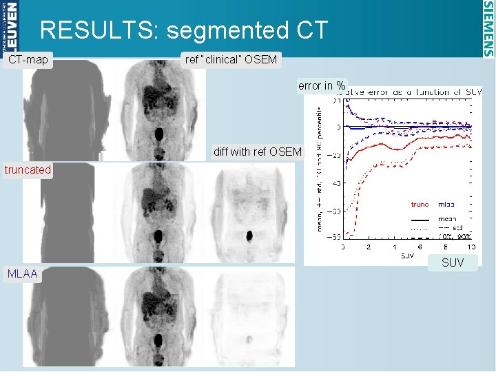
RESULTS: segmented CT CT-map ref “clinical” OSEM error in % diff with ref OSEM truncated MLAA SUV
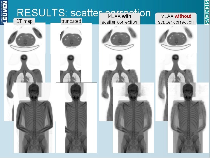
RESULTS: scatter correction MLAA with CT-map truncated scatter correction MLAA without scatter correction
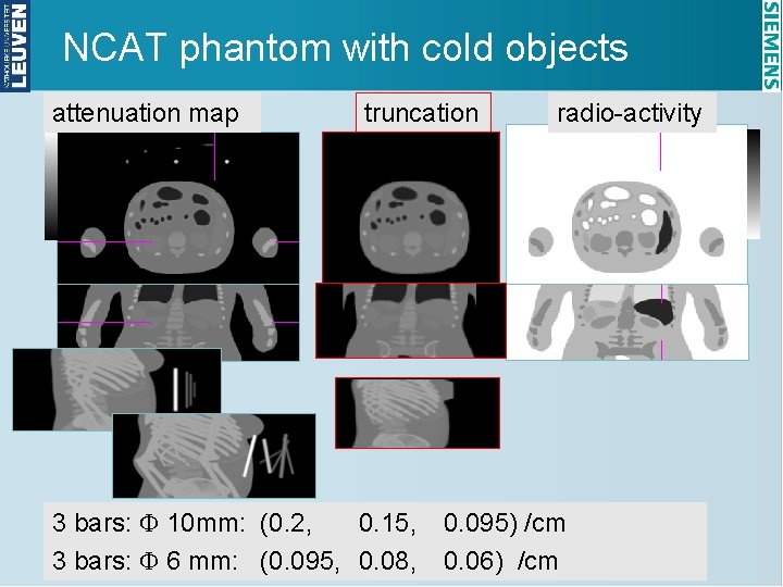
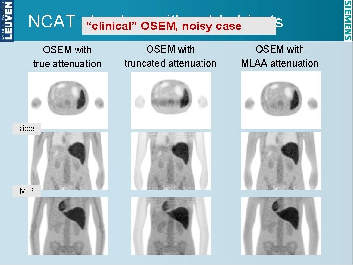
NCAT phantom with cold objects attenuation map truncation 3 bars: F 10 mm: (0. 2, 0. 15, 3 bars: F 6 mm: (0. 095, 0. 08, radio-activity 0. 095) /cm 0. 06) /cm
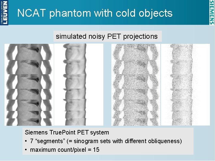
NCAT phantom with cold objects simulated noisy PET projections Siemens True. Point PET system • 7 “segments” (= sinogram sets with different obliqueness) • maximum count/pixel = 15
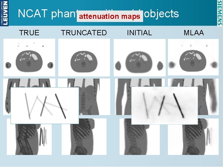
NCAT phantom with cold attenuation maps objects TRUE TRUNCATED INITIAL MLAA
NCAT phantom withnoisy coldcase objects “clinical” OSEM, OSEM with true attenuation slices MIP OSEM with truncated attenuation OSEM with MLAA attenuation
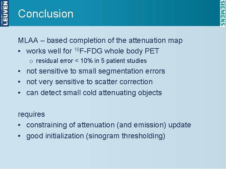
Conclusion MLAA – based completion of the attenuation map • works well for 18 F-FDG whole body PET o residual error < 10% in 5 patient studies • not sensitive to small segmentation errors • not very sensitive to scatter correction • can detect small cold attenuating objects requires • constraining of attenuation (and emission) update • good initialization (sinogram thresholding)