Complements Dr Eman Albataineh Assistant Prof Immunology College

Complements Dr. Eman Albataineh, Assistant Prof. Immunology College of Medicine, Mu’tah university Immunology, 2 nd year students

• The complement system consists of a number of small proteins found in the blood, generally synthesized by the liver, and normally circulating as inactive precursors (pro-proteins). • When stimulated by one of several triggers, activation cascade is started and lead to functional effects. • Over 25 proteins and protein fragments make up the complement system. They account for about 5% of the globulin fraction of blood serum.

• Complement was discovered many years ago as to ‘complement’ the antibacterial activity of antibody, hence the name. • Although first discovered as an effector arm of the antibody response (Adaptive), complement can also be activated early in infection in the absence of antibodies (innate).

• In the case of the complement system, the precursor are widely distributed throughout body fluids and tissues without adverse effect. At sites of infection, however, they are activated locally and trigger a series of potent inflammatory events • There are three distinct pathways through which complement can be activated on pathogen surfaces. These pathways depend on different molecules for their initiation, but they converge to generate the same set of effector molecules
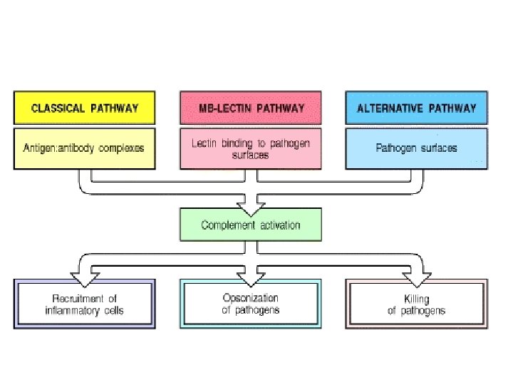

Complement pathways activation • Classical pathway; recognize antibody binding microbe as viruses or bacteria (IGG 1, IGG 3 IGA and IGM) • Alternative; recognize LPS or endotoxins (part of innate response) • Lectin pathway. The lectin pathway is homologous to the classical pathway, but with mannose-binding lectin (MBL) instead of C 1 q. and in the absence of antibody

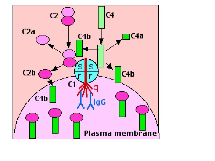
Complements • The classical pathway, so called because it was discovered first, uses a plasma protein called C 1 q to detect antibodies bound to the surface of a microbe or other structure. Once C 1 q binds to the Fc portion of the antibodies, two associated serine proteases, called C 1 r and C 1 s, become active and initiate a proteolytic cascade involving other complement proteins (C 2, C 4) to make C 3 convertase. The classical pathway is one of the major effector mechanisms of the humoral arm of adaptive immune responses.

Classical pathway • C 1 exists in blood serum as a molecular complex containing: – C 1 q – C 1 r – C 1 s • The IGM and IGG that bound by antigen, contain a binding site for C 1 q. (A single molecule of Ig. M is enough to initiate the pathway. Ig. G is far less efficient, requiring many molecules to do so. ) • Binding of C 1 q activates C 1 s and C 1 r. • Activated C 1 s (a protease) cleaves two serum proteins: – C 4 is cleaved into a large fragment • C 4 b, which binds covalently to surface of antigen (opsonisation) and • C 4 a, smaller, inactive, which diffuses away. – C 2 is cleaved into • C 2 b, which binds to a site on C 4 b, • C 2 a a smaller, inactive, fragment of which diffuses away. – The complex of C 4 b • 2 b (some books say C 4 b 2 a) is called "C 3 convertase" because it catalyzes the cleavage of C 3.

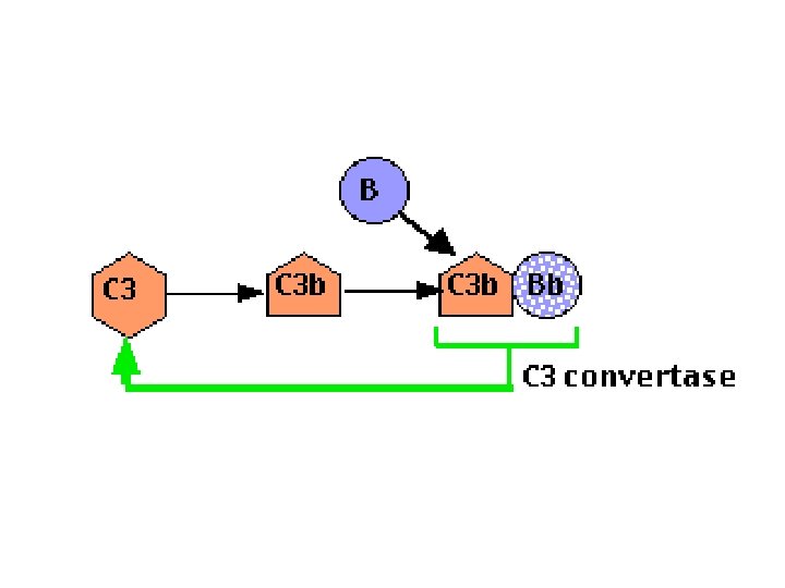
Complement • The alternative pathway, is triggered when a complement protein called C 3 simultaneously degraded to C 3 b that recognizes certain microbial surface structures, such as bacterial LPS. • C 3 b undergoes its post-cleavage conformational change, a binding site for a plasma protein called Factor B is also exposed. Factor B then binds to the C 3 b protein that is now covalently tethered to the surface of a microbial or host cell. Bound factor B is in turn cleaved by a plasma serine protease called Factor D, releasing a small fragment called Ba and generating a larger fragment called Bb that remains attached to C 3 b. • The C 3 b. Bb complex is the alternative pathway C 3 convertase,
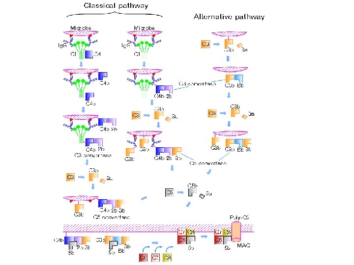

Lectin pathway • Lectin pathway. The lectin (proteins macromolecules in blood that are highly specific mannose on pathogen) pathway is homologous to the classical pathway, but with mannose-binding lectin (MBL) instead of C 1, and in the absence of antibody • This pathway is activated by binding of MBL to mannose residues on the pathogen surface, which can then split C 4 into C 4 a and C 4 b and C 2 into C 2 a and C 2 b the rest pathway is similar to classical
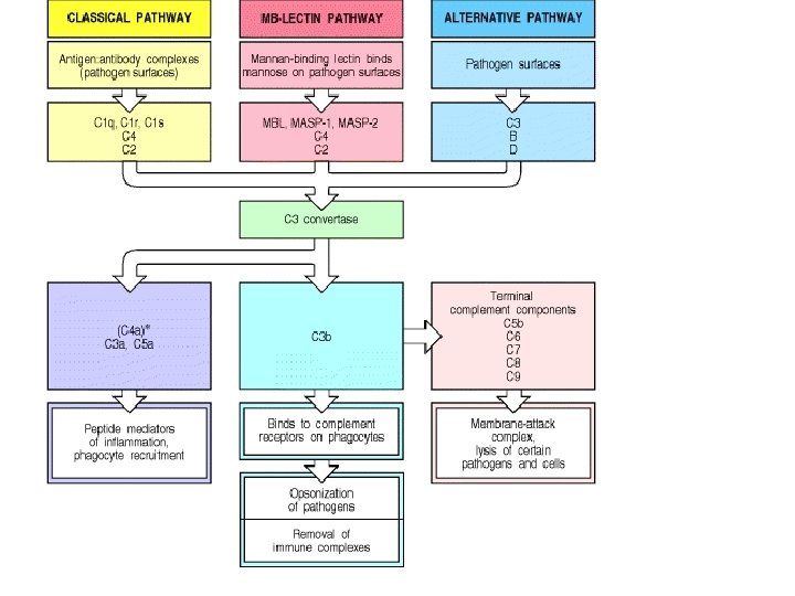

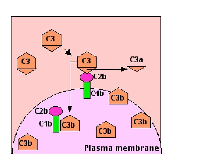
C 3 • Recognition of microbes by any of the three complement pathways results in sequential recruitment and assembly of additional complement proteins • C 3 is the most abundant protein of the complement system. Because of its abundance and its ability to activate itself (as described later), it greatly magnifies the response. – C 3 convertase cuts C 3 into two major fragments: • C 3 b, which binds covalently to glycoproteins scattered across the microbial cell surface. Macrophages and neutrophils have receptors for C 3 b and can bind the C 3 b-coated cell or particle preparatory to phagocytosis. This effect qualifies C 3 b as an opsonin. • C 3 a This small fragment is released into the surrounding fluids. It can bind to receptors on basophils and mast cells triggering them to release their vasoactive contents (e. g. , histamine). Because of the role of these materials in anaphylaxis and inflammation, C 3 a is called an anaphylatoxins. • C 3 d:

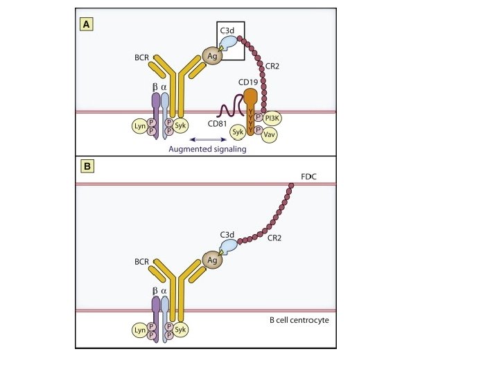
C 3 d link innate to humoral immunity • The complement system of innate immunity is important in regulating humoral immunity largely through the complement receptor CR 2, which forms a coreceptor on B cells during antigen-induced activation • antigen gets “tagged” with the appropriate C 3 d product via the classical or lectin complement pathways.

C 3 d • The CD 19/CD 21 complex is an essential B cell coreceptor that functions synergistically to enhance signaling through the B cell Ag receptor in response to T cell-dependent, complement -tagged Ags (more rapid and efficient production of antigenic peptide/class II complexes as compared with B cell Ag receptor-mediated processing alone, less concentration of antigen) • involves FDC (follicular dendritic cells) within germinal centers in which, as a result of trapping C 3 d-opsonized antigen via CR 2, they can present the antigen to previously antigenengaged B cells during the processes of selection in affinity maturation, and generation of effector and memory B cells and maintaining germinal center

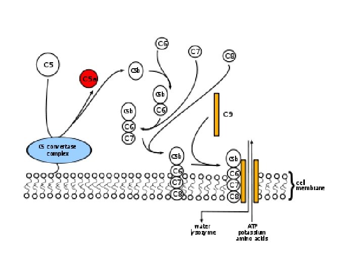
Membrane attack complex -C 5 convertase formed by joining C 3 convertase to C 3 b -C 5 – Cleavage of C 5 by the (C 3 b. Bb 3 b )and other complements (C 4 b • 2 b. 3 b)(which is thus a "C 5 convertase". ) produces: – C 5 a, which is released into the fluid surroundings where it • is a potent anaphylatoxin • is a chemotactic attractant for neutrophils – C 5 b, which serves as the anchor for the assembly of a single molecule each of • C 6; • C 7, and • C 8. • The Membrane Attack Complex • The resulting complex C 5 b • 6 • 7 • 8 guides the polymerization of as many as 18 molecules of C 9 into a tube inserted into the lipid bilayer of the plasma membrane. This tube forms a channel allowing the passage of ions and small molecules. Water enters the cell by osmosis and the cell lyses.

Summary of complement functions • Opsonization by C 3 b, i. C 3 b and C 4 b and C 5 b targets foreign particles for phagocytosis. • Chemotaxis by C 5 a, C 4 a and C 3 a attracts phagocytic cells to the site of damage. • This is aided by the increased permeability (anaphylatoxins) they cause smooth muscle contraction, vasodilation, histamine release from mast cells, and enhanced vascular permeability. mediated by C 3 a, C 5 a, C 4 a. • C 3 b ; The early complement components are also important for solubilizing antigen-antibody complexes and elimination from the body ( by binding the immune complex to CR 1 on erythrocyte). otherwise aggregation of the complexes lead to immune complex disorder (SLE, diabetes mellitus, RA) • Lysis of target cells (C 5 b-9). • Promoting antibody formation. Breakdown of C 3 b generates a fragment (C 3 d) that binds to antigens enhancing their uptake by B cells.
; bind C 3 b, and C 4")
Complement receptors • Type 1 receptor(CR 1); bind C 3 b, and C 4 b. Expressed in blood cells, erythrocytes, macrophages, neutrophil eosinophil and T lymphocytes – Do opsonization and Induce phagocytosis (with antibody) – Help to remove immune complexes from blood to liver and spleen (erythrocytes) • Type 2 receptor (CR 2), bind C 3 d, and, expressed on B lymphocytes and DC – With other proteins enhance B cell response to Tdependent antigen – Help in trapping the antibody, complement –antigen in germinal center by FDC to activate B cell affinity maturation – Receptor for epstein barr virus on B cells

• Type 3 and 4 receptors on phagocytes – bind opsonizing C 3 b and lead to phagocytosis. Found on macrophages and neutrophils

Regulation of complement activity • The explosive potential of the complement system requires that it be kept under tight control. At least 12 proteins are known that do this. Three examples: • Factor H and Decay-accelerating factor (DAC), removes Bb from the alternative pathway C 3 convertase. • Factor I inactivates C 3 b. • C 1 inhibitor (C 1 INH) binds to sites on activated C 1 r and C 1 s shutting down their proteolytic activity. • CD 59 on normal tissue cells which inhibit association of C 9 with C 5 b-8

Disorders of the complement system • With so many proteins involved, it is not surprising that inherited deficiencies of one or another are sometimes encountered in humans. Four examples: – C 3. An inherited deficiency of C 3 predisposes the person to frequent bouts of bacterial infections mainly gram negative bacteria. – C 2 , C 1, C 3 or C 4. immune complex disorders are the main problem with a deficiency of C 2 , C 1 or C 4. This emphasizes the important role of the complement system in clearing away antigen-antibody complexes. A deficiency of C 2 (or one of the other early components) is frequently found in patients with lupus erythematosus (SLE). – C 9. most people who cannot make C 9 have no problem with bacterial infections. Laboratory studies suggest that the C 5 b • 6 • 7 • 8 complex by itself is able to lyze bacteria although not as efficiently as C 9. – C 1 INH. A deficiency of C 1 INH produces hereditary angioedema. The massive release of anaphylatoxins (C 3 a, C 5 a) may cause dangerous swelling (edema) of the airways, as well as of the skin and intestine. – CD 59 deficiency in its expression lead to inadequate control of MAC assembly results in intravascular red cell lyses called paroxysmal nocturnal haemoglobinuria
- Slides: 26