Compartment Syndrome Andrew H Schmidt M D Professor


 artery Pv (Low)")
 artery Tissue ischemia arteriole l l capillary venule vein")


")


: 631 -636.")


 • Currently, the “differential pressure” is considered the most reliable indicator")
 • Currently, the “differential pressure” is considered the most reliable indicator")


- Slides: 56
Compartment Syndrome Andrew H. Schmidt, M. D. Professor, Dept. of Orthopedic Surgery, Univ. of Minnesota Chief, Department of Orthopaedic Surgery Hennepin County Medical Center April 2016
Disclosure Information Andrew H. Schmidt, M. D. Conflicts of Commitment/ Effort Board of Directors: OTA Critical Issues Committee: AOA Editorial Board: J Knee Surgery, J Orthopaedic Trauma Medical Director, Director Clinical Research: Hennepin County Med Ctr. Disclosure of Financial Relationships Royalties: Thieme, Inc. ; Smith & Nephew, Inc. Consultant: Medtronic, Inc. ; DGIMed; Acumed; St. Jude Medical (spouse) Stock: Conventus Orthopaedics; Twin Star Medical; Twin Star ECS; Epien; International Spine & Orthopedic Institute, Epix Disclosure of Off-Label and/or investigative Uses I will not discuss off label use and/or investigational use in my presentation.
Objectives • Review Pathophysiology of Acute Compartment Syndrome • Review Current Diagnosis and Treatment – Risk Factors – Clinical Findings – Discuss role and technique of compartment pressure monitoring.
Pathophysiology of Compartment Syndrome Pressure Inflexible Fascia Injured Muscle
Vascular Consequences of Elevated Intracompartment Pressure: A-V Gradient Theory Pa (High) artery Pv (Low) arteriole capillary Local Blood Flow = venule Pa - Pv R Matsen, 1980 vein
Increased interstitial pressure Pa (High) artery Tissue ischemia arteriole l l capillary venule vein Lysis of cell walls Release of osmotically active cellular contents into interstitial fluid Increased interstitial pressure More cellular injury…
Reported Causes of Compartment Syndrome • Trauma – Fracture – Crush syndrome – Soft tissue injury • Bleeding • Reperfusion Injury – Vascular Injury – Lithotomy position • • • Intensive muscle use Burns Snakebite Venous obstruction Injection injuries
Fracture Treatment Increases IMP • • • Splinting/casting Manipulation Traction Spanning Ex Fix Nailing
Incidence of ACS • • • 2 -10% tibial fractures 10% Calcaneal fractures 18% Schatzker VI plateau fractures 41% foot crush injuries 48% Segmental tibia fractures 53% Medial knee fx/dislocations
“Radiographic Predictors of Compartment Syndrome in Tibial Plateau Fractures. ” Ziran and Becher, J Orthop Trauma 2013; 27: 612– 615 > 10% Tibia Width Femoral Displacement
Consequences of Compartment Syndrome l Ischemic myonecrosis – Ischemic contracture Ischemic neuropathy l Crush Syndrome l – Rhabdomyolysis – Renal Failure Delay in dx/rx is the cause of a poor outcome
Diagnosis
Diagnosis • Traditionally based on clinical assessment of the “ 6 P’s”: – Paresthesia – Paresis – Pain on stretch – Pink Color – Pulse present – Pressure
Diagnosis • Traditionally based on clinical assessment of the “ 6 P’s”: – Paresthesia: requires detailed exam, may be deficits present upon initial presentation – Paresis – Pain on stretch – Pink Color – Pulse present – Pressure
Diagnosis • Traditionally based on clinical assessment of the “ 6 P’s”: – Paresthesia – Paresis: Difficult to grade motor strength when there is a fracture and/or splint present – Pain on stretch – Pink Color – Pulse present – Pressure
Diagnosis • Traditionally based on clinical assessment of the “ 6 P’s”: – Paresthesia – Paresis – Pain on stretch: Difficult to separate from the pain due to the fracture – Pink Color – Pulse present – Pressure
Diagnosis • Traditionally based on clinical assessment of the “ 6 P’s”: – Paresthesia – Paresis – Pain on stretch – Pink Color: These are normal findings too, so not helpful in the diagnosis of ACS – Pulse present – Pressure
Diagnosis • Traditionally based on clinical assessment of the “ 6 P’s”: – Paresthesia – Paresis – Pain on stretch – Pink Color – Pulse present – Pressure: difficult to assess reliably or to quantify
Problems with Physical Diagnosis • Literature meta-analysis found that clinical findings have poor sensitivity. – Sensitivity of 13 -19% – Positive predictive value of 11 -15%. – Specificity = 97% (3% incidence of C. S. in patients without clinical findings Ulmer, J Orthop Trauma, 16: 572
• Consecutive patients, same hospital, random surgeon call. • Incidence of CS varied from 2 -24%
• Inadequate documentation in 21 of 30 cases of CS – No date/time – Missing info about signs and symptoms – Pressures not documented
Intramuscular Pressure Measurement • Adjunct to clinical examination. • Represents the only data available in comatose or otherwise non-evaluable patient: – – Anesthesia Head Injury Sedated Intoxicated
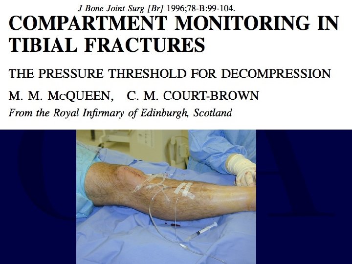
How to measure IMP Anterior Compartment: ≈ 1 cm lateral to tibial crest
How to measure IMP Lateral Compartment: Just anterior to posterior border of fibula
How to measure IMP Deep Posterior: Just behind posteromedial border of the tibia
How to measure IMP Superficial Posterior: From posterior (away from midline)
Intramuscular Pressure Measurement • Normal resting IMP 0 -8 mm Hg in adults, 13 -16 mm. Hg in children. • Elevated in CS. • Does not measure degree of tissue injury.
How to Interpret Pressure Measurements ?
The absolute pressure at which perfusion ceases varies markedly depending on the clinical situation. • • Duration of elevated pressure Systemic blood pressure Direct muscle trauma Other patient factors: – Training of individual – Muscle oxidative fiber type
Muscle Injury Related to Pressure and Time
Compartment Syndrome is also a pressure-time phenomenon • Tissue doesn’t become irreversibly damaged until it has been ischemic for 6 -8 hours. • In patients with extremity injury, you don’t know when the clock started.
Pressure Time
Pressure CS occurs X X Time
Pressure X CS doesn’t occur Time
J Bone Joint Surg Am. 1981; 63(4): 631 -636.
• Continuous monitoring associated with earlier diagnosis of compartment syndrome, earlier fasciotomy, more rapid fracture healing, and improved outcomes: Monitored Time to Fasciotomy 16 hrs Sequelae of CS 0/12 Mean time to union 17 weeks Non-monitored 32 hrs 10/11 25 weeks
1 st 12 – hours IMP > 30 IMP > 40 IMP > 50 PP < 30 53 30 4 1 2 nd 12 – hours 28 7 0 2 3 Patients had Fasciotomy No patient had any sequelae of CS at follow-up
Perfusion Pressure (∆P) • Currently, the “differential pressure” is considered the most reliable indicator of when fasciotomy is not necessary: – MAP- IMP < 45 mm Hg – DBP - IMP < 30 mm Hg BP > 30 mm HG IMP
Perfusion Pressure (∆P) • Currently, the “differential pressure” is considered the most reliable indicator of compartment syndrome: – MAP- IMP < 45 mm Hg – DBP - IMP < 30 mm Hg BP < 30 mm HG IMP
• 19 isolated LE fractures in alert patients w/out CS • Avg IMP 35. 5 ± 14 mm Hg Threshold for fasciotomy IMP > 30 mm Hg IMP > 45 mm Hg ”Delta P” < 30 mm Hg ”Delta P” < 20 mm Hg # Patients meeting criteria 95% 63% 84% 58% No patient developed CS at follow-up
“Based on our data, use of direct compartment measurements with existing thresholds and formulations to determine the diagnosis of compartment syndrome may not accurately reflect a true existence of the syndrome. A search for other quantitative measures to more accurately reflect the presence of compartment syndrome is warranted. ” Prayson et al, 2006
Intra-operative IMP • ∆P may be artifactually low due to anesthesia induced hypotension. • How to calculate ∆P in this setting?
Diastolic Blood Pressure in Patients with Tibia Fractures Under Anesthesia: Implications for the Diagnosis of Compartment Syndrome • 246 consecutive patients undergoing tibial nailing under general anesthesia • Blood pressures preoperatively, intraoperatively, and postoperatively were documented. SBP DBP Preoperative 134 ± 16 74 ± 10 Intraoperative 113 ± 12 56 ± 11 PACU 142 ± 18 73 ± 11 Tornetta et al, OTA 2006 Inpatient Floor 133 ± 16 76 ± 9
Ø Use preoperative DBP when making intraoperative decisions - Unless the patient will be under anesthesia for a long time
“ALECS is a capricious entity, insidious in onset and elusive of diagnosis. No single physical finding or clinical or laboratory findings establish the diagnosis; it is a combination of intuitive integration of available findings and above all an alertness to the possibility of the emergence of this entity that leads to early diagnosis and treatment. ” J Trauma 2007; 63: 268 -275
Diagnostic Algorithm Clinical Exam Benign Yes CS Not Present < 40 mm Hg CS Not Present > 30 mm Hg CS Not Present No, or exam not possible Measure IMP > 40 mm Hg Calculate ∆P < 30 mm Hg CS May Be Present
Treatment of Compartment Syndrome
Compartment Syndrome = If you think about it, do it! Fasciotomy
Technique of Fasciotomy • Longitudinal skin incision that extends the entire length of the compartment. • Release of fascia of involved muscle. • Skin left open.
Fasciotomy Pearls • Mark incisions ahead of time • Make long incisions • Find superficial peroneal n. in lateral compartment fasciotomy before release • Beware of saphenous n. and vein medially • Deep posterior compartment most easily identified distally where FDL is just behind tibia. Proximally, release soleus off posterior tibia.
Complications of Fasciotomy • • • Muscle Weakness Chronic venous insufficiency Tethered scars Impaired sensation Ulceration Costs
• 458 patients / 804 fasciotomies. • Higher rate of primary closure using VAC compared to wet-dry dressings (79% vs 50%). • Time-to-closure shorter in VAC group – Primary closure 7. 1 vs 9. 6 days – Secondary closure 8. 5 vs 11. 5 days
Summary – Current Dx / Rx • Both the clinical exam and pressure thresholds can reliably tell us who does not have ACS. – Perfusion pressure has well-validated thresholds for when fasciotomy is not needed (PP ≥ 30 mm. Hg). – Knowledge of pressure vs time trends further helps differentiate those who are developing ACS. • Other diagnostic modalities needed: – biomarkers
Thank You
For questions or comments, please send to ota@ota. org