Comparing types of cell division Mitosis and Meiosis


 line up on midplane �Anaphase I")

 go through mitosis")


- Slides: 21
Comparing types of cell division Mitosis and Meiosis
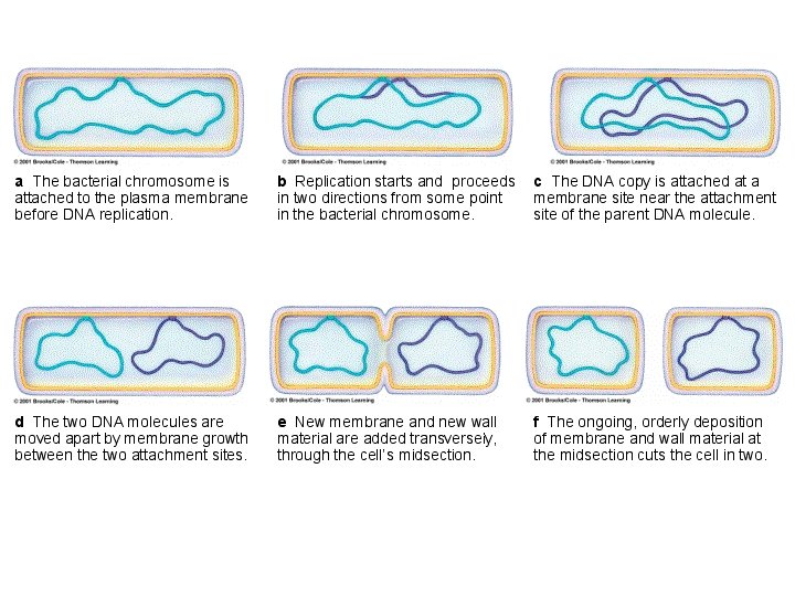
Prokaryotic Cells Divide by Fission �Most prokaryotes have 1 circular chromosome Replication begins at “ori”, moves through replication complex and ends at site called “ter” 2 “ori” regions on the 2 chromosomes attach to cell membrane; as cell grows longer, chromosomes separate �Cytokinesis begins about 20 min after replication done
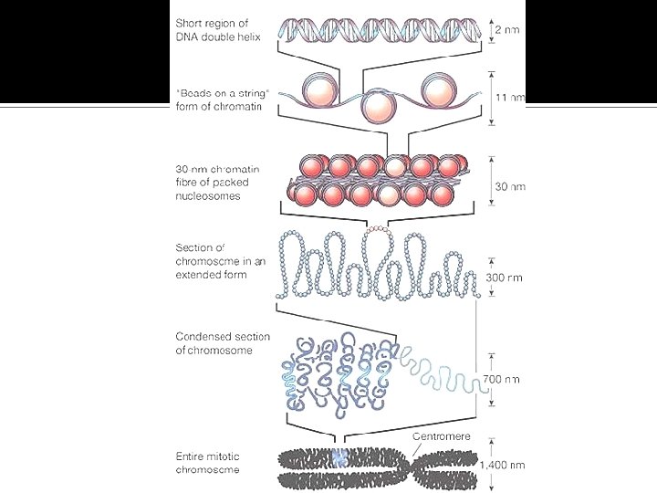
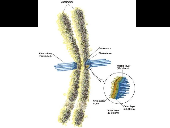
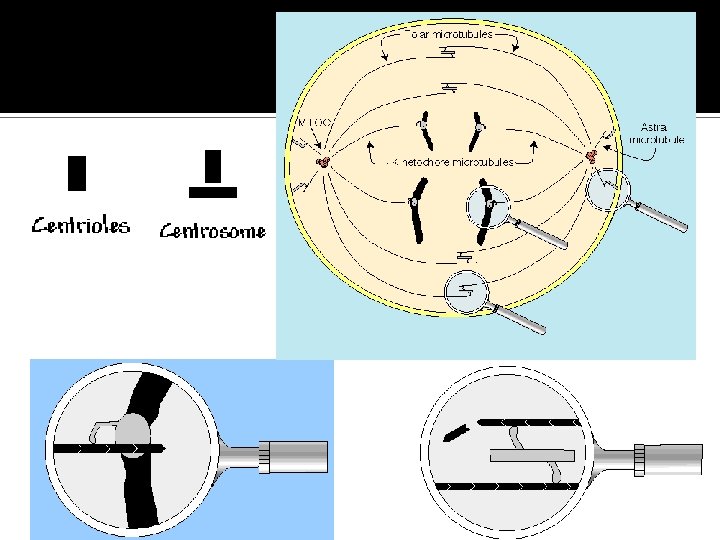
Eukaryotic Chromosomes �Made of chromatin – DNA and associated proteins �When cell isn’t dividing, chromosomes partly unraveled �During cell division, chromatin fibers condense into distinct chromosomes
� 2+ meters of DNA/human cell Must be packaged to avoid tangling �Form nucleosomes Spool that keeps DNA from tangling �Scaffolding proteins also maintain chromosome structure �During mitosis and meiosis, nucleosomes pack more tightly together
Eukaryotic Cells �Have many chromosomes Replication and segregation is more intricate �Cytokinesis is distinct from division of genetic material Differs in plants and animals
Why do cells divide? �When cells reach a certain size they either: Stop growing Divide �Volume increases faster than surface area Needed materials cannot diffuse through volume of cell efficiently
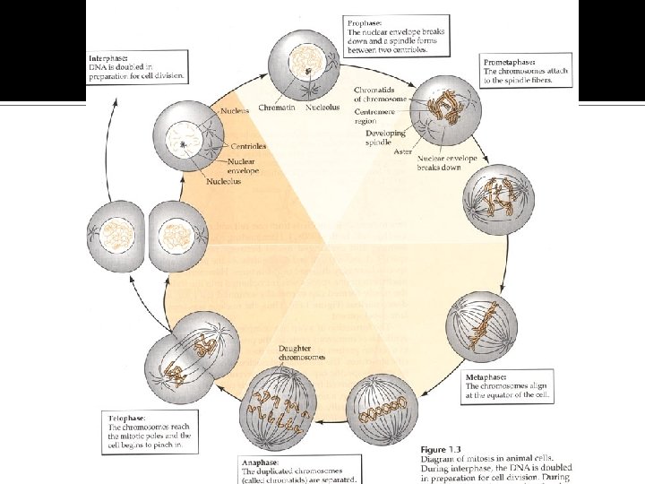
Mitosis � Nuclear Division � 4 Stages: Prophase Metaphase Anaphase Telophase � Mitosis is followed by cytokinesis Cleavage furrow in animal cells Cell Plate in plant cells � http: //www. loci. wisc. edu/outreach/bioclips/CDBio. html � http: //highered. mcgraw-hill. com/sites/0072437316/student_view 0/chapter 11/animations. html#
Sexual Reproduction �Union of 2 gametes to form single zygote �Results in genetic variation of offspring �Gametes are haploid Contain 1 of each homologous chromosome Produced by meiosis
Meiosis � Cell division that reduces the # of chromosomes � http: //www. stolaf. edu/people/giannini/flashanimat/celldivisi on/meiosis. swf � Diploid cells undergo 2 cell divisions 4 haploid cells � 4 key ways meiosis differs from mitosis: 2 nuclear and cytoplasmic divisions 4 cells DNA duplicates once, but nucleus divides twice Resulting cells are haploid Shuffling of homologous chromosomes so each haploid cell is unique
First Meiotic Division: Meiosis I Prophase I – synapsis – homologous chromosomes lie side by side Forms tetrad (4 chromatids) Proteins hold homologues together Crossing-over occurs between homologues, resulting in genetic recombination
Meiosis I, cont. �Metaphase I Chromosome pairs (tetrads) line up on midplane �Anaphase I Paired homologues separate and move to opposite poles �Telophase I Chromatids decondense some �Interkinesis, brief stage, may follow
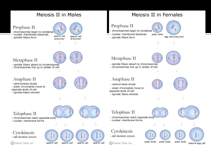
Meiosis II
Spermatogenesis in Vertebrates �Occurs in seminiferous tubules of testes �Spermatogonia (undifferentiated) go through mitosis to make 1 o spermocytes (2 n) � 1 1 o spermatocyte 2 2 o spermatocytes 4 spermatids of equal size
Oogenesis in vertebrates � Occurs in ovaries � Oogonia form during embryonic development and increase in size to become 1 o oocytes (2 n) � At birth, they are in prophase I – enter resting stage � Puberty – 1 o oocytes completes meiosis I Cytoplasm/organelles not divided evenly, form 1 2 o oocyte and first polar body (may divide)
Oogenesis, cont. � 2 o oocyte begins 2 nd meiotic division, but remains in metaphase II until fertilized �After fertilization, meiosis II continues, forming 1 ovum and 2 nd polar body �Polar bodies eventually disintegrate Provide a place for chromosomes to go