Common Foot and Ankle Disorders Abdulaziz Almaawi MD

Common Foot and Ankle Disorders Abdulaziz Almaawi. MD, MSc, FRCSC Consultant Orthopedic Surgeon, King Khalid University Hospital Assistant Professor, King Saud University

Objectives Anatomy in Foot and Ankle. Common Foot and Ankle disorders. To differentiate from simple disorders and serious ones. To learn about initial management and prognosis.
 of the")
Importance of Foot and Ankle • Subject to most weight bearing (Loading) of the body. • Important proprioception function. • Deformity is easily noticeable. • Improper shoe wear can cause symptoms. • With advancing age; deformity becomes more symptomatic.

Foot Anatomy

X-ray standing

Common Disorders Flat Foot. Hallux Valgus. Plantar Fasciitis Ankle Sprains and Ankle Instability. Osteochondral lesions of Talus. Diabetic Foot. Charcot Foot.

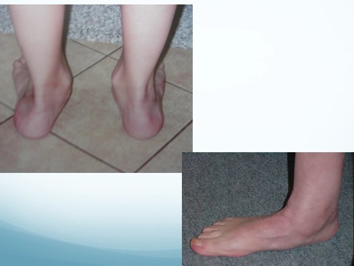
Flat Foot • Reduction of longitudinal arches of the foot. • Most cases are Developmental: i. e. arches do not develop normally. • Usually painless. • Acute flat foot? • Rigid flat foot?

X-ray standing

Flat Foot Flexible Ligamentous laxity Rigid Tarsal coalition: Fibrous or Bony union between bones of the foot Standing on tip-toes: Heels move inward Subtalar motion (inversion/Eversion): Normal Standing on tip-toes: Heel does not move inward Subtalar motion (inversion/Eversion): Reduced or absent

Flat Foot Flexible Rigid

Flat Foot management • Foot exercises: Strengthen muscles but will not correct deformity • Orthotics/Insoles: Protective, correct Malalignment • Good Shoes: Beneficial on the long run • Surgery: Painful flatfoot with complication Rigid flat foot Acute flat foot

Hallux Valgus Lateral deviation of big toe. at MTP joint Mostly painless. Bunion: Swelling and protrusion at the medial aspect of big toe. Severe HV: Interferes with shoe wear

Hallux Valgus measurements

Hallux Valgus Measurements Hallux Valgus Angel: angle between line extending along 1 st metatarsal and a line extending along proximal phalanx. Normal : < 15° Mild HV: 16 -25° Moderate HV: 26 -35° Severe HV: > 35°

Hallux Valgus Measurements 1 st intermetatarsal Angle between 1 st metatarsal long axis and 2 nd metatarsal N < 10° Hallux interphalangeus Angle between long axis of proximal and distal phalanges N < 8°

Hallux Valgus Management Correct and suitable shoe wear. Avoidance of tight shoes. Protection to the bunions. Surgery is reserved for symptomatic cases. Following surgery; patient has to continue proper shoe wear.

Hallux Valgus Surgery

Plantar Fasciitis • Common disorder at middle age and elderly. • Insidious in onset; • unilateral or bilateral. • Localized tenderness to insertion of plantar fascia into calcaneum. • Plain lateral X-ray of heel frequently shows calcaneal spur (prominence or ossification at the site of anterior calcaneum at plantar fascia insertion site)

Plantar fasciitis Commonly associated with flat feet. No visible heel swelling, no skin changes and no increase in local temperature. Inflammatory process is at site of pain; i. e. at plantar fascia insertion into calcaneum. Heel pain like stabbing pain when patient puts foot to the ground first step in the morning; and gets less after some walking.
")
Calcaneal spur (Early)
")
Calcaneal Spur (Advanced)

Management NO easy or simple management is available. Mainly conservative. Includes stretching exercises to plantar fascia: active and passive. Use of soft heel insoles (Silicone) may be helpful. Shock wave therapy (SWT) may be effective. Local steroid injections are helpful sometimes. Surgery: Last solution, very rare

Ankle Sprains • One of most common injuries. • Usually occurs during sports activities. • May occur at home or street. • The injury is partial or complete ligament rupture.

Clinical picture of Ankle Sprains • History of twisting injury. • Pain, swelling and bruising at and around ankle. • No tenderness of lateral malleolus; but tenderness anterior, posterior or inferior to it i. e. over ligaments. • Dorsi-flection and plantar flexion possible; but inversion and eversion very painful. • X-Rays : NO fracture.
")
Ankle Ligaments (Lateral)

Ankle Sprain Most commonly injured ligament is the Anterior Talo. Fibular Ligament. Ankle anterior drawer test is positive. Compare with normal side Other ligaments are Posterior Talo-Fibular Ligament and Calcaneo-Fibular ligament.

Management of Ankle Sprain RICE: Rest, Ice, compressors, Elevation. Apply Back-slab splints for few days: if not able to weight bear. Might use protection with brace Early physiotherapy and strengthening. Mostly heal with no surgery. Surgery: if physio fail and there is clear instability
 • Damage at localized areas of Talar articular cartilage")
Osteochondral Defects of talus (OCD) • Damage at localized areas of Talar articular cartilage • Lack of blood supply is often post traumatic, but occasionally No cause can be found. • A local cartilage & varying depth of underneath bone are involved and may separate of main talus inside the ankle joint. • Usually postero-medial part of dome of talus. • Localized pain on weight bearing and even at rest may present.

Plain AP X-ray : lesion is suspected

CT Coronal view; lesion highly suspected

MRI: lesion is confirmed

Management of OCD Depends on: i. Symptoms: Pain and recurrent swelling ii. Size OCD: large and iii. Loose fragment Arthroscopic debridement: of the lesion and drilling of its base Fixation with headless screw: large OCD with large bony part
 i. Long term diabetes or ii. Failure to")
Diabetic Foot • Neuropathy (nerve damage) i. Long term diabetes or ii. Failure to control diabetes numbness, tingling and reduced sensation of the feet. Associated with Decreased circulation (neuropathy, calcification of vessels, CAD) may result in delayed healing, infections, Gangrene and Amputations

Care of Feet in DM • Primary target: Prevention i. Blood sugar control (best indicator) ii. Daily self inspection of feet is mandatory iii. Member of the family or assistant should do it. iv. Regular inspections by healthcare personnel should be arranged v. A visit to a doctor should take place immediately whenever any complication occurs.

Surgery in Diabetic Foot Skilled care of wounds and ulcers in diabetic foot is required. Wound debridement, antibiotics and repeated dressing should be done. Amputations may become necessary when there is Gangrene. Toe amputation or ray amputation, forefoot amputation, below or above knee amputation.

Charcot Foot Significant nerve damage to the foot leads to i. Bones of the foot become weak ii. Joints inflamed, swollen and lax walking on the foot leads to disintegration and collapse of the joints and Deformity: such as Rocker- bottom deformity.

Charcot Foot Causes Any disorder which lead to Neuropathy. There is decreased sensation and decreased ability to feel temperature, pain and trauma.

Clinical picture Look: Foot is red or dusky in color. Swelling in the area. Deformity Feel: No Pain or soreness, Warmness of foot. Move: decreased ROM X-rays changes are important to detect and interpret: i. Early X-rays: show NO changes. ii. Later X-rays: haziness, osteopenia, irregular joint destruction, subluxation or even dislocation.

Diabetic foot 04/03/1428

Advanced Case of Charcot

Diagnosis of Charcot Foot • Good history and clinical examination. • Awareness. • Exclusion of other causes which may give similar picture: like infection or tumour. • MRI, bone scans can help.

Management of Charcot Foot • Immobilization • Custom Shoes and Bracing • Activity modification

Surgery in Charcot Foot

Amputation in Charcot foot • May be indicated as a last option. • Mainly when there is severe instability which cannot be controlled by surgery or orthosis. • Also when surgery fails to achieve stability. • Recurrent infection increase the possibility of amputation.

Questions Thank you
- Slides: 46