Common Cutaneous Infections and Infestations Mohammed AlHaddab MD

Common Cutaneous Infections and Infestations Mohammed Al-Haddab, MD, FRCPC Assistant Professor, consultant dermatologist and dermatosurgeon

Objectives • To be familiar with common bacterial, viral, mycobacterial, fungal, and parasitic infections of the skin. • To be familiar with presentation and management of common skin infections. • To be familiar with skin infestation, their clinical features, prevention and treatment. • The main reference is the TEXTBOOK. .

Introduction • Human body contains > 10 times more microbial cells than human cells. • Microbial colonization of skin is more dense in humid intertriginous areas. • An intact stratum corneum is the first barrier against pathogenic invasion. • Most common bacterial infections are caused by staph. Aureus an streptococcus group A

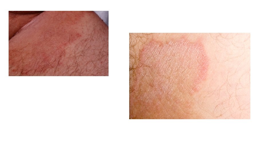
Erythrasma • Light brown sharply marginated patches in intertriginous skin like axillae, groins, toe web spaces. • Woods lamp shows coral red fluorescence. • Web space is the most common site. • DDx: inverse psoriasis, tinea, Hailey-Hailey disease. • Rx: benzoyl peroxide, clindamycin and erythromycin lotion.
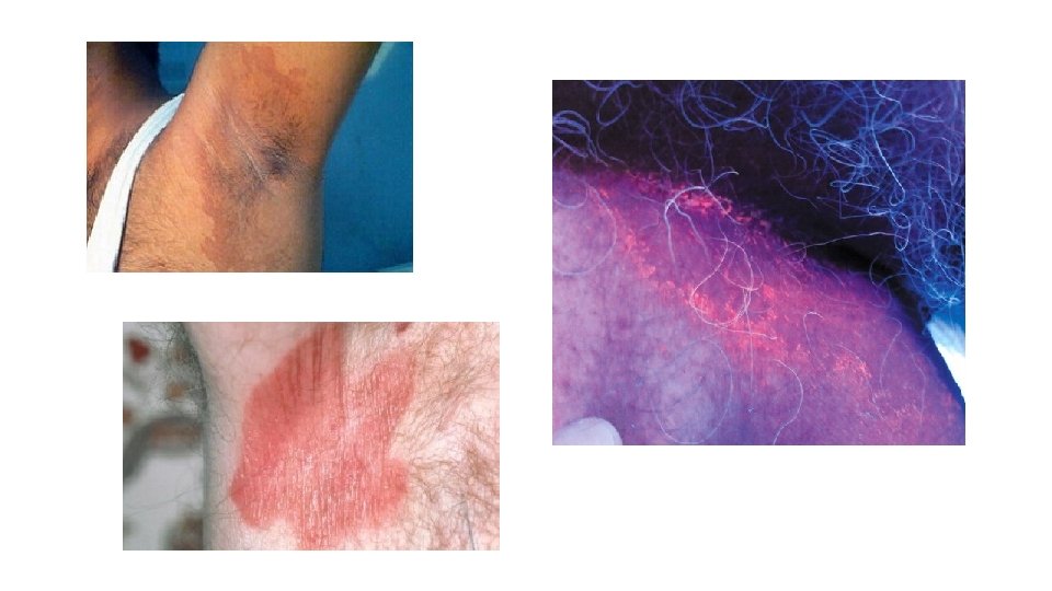

Intertrigo • Inflammation of body folds. • May represent inflammatory dermatosis or superficial colonization or infection. • Infectious Intertrigo could be caused by staph aureus, streptococcus group A, B, G, or pseudomonas aeruginosa. • Usually asymptomatic, sometimes itchy. • DDx: inverse psoriasis, Erythrasma, Hailey-Hailey disease, tinea, Langerhans Histiocytosis. • Rx: treat the underlying cause. • Topical mild corticosteroids with topical anti bacterial or anti fungal.

Impetigo • Mostly caused by staph aureus and streptococcus group A. • Could be primary mostly in children. • Secondary that arise next to area of colonization, wounds or site of trauma, eczematous lesions and other skin dermatosis (impetiginization). • Honey colored crusted erosions. Mostly asymptomatic. • Bullous impetigo with superficial bullae with erythematous halo mostly in intertriginous areas. • Ecthyma is ulceration with thick adherent crust at occluded sites in patients with poor hygiene.
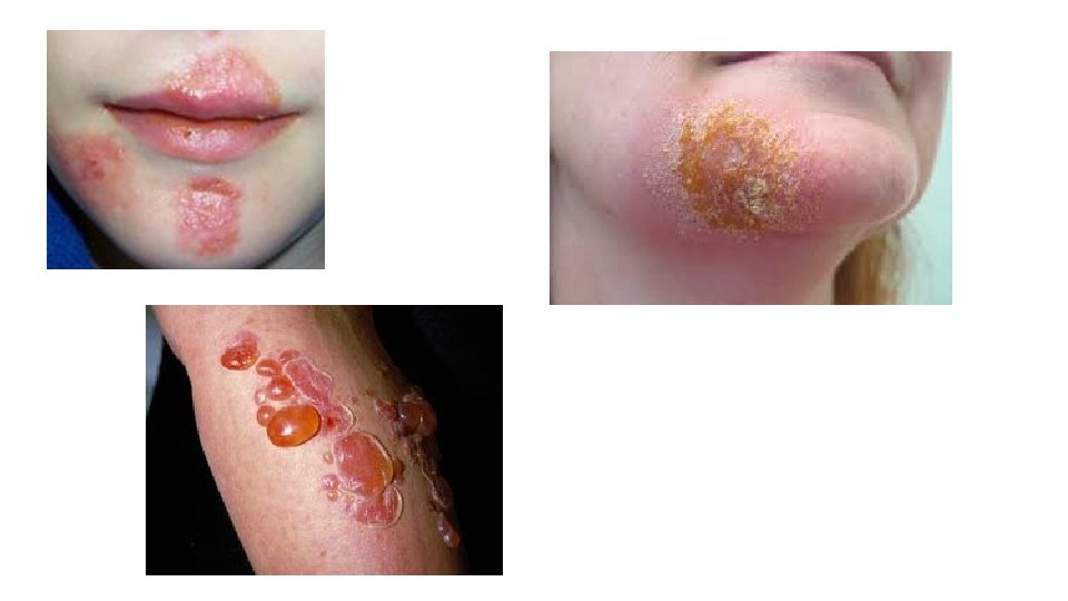

Impetigo • DDx: excoriation, contact dermatitis, herpes simplex, tinea, and scabies. • Diagnosis confirmed by culture but not usually necessary. • If untreated it becomes more extensive and may develop into ecthyma or cellulitis. • Ecthyma might heal with scarring. • Rx: topical fucidic acid or mupirocin. • Systemic antibiotics in severe cases.

Folliculitis and furuncles • Deep infection of the skin either through trauma or extension of superficial skin infection into hair follicle. • Mostly caused by staph aureus. • Folliculitis can be caused by fungi, viruses, and mites. • Shaving, occlusion of hairy regions, topical corticosteroids, diabetes, immunosuppression. • If it involve the deep parts of hair follicle will lead to furuncle. • Gram negative folliculitis usually occur in patients with acne treated by long term systemic antibiotics. • Hot tub folliculitis on trunk caused by pseudomonas aeruginosa.
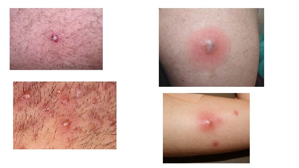

Folliculitis and furuncles • DDx folliculitis: acne, rosacea, HIV associated, eosinophilic folliculitis, acneiform drug eruption, corticosteroid acne, pseudofolliculitis barbae. • Rx: dicloxacillin, cephalosporin, clindamycin for one week. Minocycline, trimethoprim-sulfamethoxazole, and quinolones secondary options. • Gram negative folliculitis Rx to stop offending agent, Isotretinoin. • Furuncle and abscess treated by incision and drainage with antibiotics. • Recurrent folliculitis and furuncles need prolonged antibiotic therapy such as clindamycin and rifampicin for 8 weeks, with topical mupirocin for body folds and nares to decrease the bacterial colonization.

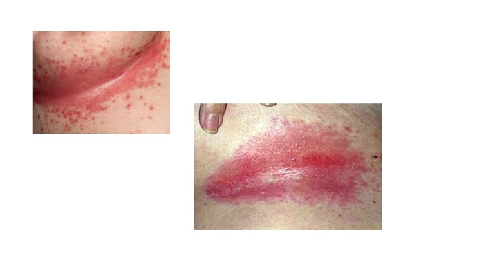
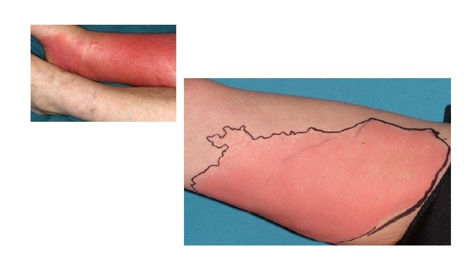
Cellulitis • Staph aureus, group A streptococcus. • Start as spreading and expanding erythematous lesion at the site of entry like trauma, tinea pedis, leg ulcers. • Fever, chills can develop before cellulitis, local pain, tenderness. • Higher fever and chills are usually associated with streptococcus infection. • Presents as red, hot, edematous plaque with sharp irregular borders, sometimes with vesicles and erosions. • Lower legs most commonly in adults.

Cellulitis • Erysipelas is caused by streptococcus group A. • Spread rapidly along superficial cutaneous lymphatic vessels. • Tender red edematous expanding plaque on leg or face. • Influenza in children < 2 year old, cheek periorbital area. • Aeruginosa cause ecthyma gangrenosum a necrotizing soft tissue infection in immunocompromised patients mostly in axilla, groin or perineum.

Cellulitis • DDx: DVT, urticaria, insect bite, fixed drug eruption, erythema nodosum, erythema migrans. • Diagnosis is clinical, culture is positive in 30% only. • Skin biopsy could help. • Rx: systemic antibiotics
 • Exfoliative toxin induced. • Macular scarlitinform rash or")
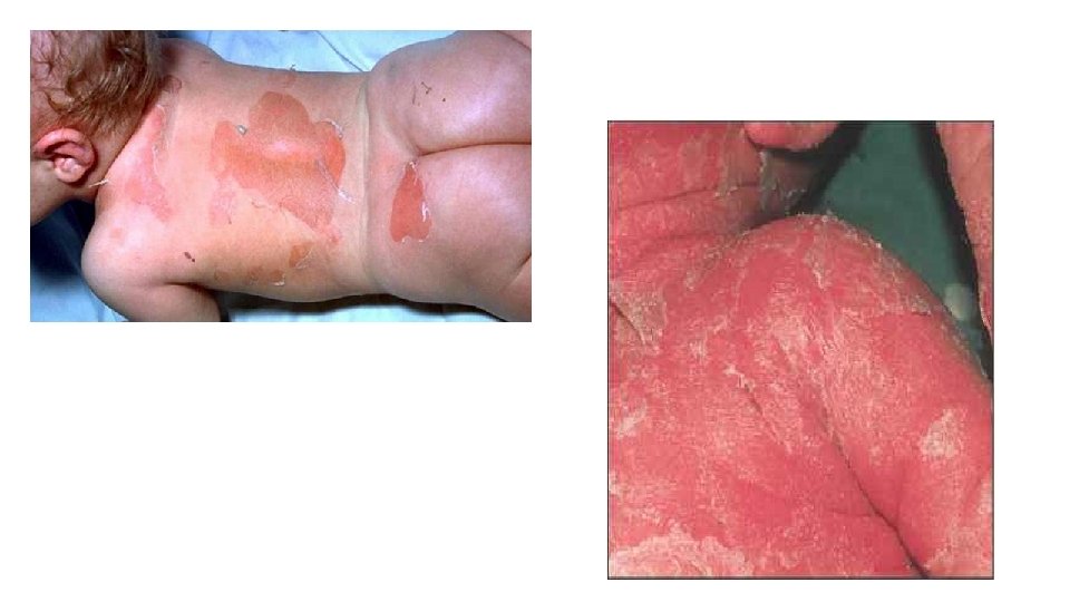
Staphylococcal Scalded Skin Syndrome (SSSS) • Exfoliative toxin induced. • Macular scarlitinform rash or diffuse ill defined erythema and a fine sandpaper rash initially that rapidly become more erythematous tender. The skin can be removed by gentle pressure (Nikolsky's sign). • Desquamation occurs with healing with no scarring. • No mucus membrane involvement. • DDx: Kawasaki syndrome, drug eruption, scarlet fever. • Rx: systemic antibiotics.

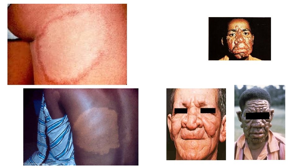
Leprosy • Mycobacterium liprae. • Incubation period 5 -7 years. • Affects skin, peripheral nerves, upper respiratory tract, eyes, testes. • Tuberculoid: localized skin and nerve involvement. • Lepromatous: generalized involvement of skin and other tissues. • Borderline: with different skin manifestations and many bacilli in histopathology. • Inverse relationship between skin color and severity. • Mode of transmission is unknown, close contact, respiratory droplets.

Leprosy • Immune responses: • Type 1 acute tenderness and pain along affected nerves with loss of function and skin lesions become inflamed and may ulcerate. Edema on face, hands, and feet. • Type 2 erythema nodosum leprosum which is seen in 50% of patients with Lepromatous leprosy (LL) after starting treatment. With painful red skin nodules on the face and extensor limbs. • Type 3 Lucio reaction in Mexico, manifests as erythematous plaques with shallow large polygonal sloughing ulcerations on the legs in patients with LL. • Onset of leprosy is insidious and painless, affects nerve initially with numbness, paraesthesias, eventually muscle weakness, atrophy and contracture.

Leprosy • Tuberculoid Leprosy TL: multiple well defined hypopigmented annular plaques with raised borders and anesthetic center. May resolve spontaneously and is not associated with immune reaction. Nerve thickening ulnar, posterior auricular, perineal, and posterior tibial. • Borderline Leprosy BL: has both features from Tuberculoid and Lepromatous, anesthesia and decreased sweat is prominent, does not heal spontaneously and could be associated with type 1 immune reaction. • Lepromatous Leprosy LL: skin colored papules, nodules, might coalesce with diffuse dermal infiltration with loss of hair of lateral brows and eyelashes and leonine face. Nerve involvement is more extensive than TT, upper respiratory tract, eyes, testes might be involved.

Leprosy • URT involvement: chronic nasal congestion, epistaxis, saddle nose deformity. • Eyes involvement: cranial nerve palsies, lagophthalmos, corneal insensitivity, uveitis, glaucoma and cataract. • Testes involvement may lead to hypogonadism. • DDx: sarcoidosis, Leishmaniasis, syphilis, granuloma annulare, lymphoma. • Dx: slit skin smears from earlobes, elbows, knees, a slit is made then tissue fluid is stained by Ziehl-Neelsen stain. PCR. No culture. • Skin biopsy: TL shows epitheliod granuloma around dermal nerves with sparse or absent bacilli. LL shows extensive cellular infiltrate, no skin appendages, macrophages filled with M. leprae Virchow cells.

Leprosy Management • TL: dapsone+ rifampicin • LL: dapsone+rifampicin+ clofazimine • Immune reactions: prednisolone, thalidoamide. • Prevention and rehabilitation after nerve damage. • Patient rehabilitation in society after treatment.

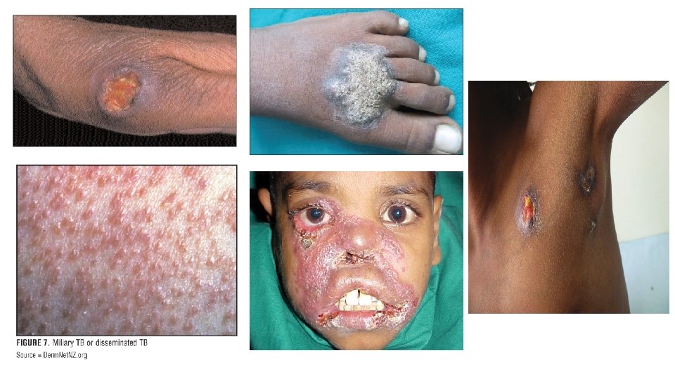
Cutaneous Tuberculosis • Mycobacterium tuberculosis. • Tuberculous chancre: exogenous inoculation into skin in non immune patient. • Tuberculosis verrucosa cutis: exogenous inoculation into skin in immune patient. • Scrofuloderma: direct extension from joints, bone or lymph nodes. • Lupus vulgaris: lymphatic spread to skin. • Acute military TB: hematogenic dissemination.

Cutaneous tuberculosis • Tuberculous chancre: starts as papule at inoculation site after 2 -4 weeks then enlarge to form a painless ulcer and regional lymph node. • Tuberculosis verrucosa cutis: papule with violaceous border that evolve to hyperkeratotic warty plaque. Usually single on hands in adults, and on legs, knees in children with no lymph nodes. • Lupus vulgaris: papules evolve to well defined irregular scaly plaque, that is soft and friable, mostly on nose, ear and scalp. Scarring is prominent. • Scrofuloderma: firm subcutaneous nodule that liquefies and perforate leading to undermined ulcer with pus or caseous discharge. On the parotid, submandibular, supraclavicular, lateral neck. • Acute miliary TB: disseminated minute macules and papules or purpuric lesions allover body especially trunk.

Cutaneous tuberculosis • Dx: PPD, skin biopsy, PCR, culture. • Rx: isoniazid+rifampicin • Tuberculosis verrcuosa cutis if small surgical excision.

Fungal Infections • superficial: fungi that affect skin, hair, nail and mucosa, like candida, malasssezia, and dermatophytes. • Deep chronic cutaneous: after percutaneous inoculation like eumycetoma, chromoblastomycosis and sporotrichosis. • Systemic with cutaneous dissemination like cryptococcosis, histoplasmosis, blastomycosis, coccidioidomycosis.

Candidiasis • Cutaneous candidiasis: intertriginous and occluded skin. • Mucosal candidiasis: oropharynx and genitalia. • Disseminated candidemia: host defense defects especially neutropenia. Usually after invasion of GIT. • Candida albicans, tropicalis, parapsilosis, krusei, kefyr, glabrata. • 20% of healthy individuals are colonized. • 10% of women colonized vaginally which is increased by systemic antibiotics, pregnancy, IUD, OCP. • Immunocompromised, DM, obesity, hyperhidrosis, warm climate and maceration, systemic corticosteroids.

Candidiasis • Candidal Intertrigo: sharply demarcated, polycyclic, erythematous, eroded patches with satellite pustules affect body folds. • Interdigital: in obese elderly, webspace between 3 rd and 4 th toes. • Diaper dermatitis: beefy red plaques with papular and pustular lesions, erosions on genital and perianal area, inner thighs and buttocks.

Candidiasis • Prevention: keep intertriginous area dry, anti fungal powder. • Topical anti fungal: nystatin, azoles, imidazole. • Systemic anti fungal: fluconazole, itraconazole, voriconazole.
 Versicolor • Superficial overgrowth of Malassezia furfur and M. globosa, both resident")
Pityriasis (Tinea) Versicolor • Superficial overgrowth of Malassezia furfur and M. globosa, both resident cutaneous flora. • Malassezia changes from a yeast to hyphal form leading to inflammation of the skin. • Not contagious. More with sweating, warm weather, oily skin • Asymptomatic tan-light brown macules with fine scales, on dark skin could be hypopigmented macules mostly on trunk, also on neck, upper arms axillae and groin. • Woods lamp: blue green fluorescence of scales. • Hypopigmentation is caused by dicarboxylic acid produced by Malassezia species which inhibits tyrosinase in melanocytes.
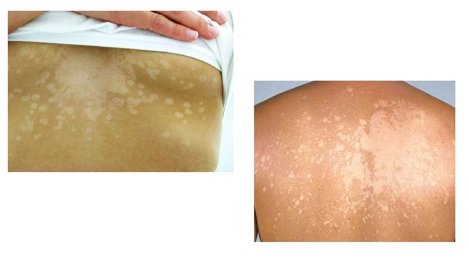

Pityriasis Versicolor • DDx hypopigmented macules: vitiligo, pityriasis alba, post inflammatory hypopigmentation. • DDx Scaly macules: tinea corporis, seborrheric dermatitis, mycosis fungoides. • KOH: filamentous hyphae and globosa yeast forms (spaghetti and meatballs) • Histopath: budding yeast, and hyphal forms in the stratum corneum by PAS stain, hyperkeratosis, hyperplasia and chronic inflammation. • Rx: ketoconazole shampoo, azole creams, terbinafine cream • Systemic fluconazole 300 mg weekly for 3 weeks or itraconzole 400 mg single dose.

Dermatophytosis • Dermatophytes are fungi that infect keratinized cutaneous structures like stratum corneum, nails, and hair. • 3 genera of dermatophytes: trichophyton, microsporum, and epidermophyton. • Trichophyton rubrum is the most common cause of epidermal dermatophytosis. • Children have scalp infections, adults have intertriginous infections. • Transmission from person to person and from animals like dogs, cats, rabbits.

Tinea Pedis • Dermatophytic infection of the feet. • Hot, humid, occlusive footwear, hyperhidrosis. • Usually asymptomatic, with hyperkeratotic macerated plaques between toe webs. Could be seen with tinea unguium. • Interdigital type: dry scaling or maceration and fissuring of toe webs, hyperhidrosis is common most commonly between 4 th and 5 th toes. • Moccasin type: well demarcated scaling with erythema and minute papules on margin, fine white scales and hyperkeratosis on soles, heels and lateral border of feet. Bilateral involvement is common. • Inflammatory type: vesicles or bullae, may be associated with id reaction. • Ulcerative type: extension of interdigital tinea onto plantar and lateral foot ? Secondary bacterial infection.
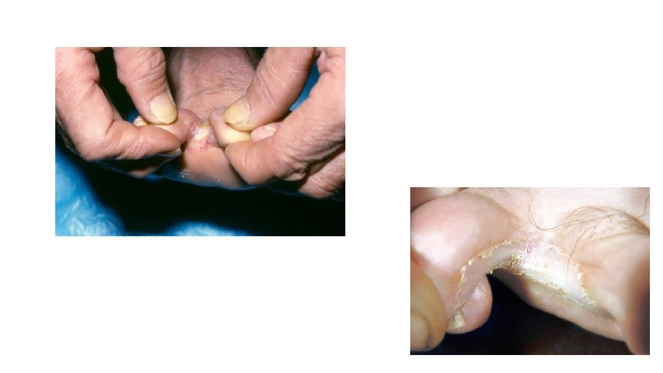

Tinea Pedis • DDx interdigital type: Erythrasma • DDx moccasin type: psoriasis, chronic eczema • DDx inflammatory type: bullous impetigo, allergic contact dermatitis, dyshidrotic eczema, bullous disorders

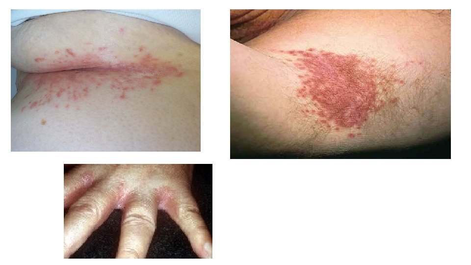
Tinea Cruris • Usually associated with tinea pedis. • Large scaling well demarcated erythematous-brown plaques, with papules and pustules at the periphery in groin, thighs and buttocks. • DDx: Erythrasma, candida Intertrigo, inverse psoriasis, Pityriasis Versicolor. • Rx: topical antifungal imidazole, allylamines, naphthionates and pyridine. • Rx: if severe ? Systemic antifungal, terbinafine, itraconazole, fluconazole.

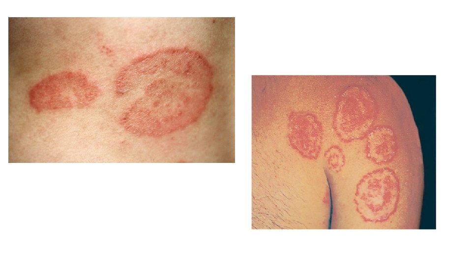
Tinea Corporis • Mostly by T. rubrum on trunk and extremities. • Well defined scaly erythematous plaques with advanced border and central clearing, sometime it coalesce to form large polycyclic annular and concentric rings. • DDx: allergic contact dermatitis, atopic dermatitis, annular erythema, psoriasis, Pityriasis rosea, Pityriasis Versicolor, subacute lupus, mycosis fungoides.

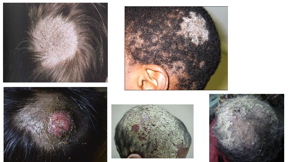
Tinea Capitis • 6 -10 years of age most commonly affected. • T. tonsurans, M. canis, M. audouinii, T. violaceum, T. schoenleinii. • Ectothrix infection: occurs outside the hair shaft with cuticle destruction. Caused by M. audouinii, M. canis. • Endothrix infection: occurs within the hair shaft without cuticle destruction. Caused by T. tonsurans, T. violaceum. • Black dot: resembling seborrheric dermatitis. • Kerion: with boggy inflammatory plaques. • Favus: endemic in middle east and Africa.

Tinea Capitis • Non inflammatory infection: gray patch type, with scaling diffuse or circumscribed alopecia. Circular alopecia with broken off hairs, fine scales, no or minimal inflammation. M. canis may show green fluorescence under woods lamp. • Black dot: broken off hairs near scalp, diffuse poorly circumscribed, usually caused by T. tonsurans. • Kerion: inflammatory mass with loose hairs, boggy, painful, purulent, inflamed nodules and plaques oozing pus with lymphadenopathy. Caused by T. verrucosum, T. mentagrophytes. Heal with scarring. • Favus: thick yellow adherent crust (scutula) composed of skin debris and hyphae pierced by remaining hair shaft. Often results in scarring.

Tinea Capitis • Griseofulvin 15 -20 mg/kg/day for 4 -6 weeks. • Terbinafine 3 -6 mg/kg/day for 4 -6 weeks • Itraconazole 3 -5 mg/kg/day for 4 -6 weeks. • Fluconazole 3 -6 mg/kg/day for 4 -6 weeks

Viral Infections • Primary infections with many viruses cause acute systemic febrile illnesses and exanthems, are usually self-limited, and convey lifetime immunity. • Herpes virus infections often have asymptomatic primary infection but lifelong latent infection. With immunosuppression the can become active and cause disease.

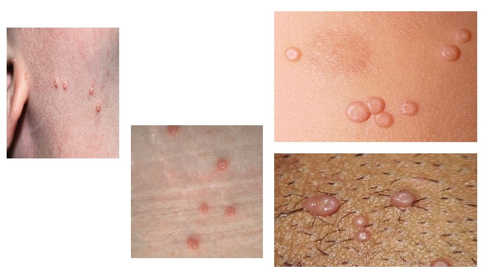
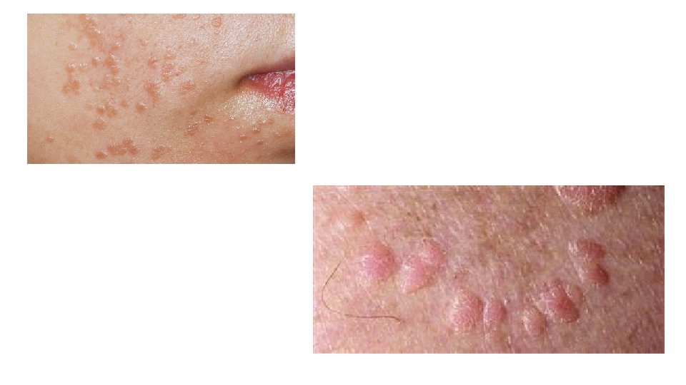
Molluscum Contagiosum Caused by Molluscum Contagiosum virus. Self limited viral infection, transmitted by skin to skin contact. More common in children, in adults mostly sexually transmitted. Multiple scattered skin colored umbilicated papules and nodules. Autoinoculation can occur by scratching or touching the lesions. Can be widespread in patients with atopic dermatitis. Histopathology: infected cells contain large intracytoplasmic inclusions called Henderson-Pattterson bodies. • Often persists for 6 months then undergo spontaneous regression without scarring. • Rx: curettage, cryotherapy, electrodessication, and cantharidin. • •
 infections • Double stranded DNA viruses that infect squamous epithelia")
Human Papilloma Virus (HPV) infections • Double stranded DNA viruses that infect squamous epithelia of skin and mucus membrane. • Clinical lesions are determined by HPV type. • Transmitted by skin to skin contact. • Immunosuppression, organ transplantation and HIV, increase the incidence and severity of warts. • Genital warts in mothers during delivery can be transmitted to neonates resulting in genital warts and respiratory papillomatosis. • HPV 16, 18, 31, 33 has high malignant potential and could lead to cervical carcinoma in women.

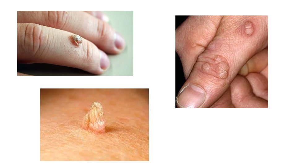
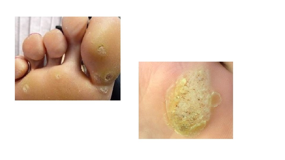
Verruca Vulgaris • Firm papules from 1 -10 mm hyperkeratotic, clefted surface, wit vegetations (warty), with characteristic red or brown-black dots. • Inoculation by scratching lead to linear arrangement. • Annular warts at sites of previous therapy. • Filiform warts with small bases. • Butcher warts, large cauliflower-like lesions on hands of meat handlers. • Mostly caused by HPV 1, 2. • DDx: Molluscum Contagiosum, seborrheric keratosis, actinic keratosis, SCC.

Plantar Warts • Hyperkeratotic plaques with brown dots on plantar surface, that could be tender when walking. • Mosaic warts if multiple papules coalesce to form large plaque, difficult to treat. • HPV 1, 2 • DDx: callus, corn.

Flat Warts • Small skin colored or light brown flat papules 1 -5 mm in size mostly on face, beard area, dorsum of hands. • HPV 3, 10 • DDx: syringoma, Molluscum Contagiosum

Epidermodysplasia Verruciformis • Autosomal recessive. Usually first occur at 5 -7 years. • Flat topped papules, skin colored to light brown, could be numerous on face, dorsa of hands, arms, legs, anterior trunk. • HPV 5, 8 • Premalignant and malignant lesions mostly SCC usually on the face in 3050% of patients. • DDx: Pityriasis Versicolor, actinic keratosis, seborrheric keratosis, BCC, SCC.


Warts • Histopathology: acanthosis, papillomatosis, hyperkeratosis. Vacuolated cells, vertical tiers of parakeratotic cells, and foci of clumped keratohyaline granules. • Warts in immunocompetent individuals usually resolve spontaneously. In immunocompromised patients they persists and tend to resist therapy. • Rx: salicylic acid 27 -40%, cryotherapy, Electrocautery, laser, surgery. • DPCP, candida antigens, bleomycin injection.

Genital Warts • HPV 6, 11 are the most common, 16, 18, 31, 33 has high malignancy potential. • Incubation period 3 -30 weeks. Mostly 10 weeks. • Risk factors: number of sexual partners, infection with other STDs. • Transmission: sexual contact, mother to neonates. • Usually asymptomatic, four clinical types: small papular, cauliflowerfloret, keratotic, flat topped papules most common on cervix. • Laryngeal papillomas associated with HPV 6, 11. • Pap smear for women with warts. Screening for STDs for all patients.
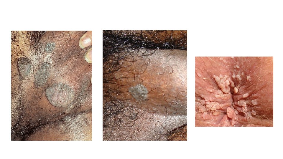

Genital Warts • Highly infectious, if left untreated my resolve on their own, remain unchanged, or grow. • After regression subclinical infection may persist for life. • In pregnancy warts may increase in size and number. • Prevention: HPV vaccine. • No therapy will eradicate HPV. • Rx: imiquimod 5% cream, podophylox 0. 5% solution, podophyllin 1020% (applied at the clinic), cryotherapy, Electrocautery, surgery.

Rubella RNA togavirus. Children <15 years old. Transmission through inhalation of aerosolized droplets. Prodrome usually absent. Low grade fever, malaise, headache, mild URT symptoms. • Pink macules, papules initially on forehead spreading to face and trunk that fades after 3 days. Petechiae on soft palate and lymphadenopathy. • Arthritis in adults, splenomegaly. • Congenital rubella syndrome: congenital heart defects, cataracts, microphthalmia, microcephaly, hydrocephaly, and deafness. • •
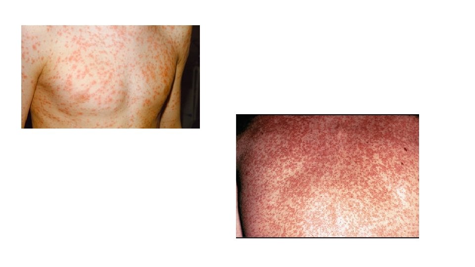

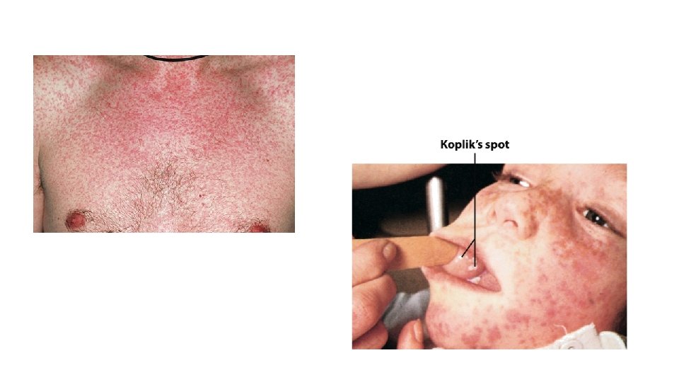
Measles • • • RNA morbillvirus. Transmission through respiratory droplets. Incubation period 10 -15 days. Fever, malaise, coryza, vomiting, diarrhea, photophobia, conjunctivitis and periorbital edema. These symptoms subside with appearance of rash. On the 4 th day, erythematous macules and papules appear on the forehead, behind the ears then face, trunk, extremities, reaching feet by the 3 rd day and resolve in 6 days. Tiny bluish-white spots on red background on the 2 nd day of fever on the buccal mucosa (Koplik spots). Generalized lymphadenopathy, splenomegaly. Acute complications in 10%: otitis media, pneumonia, diarrhea, encephalitis, and thrombocytopenia. Chronic complication: subacute sclerosing pan encephalitis.

Hand Foot and Mouth Disease • Coxsackievirus A 16 and E 71. • First decade of age, late summer and early fall. • Multiple painful mouth ulcers, macules and papules that evolve to vesicles on palms and soles, sides of fingers, toes and buttocks. • Vesicles can rupture with formation of erosions and crusts. • May be associated with fever, malaise, diarrhea, and arthralgia. • Self limited, symptomatic treatment. • E 71 infections have higher morbidity from CNS and pulmonary edema.


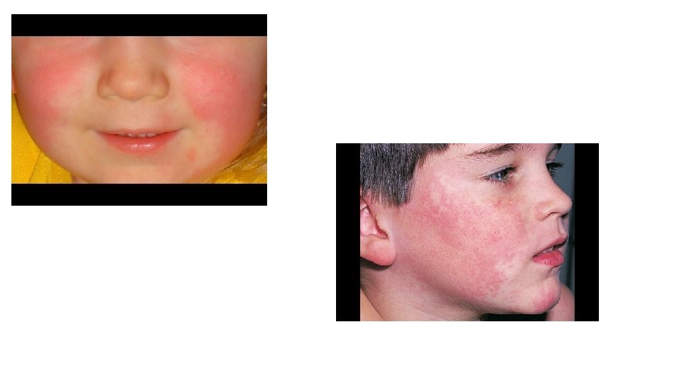
Erythema Infectiosum • Human parvovirus B 19. • Transmission by respiratory droplets. • Viremia develops 6 days after intranasal inoculation of the virus, significant bone marrow suppression (aplastic crisis) can occur at this time. • Exanthem begins 18 days after with arthralgia. • Fever and adenopathy are more severe in adults. • Numbness and tingling of fingers. • Diffuse erythematous patches on cheeks (slapped cheek) fades over 1 -4 days, this is absent in adults. • Erythematous macules, papules that become confluent with reticulated appearance on extensor arms, trunk and neck fade in 5 -9 days.

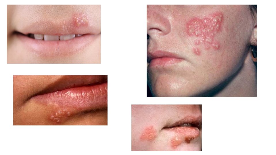
Herpes Simplex • • HSV 1 mostly herpes labialis & 2 mostly genital herpes. Transmission through skin-skin or skin-mucosa contact. After primary infection, HSV persists in sensory ganglia for life. Recurrence with skin or mucosa irritation, UV radiation, menstruation, fever, common cold, immunosuppression. After exposure, the virus replicate in epithelial cells, causing lysis of infected cells, vesicle formation and local inflammation. Recurrent viral shedding can be with or without lesions, and transmission usually occur while shedding. Dx: Tzanck smear: fluid from vesicles stained with Giemsa stain will show or multinucleated giant acantholytic keratinocytes. DFA will differentiate between type 1&2. Viral culture, serum antibodies for previous infections, seroconversion will take 2 -6 weeks.

Non Genital Herpes Simplex • Primary infection is often asymptomatic. • Primary herpetic gingivostomatitis is the most common in children. • Grouped vesicles on erythematous base that rupture easily forming erosions and crusts with regional adenopathy. • Recurrent herpetic infection has prodrome of tingling, itching, or burning sensation 24 hours prior to skin lesions. • Herpetic whitlow: infection of the tip of finger or thumb with painful neuritis in affected finger. • Eczema herpeticum: HSV infection in atopic patients with widespread vesicles not grouped, other dermatoses with risk of HSV infection are Darier disease, burns, Hailey-Hailey, bullous disorders, ichthyosis.

Non Genital Herpes Simplex • For primary herpes: Acyclovir 400 mg tid for 7 -10 days. • Valacyclovir, prodrug with better bioavailability 1 g bid for 7 days. • Famciclovir 250 mg tid for 7 days. • For recurrence: same doses for acyclovir and valacyclovir for 5 days. • Famciclovir 1 g bid for one day for recurrent genital HSV, 1500 mg one dose for recurrent herpes labialis. • Maintenance therapy in patients with > 6 attacks/year: valaciclovir 500 mg daily, acyclovir 400 mg bid, famciclovir 250 mg daily.

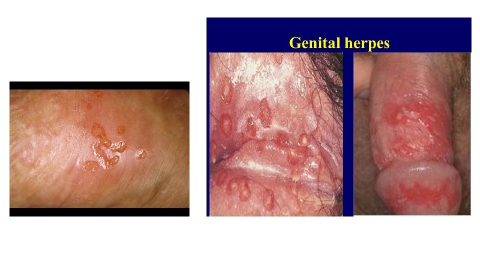
Genital Herpes Simplex • HSV 2 mainly, transmitted by skin-skin contact. With 70% of transmission during times of asymptomatic HSV shedding. • Most clinical lesions are minor breaks or erosions, abrasions. • Primary infections mostly asymptomatic, erythematous papules evolving to vesicles and erosions that heal in 2 -3 weeks. • Recurrent genital herpes my present with itching, burning, redness prior to eruption of vesicles. • Inguinal lymph nodes may enlarge.

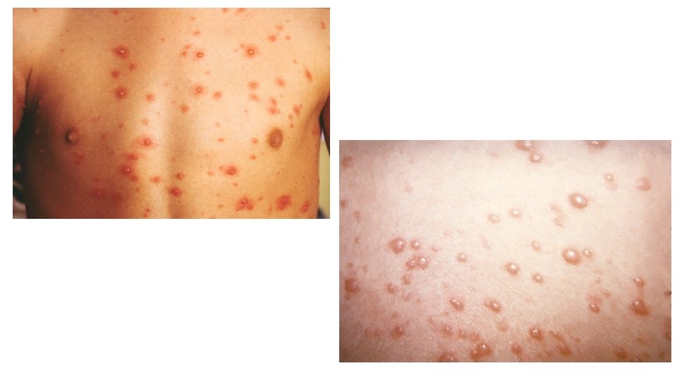
Varicella Virus • Without immunization 90% of cases before age 10. • Transmission by airborne droplets and direct contact. • Patients are contagious few days before the rash and until the last crop of vesicles dry out. • Vesicular lesions occur in successive crops. Few vesicles in children but more numerous in adults. • First lesions are papules that quickly evolve to vesicles with surrounding erythema. Vesicles evolve to pustules and crusted erosions after few hours that heal in 3 weeks leaving punched out scars. • Lesions start on face then trunk and extremities, highest densities on back. • In adults pneumonitis, cerebellar ataxia and encephalitis may occur.

Varicella Virus • Vaccination is 80% effective. • Rx is usually symptomatic. • If oral antiviral therapy is given in the first 24 hours the severity will be decreased. • Neonates: acyclovir 10 mg/kg/q 8 h for 10 days. • Children: acyclovir 20 mg/kg/q 6 h or valacyclovir 20 mg/kg/q 8 h for 5 days. • Immunocompromised adults: acyclovir 800 mg 5 times/day, or valacyclovir 1 g tid, or famciclovir 500 mg tid for 7 -10 days.

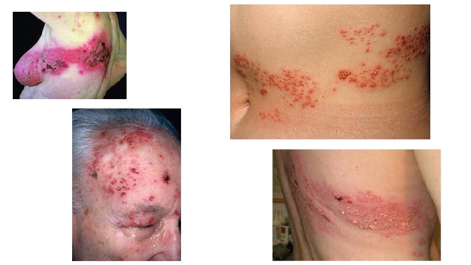
Herpes Zoster Virus • Reactivation of varicella virus in those ganglia in which varicella virus has achieved the highest density. • Triggered by immunosuppression, trauma, tumor, or irradiation. • Prodrome with pain, tenderness and paresthesia in the involved dermatome. Flu like symptoms can occur. • Papules evolve to vesicles and pustules in 3 -4 days then crusts after 10 days with new vesicles appearing over one week. All on erythematous base with regional adenopathy. Crusts resolve totally after 3 -4 weeks. • Thoracic area 50%, trigeminal 20%, lumbosacral 10%. • Ophthalmic zoster: nasociliary involvement of ophthalmic branch of trigeminal nerve, early lesions vesicles on tip of nose, may lead to keratitis, conjunctivitis, retinitis, optic neuritis. Has the highest incidence of post herpetic neuralgia. • Post herpetic neuralgia: constant severe stabbing or burning pain in 40% of patients above 60 often resolves in 6 months but may persist for years. • Sometimes scars and PIH persists for years.

Herpes Zoster Virus • Vaccine against VZV reduces the incidence of zoster by 50%. • Famciclovir 500 mg/8 h/7 days. • Valacyclovir 1 g/8 h/7 days. • Acyclovir 800 mg/5 times daily/7 -10 days. • In immunocompromised: acyclovir 10 mg/kg IV Q 8 h for 10 days. • In acyclovir resistant: IV foscarnet 40 mg/kg IV Q 8 h till resolution. • For post herpetic neuralgia: gabapentin, pregabalin, tricyclic anti depressant, analgesics, nerve block.

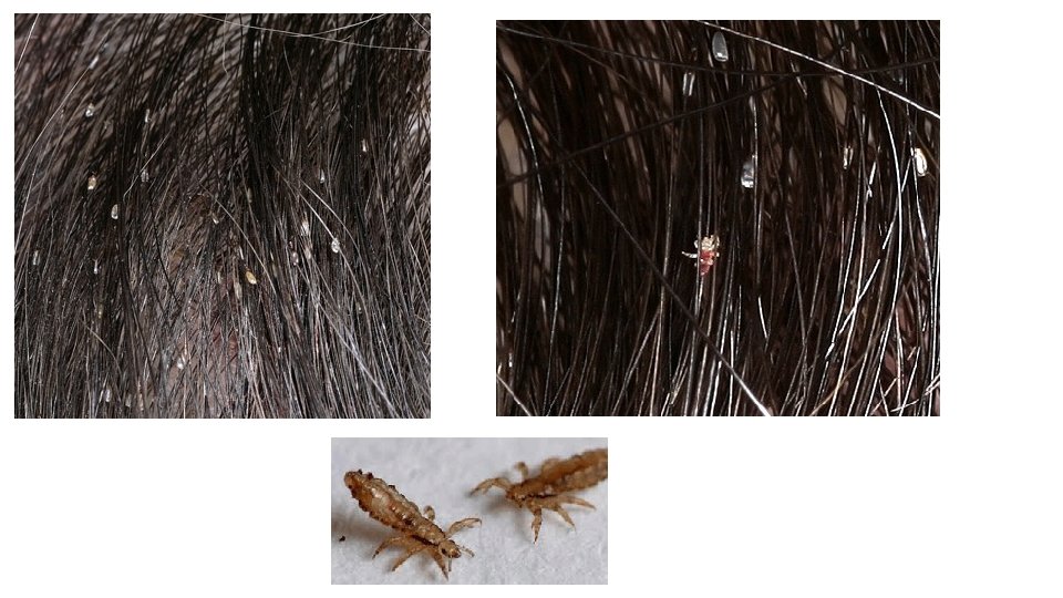
Pediculosis Capitis • • Infestation by Pediculus Humanus Capitis 1 -2 mm in size. Can crawl 23 cm/day. lay nits 1 -2 mm from scalp. Lice hatch within 1 week, mature into adult within 1 week. One female can lay 100 ova in 2 weeks lifetime. Can survive only for few hours off scalp. Transmission: head to head contact, shared hats, comb, brushes, pillows. Present as pruritus of the back and side of scalp with secondary bacterial infection and regional adenopathy. New viable eggs are creamy-yellow, empty eggshells are white. Rx: topical Permethrin, malathion, pyrethrin, dimethicone, and Ivermectin(topical or systemic in severe cases)

Scabies Epidermal infestation by mite Sarcoptes scabiei var. hominis. Transmission: skin-skin contact and fomites. Mites burrow into epidermis shortly after contact no deeper than stratum granulosum. Female life span 4 -6 weeks, lays 3 eggs daily, eggs hatch in 4 days. Leave burrow at night and lay eggs during the day. can be alive for 2 days on clothing or bedding. • Intense pruritus is the main symptom, hypersensitivity of immediate and delayed type are responsible for other lesions other than the primary burrows. • Burrows are skin colored ridges 1 cm long with minute vesicle or papule at the end of the tunnel. Mainly on interdigital webs, wrists, penis, buttocks, scrotum. And in infants might involve head and neck. Sometimes there are nodules on scrotum, penis, axillae, and buttocks. • Secondary changes: excoriations, lichen simplex chronicus, prurigo nodules, PIH, secondary infections, id reaction. • • •
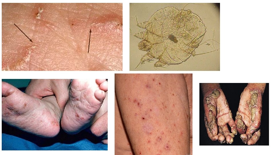

Scabies • Dx: microscopy by putting a drop of mineral oil on the burrow, then the burrow is scraped off then placed on microscopic slide to look for mites, eggs and fecal pellets(scybala). • Histopathology: female mite with eggs, Spongiosis and vesicle formation. Eosinophilic infiltrate in dermis. • Rx: treatment of patient and close contacts and house, bed cleaning. • Permethrin 5% cream to whole body for 8 hours or overnight. • Crotamiton, sulfur, benzyl benzoate, malathion, Lindane, Ivermectin. • Oral Ivermectin 0. 2 mg/kg single dose. • Post scabietic itching may persist after successful ttt and represent hypersensitivity to remaining dead mites and its products.

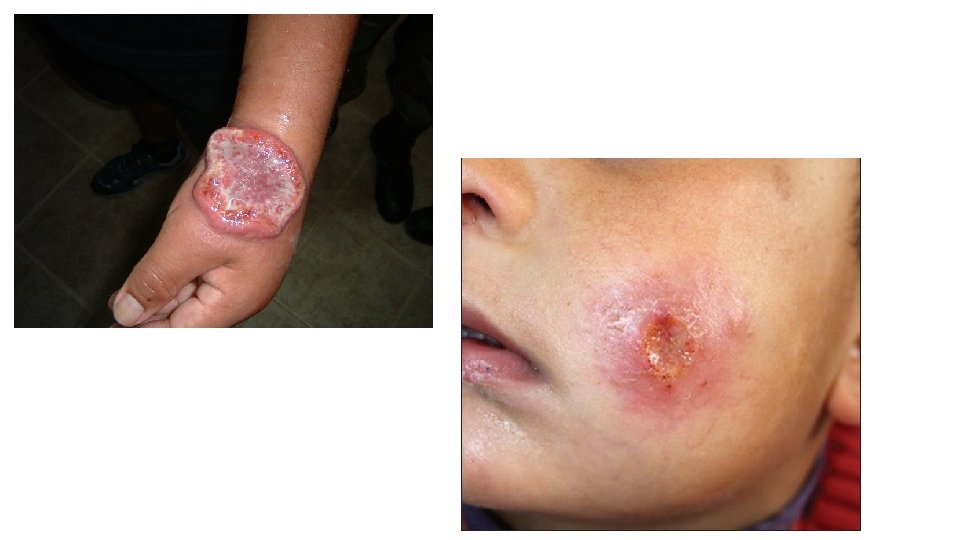
Leishmaniasis • Single or multiple asymptomatic cutaneous papules and nodules at the site of a sand-fly bite that may evolve over weeks to ulcer which heal spontaneously with a depressed scar. • Common in Alkharj, Alahsa, Alqaseem, Hail, Gizan. • Leishmania tropic major Incubation period 1 -4 weeks. • Histopathology: Dermal granulomatous inflammation with prominent lymphocytes, histiocytes with intracellular non-flagellated amastigote. • Rx: sodium stibogluconate intralesional if single lesion every week till resolution. Or systemically IM 15 mg/kg/d for 21 days. • Itraconazole, amphotericin B, paromomycin, miltefosine.

Syphilis • Chronic systemic infection by spirochete T. pallidum. • Transmission: sexual contact, perinatal in congenital syphilis. • Primary syphilis is the most contagious stage. • Later syphilis is a vascular disease , lesions are secondary to obliterative endarteritis of terminal arterioles and small arteries with the resulting inflammatory and necrotic changes. • Dx: dark field microscopy positive in primary chancre and papular lesions of secondary syphilis(condylomata lata), DFA test in exudate. • Serologic Test for Syphilis STS: always positive in secondary syphilis, VDRL is non reactive in 25% of primary syphilis.

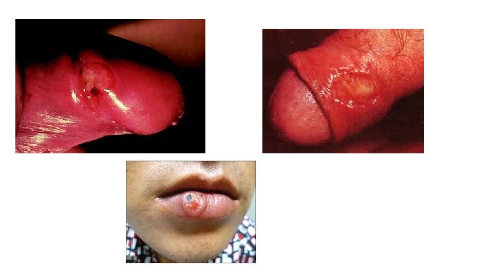
Primary Syphilis • Chancre is single button like papule develops at site of inoculation into a painless erosion and then ulcerate with raised border and minimal exudate on penis or vulva, vagina, cervix in women. Lymph nodes are discrete, firm, nontender and unilateral appear within 7 days. • Rx: IM benzathine penicillin G 2. 4 million units single dose. • Doxycycline 100 mg twice daily for 2 weeks.

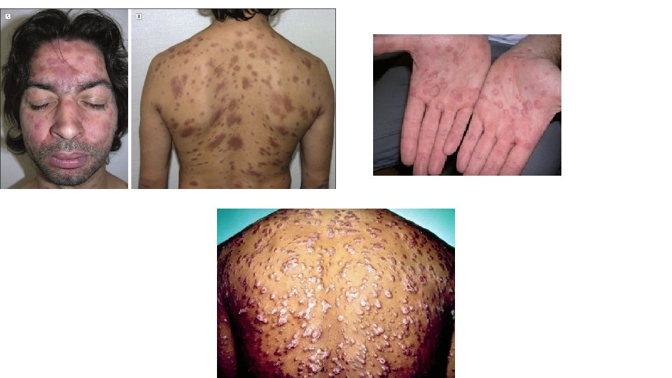
Secondary Syphilis • Appear 2 -6 months after primary infection. • Fever, sore throat, weight loss, malaise, headache, and meningismus. • Oval pink-brownish macules and papules, scaly papules, pustules, that could be annular or polycyclic, usually scattered and symmetric. • Condylomata lata in the anogenital area and mouth. • Diffuse hair loss, patch moth-eaten alopecia, loss of later eyebrows. • Small asymptomatic flat-topped papules on the oral and genital mucosa, split papules at the angle of the mouth. • Generalized lymphadenopathy. • Periosteitis of long bone mostly tibia, arthralgia, • Acute iritis, optic neuritis, uveitis. • Diffuse pharyngitis, hypertrophic gastritis, hepatitis, ulcerative colitis. • Glomerulonephritis, nephrotic syndrome, cystitis, prostatitis.

Secondary Syphilis • Histopathology: epidermal hyperkeratosis, capillary proliferation with endothelial swelling, perivascular infiltration by monocytes, plasma cells, and lymphocytes. Spirochete is present in many tissues like skin, eye, CSF(abnormal in 40% of patients). • Elevated liver enzymes. • Renal function: immune complex-induced membranous glomerulonephritis. • Rx: as primary syphilis.

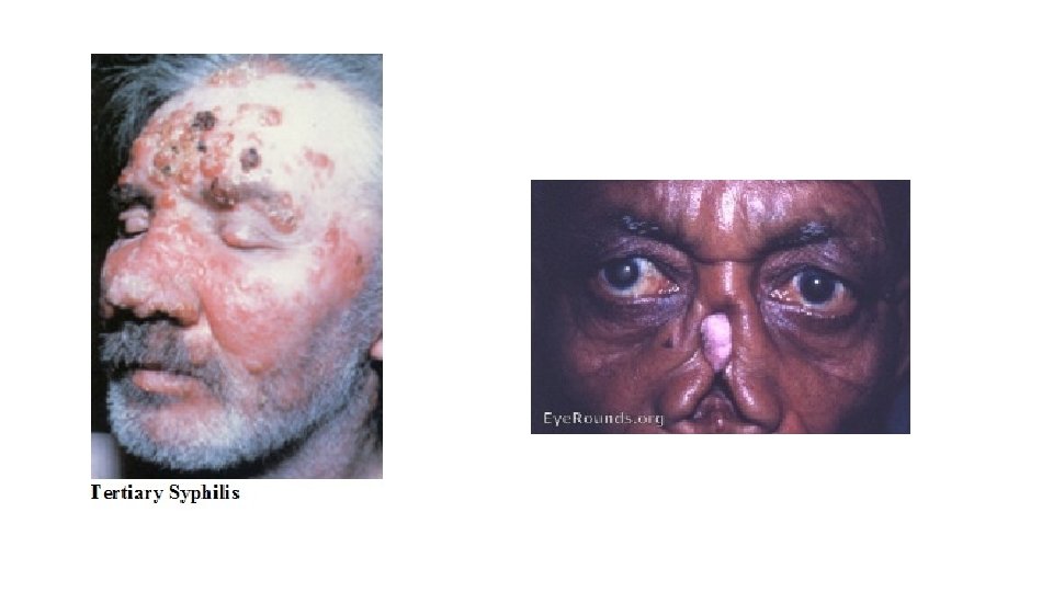
Tertiary Syphilis Gumma: nodular or scaly plaques that may ulcerate and form circles. May expand rapidly causing local destruction and heal by scarring. Could be in skin, bones, liver, stomach, upper airway. Nodular ulcerative syphilides: like gumma but flatter with central healing. Meningeal syphilis: headache, nausea, vomiting, stiff neck, cranial nerve palsies, seizures. • General paresis: after 20 years of infection, hyperactive reflexes, Argyll. Robertson pupil, delusions, hallucinations. • Tabes dorsalis: 25 -30 years after infection, ataxic wide based gait, foot slap, paresthesia, bladder dysfunction, impotence, optic atrophy • Cardiovascular syphilis: endarteritis obliterans. • • •
- Slides: 100