COMMON ADULTS FRACTURES OBJECTIVES CLAVICAL FRACTURE HUMERUS PROXIMAL

COMMON ADULT’S FRACTURES
 BOTH BONE FOREARM FRACTURS DISTAL")
OBJECTIVES • • CLAVICAL FRACTURE HUMERUS (PROXIMAL & SHAFT) BOTH BONE FOREARM FRACTURS DISTAL RADIUS FRACTURE HIP FRACTURE FEMUR SHAFT FRACTURE TIBIAL SHAFT FRACTURE ANKLE FRACTURE

CLAVICLE FRACTURE • Clavicle is S shape bone • It is anchored to scapula via ACJ. • It is anchored to trunk via SCJ • Most of fracture occurs as result from fall onto shoulder.

• Fracture is classified into: proximal, middle and lateral third fractures. • Most of fractures are of middle third.

• Clinical findings: – Check the skin • Injury to brachial plexus and subclavian artery/vein may be present • Rarely, Pneumothorax can occur.

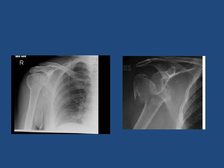
• X-rays: • AP chest and Clavicle special view.

• Treatment: – Most of clavicle fractures are treated with a sling. – Few fractures should be treated surgically with open reduction and internal fixation • Skin is tented • Severe displacement

PROXIMAL HUMERUS ANATOMY • Proximal humerus has four anatomic parts: – – Head Greater tubrosity Lesser tubrosity Shaft • Anatomic neck v. s surgical neck.

PROXIMAL HUMERUS FRACTURE • In younger patients: violent trauma. • In older patients: minor trauma. • Most fractures are minimally displaced.

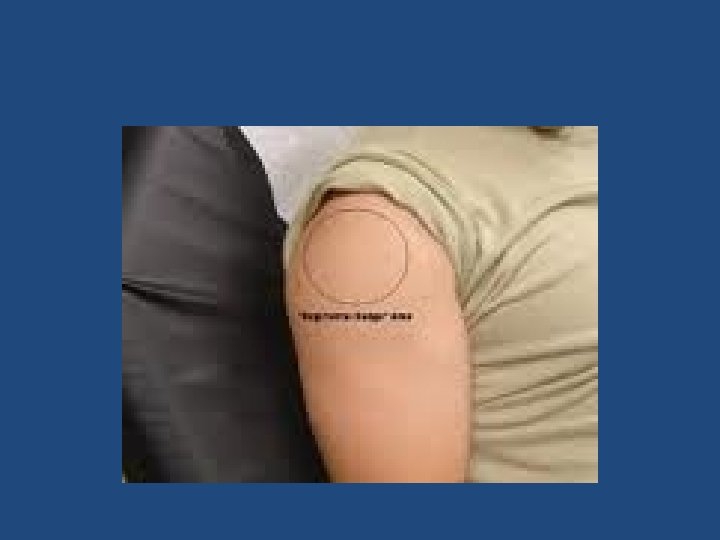
PHYSICAL EXAM • • • Expose the shoulder very well. Look for fracture signs Check the skin. Peripheral N/V exam. Axillary nerve: lateral skin patch. Examine cervical spine.

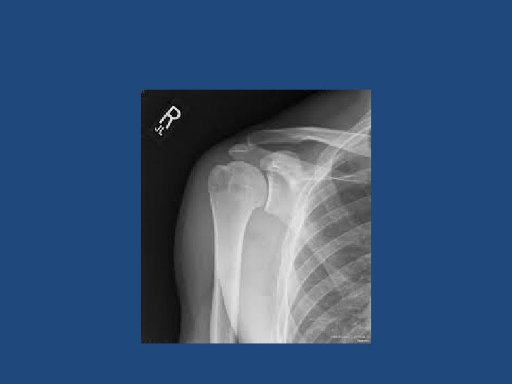
X-rays • AP, lateral and axillary views.

X-rays • Fracture is defined by the fragments displaced. • Displacement: more than 1 cm.

• If fracture is not displaced: – Treatment with sling and NWB of UE for 6 -8 weeks. – Early ROM exercises after 2 -4 weeks. – Normal function can be resumed after 3 -4 months.

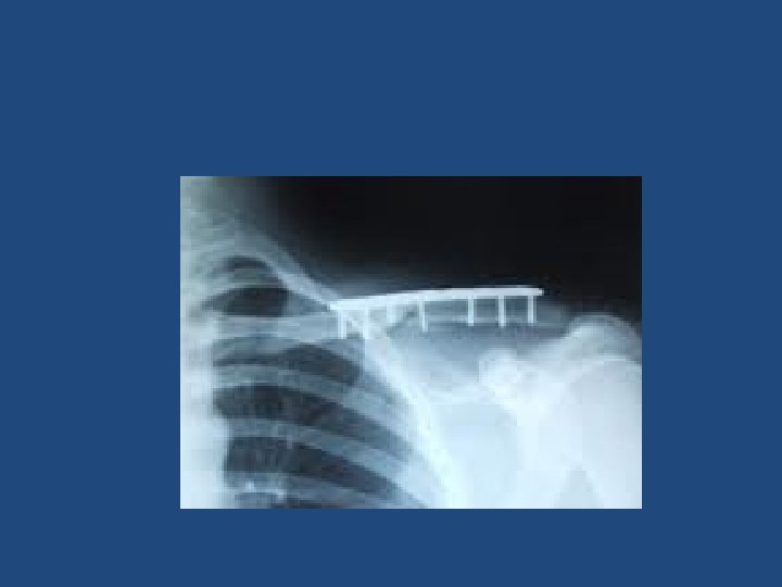
• If the fracture is displaced: – Surgery is indicated. – ORIF is indicated (plate and screws). – Shoulder hemiarthroplasty is indicated in some cases.

HUMERUS SHAFT FRACTURE • It can be classified based on location of fracture. (proximal, middle and distal) • Fracture symptoms. • On exam: – Skin – N/V – Compartment • Watch for radial nerve palsy.

X-rays

• Almost all humerus shaft fracture can be treated non-surgically. – Close reduction – Functional brace x 4 -6 weeks + NWB – Early ROM of elbow and shoulder.

• Surgery is indicated for specific conditions like: – – – Segmental fracture Open fracture Obese patient Bilateral fracture Floating elbow ( forearm and humerus) • Surgery: ORIF with plate and screws.

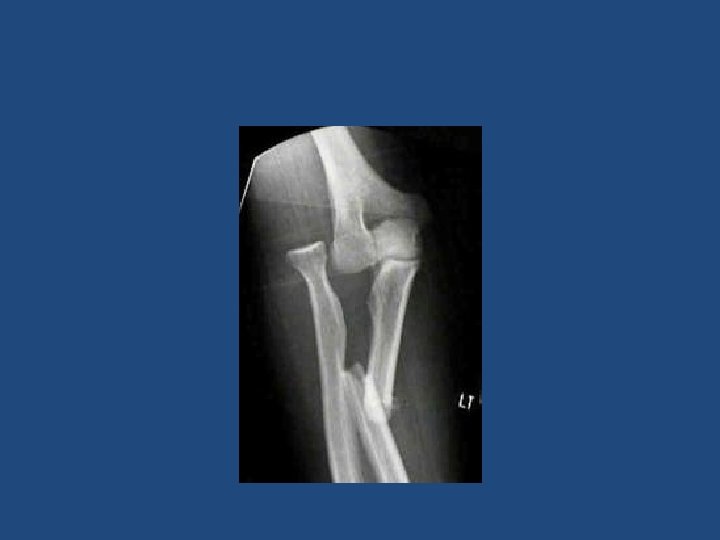
BOTH BONES FOREARM FRACTURE • Forearm is complex with two mobile parallel bones. • Radius and ulna articulate proximally and distally. • It very unlikely to fracture only one bone without disruption of their articulation: – Both bone fracture – Monteggia fracture – Galeazzi fracture.

• Fractures are often from fall or direct blow. • Both bones fracture: – Means radius and ulna are broken. • Monteggia fracture: – Means proximal or middle third ulna shaft fracture with dislocation of radius proximally (at elbow) • Galeazzi fracture: – Means distal third shaft radius fracture with disruption of DRUJ.

Monteggia

Galeazzi

Galeazzi

CLINICAL Symptoms and signs of fracture Check the skin Check the compartments of forearm Check Ulnar, median and radial nerve (PIN, AIN) • Check vascularity: color, temperature, capillary refill and pulse. • •

Images • 2 orthogonal views
 – Are")
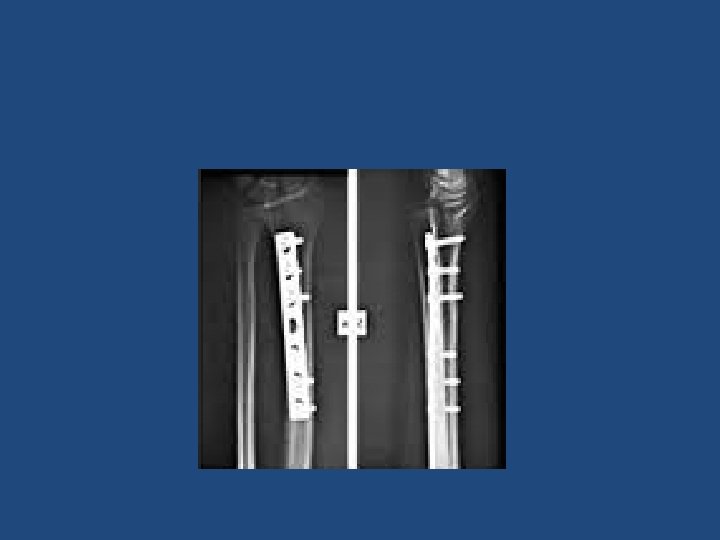
Treatment • Both bone fracture: – Reduce and splint at ER/clinic (temporary) – Are treated almost always with ORIF: (plate and screws) • Monteggia fracture: – ORIF ulna and close reduction of radial head • Galeazzi fracture: – ORIF radius and close reduction of DRUJ.

DISTAL RADIUS FRACTURE • Most common fracture of upper extremity. • Most frequently are seen in older women. • Young adults fractures are most commonly secondary to high energy trauma.
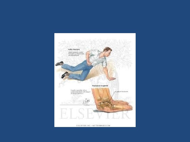

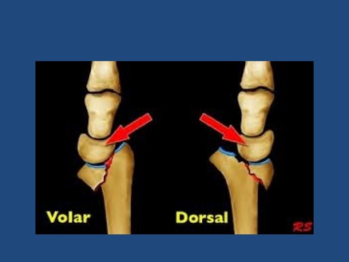
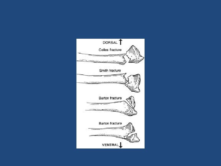
• Extra-articular: – Colles’ Fracture: dorsal angulation, shortening and radial deviation – Smith’s fracture: shortening and volar angulation. (reverse Colles’) • Intra-articular: – Barton’s fracture: volar or dorsal – others

Colles’

Smith’s

Clinical

X-rays Colles’ Smith’s

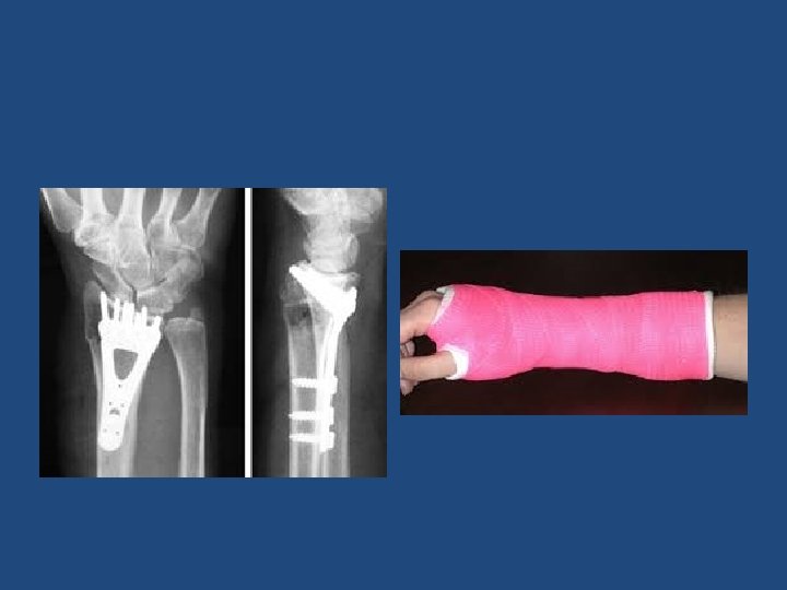
• Extra-articular fractures: – Close reduction and cast application. – Immobilization for 6 -8 weeks. – ROM exercises after cast removal. – Surgery: if reduction is not accepted • Intra-articular fracture: – a step more than 2 mm is an indication for surgery. – ORIF with plate and screws.

LOWER EXTREMITY
 • It is the most common fracture of LL. •")
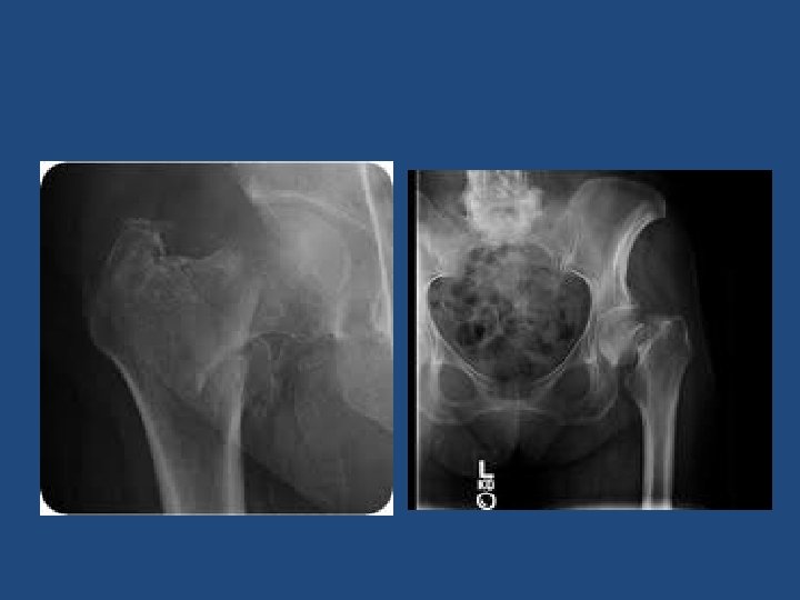
HIP FRACTURE (old patients) • It is the most common fracture of LL. • It is associated with osteoporosis. • Most common mechanism is a fall from standing height. • Other causes of fall (stroke, MI) should be rolled out during clinical evaluation. • It is a life changing event.

• Fractures can be classified – Intra-capsular – Extra-capsular – Displaced vs not displaced

• Intra-capsular: – Subcapital – Trans-cervical • Extra-capsular: – Basicervical – Intertrochanteric • AVN risk is higher with intra-capsular fracture.

Clinical • Full detailed history of mechanism of injury. • R/O syncope, chest pain, weakness etc. • A detailed systemic review. • Deformity: Abduction, External rotation and shortening. • Assess distal N/V status

• 3 views are needed: – AP pelvis – AP hip – Lateral hip

Treatment No close reduction is needed. No traction is needed. Patient needs surgery ideally within 48 hrs. The goal is to ambulate patient as soon as possible. • Be sure that DVT prophylaxis is started. • Be sure that patient will be evaluated for osteoporosis after discharge. • •

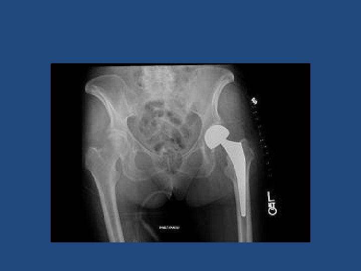
Treatment • If fracture is intracapsular: – Displaced: hemiarthroplasty – Not displaced: close reduction and Screw fixation. • If fracture is Extracapsular: – Close reduction and DHS or IM nail fixation

DHS IM nail
 • It is a completely different entity from similar fractures")
HIP FRACTURE (young patients) • It is a completely different entity from similar fractures in elders (>60 years). • High energy mechanism. • Patient should be taken to operative room for ORIF within 6 hours.

FEMUR SHAFT FRACTURE • High energy. • Associated injuries. • Early fixation to avoid pulmonary complications. • Skin/ skeletal traction while waiting, • IM nail within 6 -12 hrs.

TIBIA SHAFT FRACTURE • High energy mechanism • It carries the highest risk of compartment syndrome. • Carefully examine the skin. • Splint patient after reduction. • IM nail fixation unless it is not displaced

ANKLE FRACTURE
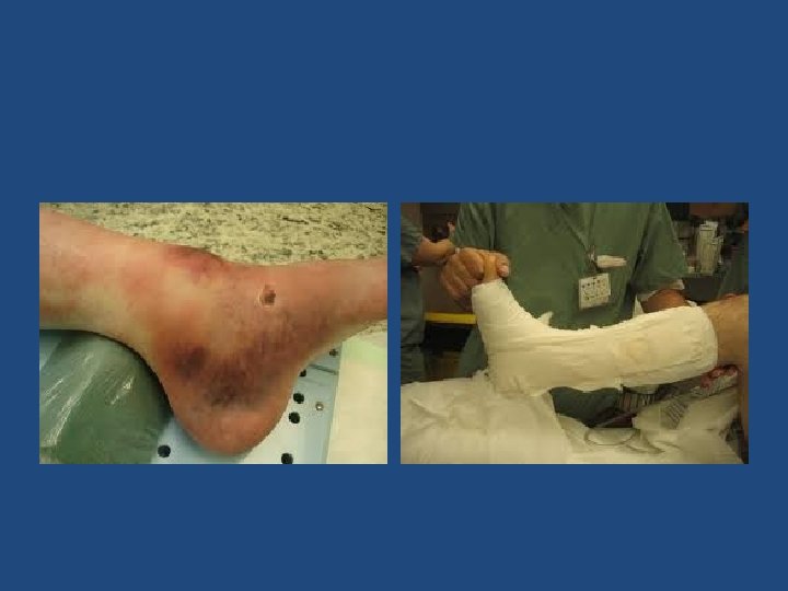
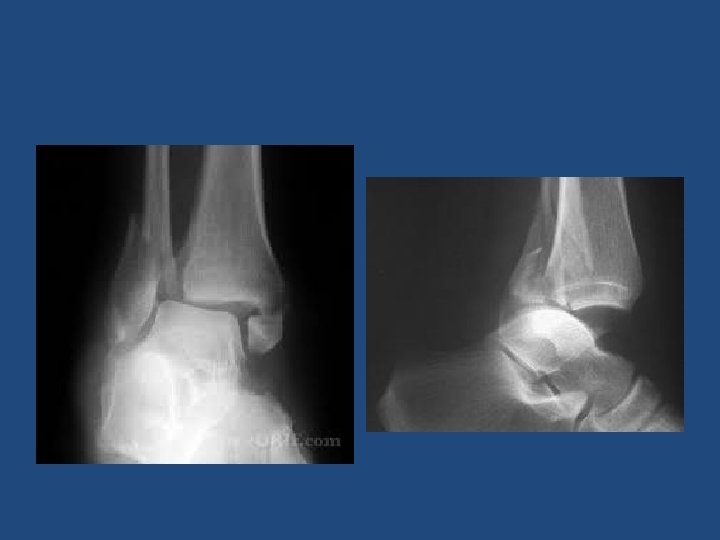

• Intact medial malleolus: – Weber A: • splint + NWB X 6 weeks. • Early ROM. – Weber B/C: • If medial joint line widen: ORIF. • If not: ? – If both malleoli are broken: • ORIF

THANKS
- Slides: 62