COLON CANCER ABORAJOOH EMAD AREF MD General Surgery

COLON CANCER ABORAJOOH EMAD AREF MD, General Surgery GI and Minimally Invasive Surgery IMRCS JB and AB 1
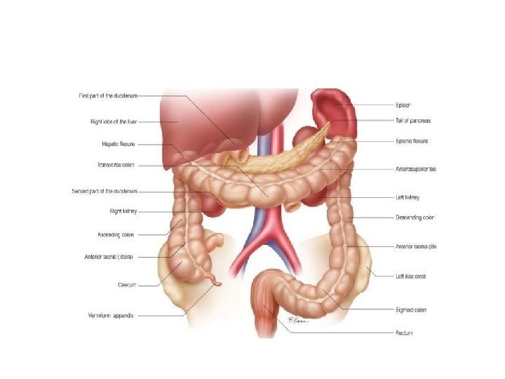

Blood Supply

Blood Supply

• Adjacent branches of the superior and inferior mesenteric arteries anastomose so there is usually a complete vascular supply along the colon named the ‘marginal artery of Drummond’ • Venous and lymphatic drainage of the colon follows the arterial supply and as for the small intestine system, venous drainage is into the portal system. • The parasympathetic nerve supply to the right and transverse colon is through the vagus nerve. • While the distal colon and the rectum are supplied by the nervi erigentes (the pelvic splanchnic nerves) from S 2, 3, 4. • The sympathetic system supplies the blood vessels through the greater and lesser splanchnic nerves.

Lymphatic Drainage 1 -Epicolicnodes on the bowel wall 2 -paracolic nodes between the marginal artery and the bowel 3 -Intermediate nodes on the main vessels 4 -Principal nodes alnogside the superior and inferior mesenteric vessels.

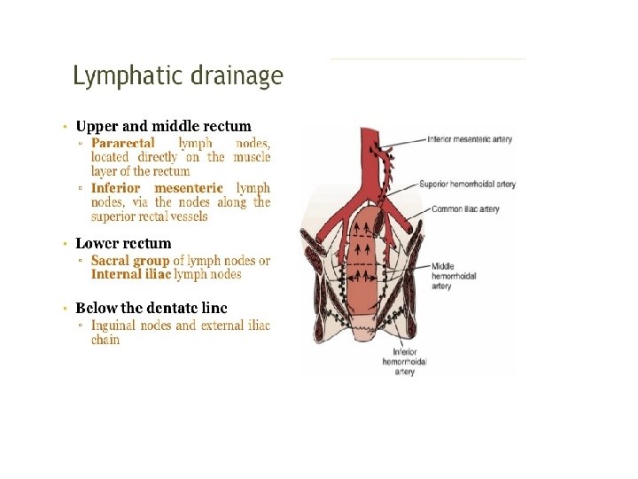
Rectum Arterial supply • Superior rectal A – fr. IMA; supplies upper and middle rectum • Middle rectal A- fr. Internal iliac A. (supplies lower rectum) • Inferior rectal A- fr. Internal pudendal A. Venous drainage – Superior rectal V- upper & middle third rectum – Middle rectal V- lower rectum and upper anal canal – Inferior rectal vein- lower anal canal Innervations • Sympathetic: L 1 -L 3, Hypogastric nerve • Para. Sympathetic: S 2 -S 4

Function of The Colon • Absorption of water; 1000 m. L of ileal contents enter the caecum every 24 hours of which only about 150– 250 m. L is excreted as faeces. • Sodium absorption is efficiently accomplished by an active transport system, while chloride and water are absorbed passively following gradients established by the sodium pump • Fermentation of dietary fibre in the colon by the normal colonic microflora leads to the generation of short chain fatty acids. • Some absorption of nutrients including glucose, fatty acids, amino acids and vitamins can also take place in the colon. • In general, faecal residue reaches the caecum 4 hours after a meal and the rectum after 24 hours. • Passage of stool is not orderly, however, because of mixing within the colon. It is thus common for residue from a single meal to still be passed 4 days later.

Classification of intestinal polyps

Risk of Malignancy • A 10 % risk of cancer in a 1 -cm diameter tubular adenoma, whereas with villous adenomas over 2 cm in diameter, there may be a 15 % chance of carcinoma. • Almost one-third of large (>3 cm)colonic adenomas will have an area of invasive malignancy within them at the time of resection. • Adenomas larger than 5 mm in diameter are usually excised because of their malignant potential. • Colonoscopic snare polypectomy is usually possible for colonic polyps, but larger sessile polyps can require endoscopic mucosal resection (EMR) • Larger rectal adenomas may require transanal resection or, where the adenoma is too high for safe conventional access, transanal endoscopic microsurgery (TEMS)

Colorectal Cancer • colorectal cancer is the second most common cause of cancer death. • Globally 800, 000 new CRCs occur each year, accounting for 10% of all incident cancers with 450, 000 deaths/year • Incidence : 35. 8/100, 000 (USA)

Jordan Cancer Registry

Ten most common cancers among Jordanians both genders, 2012

Aetiology § § § • • § § § • § Environmental & dietary factors Male sex Family history of colorectal cancer Personal history of colorectal cancer, ovary, endometrial, breast Excessive BMI Red meat , animal fat, smoking and alcohol Protective effect of dietary fibre Low folate consumption Neoplastic polyps. IBD Cholecystectomy Ureterosigmoidostomy. Adenoma– carcinoma sequence Hereditary Conditions (FAP, HNPCC)

Adenoma– Carcinoma Sequence Normal Hyperplasia Early Adenoma Intermedia te Adenoma Late Adenoma Metastases Carcinoma

Adenoma– Carcinoma Sequence APC Loss/mutation Ch. 5 q Normal Epithelium Loss of DNA methylation Hyperproliferation K-ras Mutation Ch. 12 p (50%) Early Adenoma DCC loss Ch. 18 q DPC 4 Ch. 4 Intermediate Adenoma p 53 Loss Ch. 17 p Late Adenoma Cancer

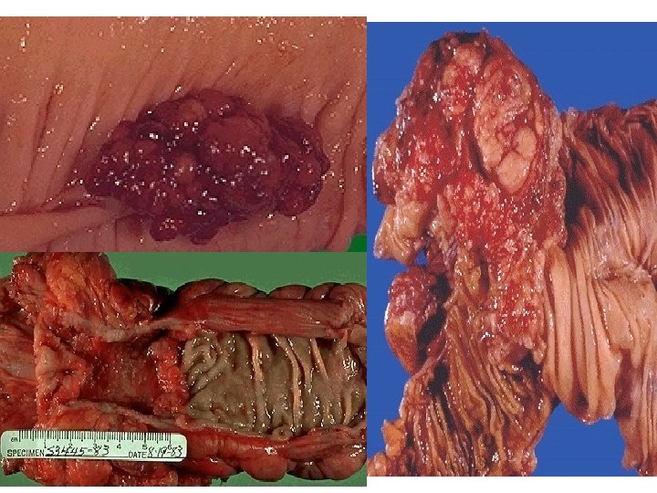
1. FAP : - It is a rare disease. - Autosomal dominant inheritance. - Accounts for about 0. 5% of all colon cancers. - Associated with alterations in APC gene which is located on 5 q. In addition to that alterations in APC gene appear to play a role in sporadic colon cancer. - Phenotypically characterized by hundreds to thousands of adenomas in the colon. - A very small proportion of those adenomas will progress. into carcinoma but eventually all patients with FAP will develop colon cancer.
 • • Inherited as an autosomal dominant Mutations in the")
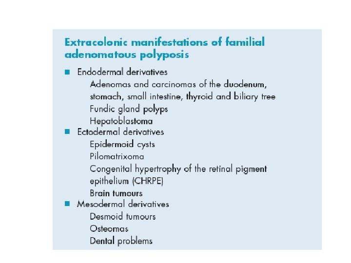
Familial Adenomatous Polyposis (FAP) • • Inherited as an autosomal dominant Mutations in the adenomatous polyposis coli (APC) gene Presence of more than 100 colorectal adenomas Characterised by duodenal adenomas and multiple extraintestinal manifestations. Accounts for 1 % or less of all colon cancer The risk of colorectal cancer is 100 % Associated with benign mesodermal tumours such as desmoid tumours and osteomas 50 % of patients have congenital hypertrophy of the retinal pigment epithelium (CHRPE), which can be used to screen affected families if genetic testing is unavailable.
 • Characterised by increased risk of colorectal cancer")
Hereditary non-polyposis colorectal cancer (Lynch syndrome) • Characterised by increased risk of colorectal cancer and also cancers of the endometrium, ovary, stomach and small intestines. • Accounts for about 5 – 10 % of all colon cancers. • Autosomal dominant condition caused by a mutation in one of the DNA mismatch repair genes( MLH 1 , MSH 2). • The lifetime risk of developing colorectal cancer 80 %, and the mean age of diagnosis is 45 years. • Most cancers develop in the proximal colon. • 30– 50 % lifetime risk of developing endometrial cancer. • Diagnosed by genetic testing or the Amsterdam II criteria. • Patients with HNPCC are subjected to regular (every one to two years) colonoscopic surveillance.

Amsterdam II criteria • Three or more family members with an HNPCCrelated cancer (colorectal, endometrial, small bowel, ureter, renal pelvis), one of whom is a first -degree relative of the other two • Two successive affected generations • At least one colorectal cancer diagnosed before the age of 50 years • FAP excluded • Tumours verified by pathological examination.

IBD • The risk of cancer in ulcerative colitis increases with duration of disease. • At ten years from diagnosis, it is around 1 %. This increases to 10– 15 % at 20 years and may be as high as 20 % at 30 years.

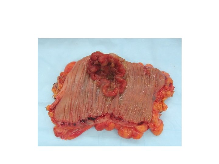
Pathology

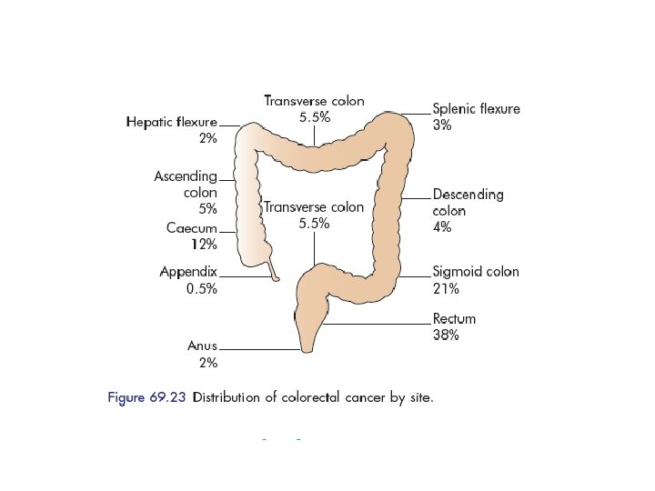
Clinical features • Occurs in patients over 50 years of age and is most common in the eighth decade of life • 20% of cases present as an emergency with intestinal obstruction or peritonitis. • left sided colonic tumors which are far more common usually present with a change in bowel habit or rectal bleeding • While more proximal lesions typically present later with iron deficiency anaemia or a mass. • Patients may present for the first time with metastatic disease. • Lesions of the flexures may present with vague upper abdominal symptoms for many months before other, more specific symptoms suggestive of colonic disease appear.

• Signs – Pallor – Abdominal mass – PR mass – Jaundice – Nodular liver – Ascites

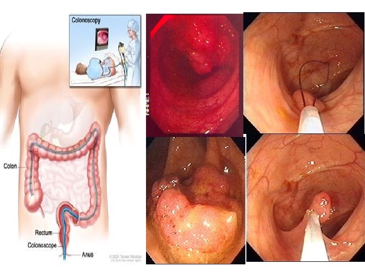
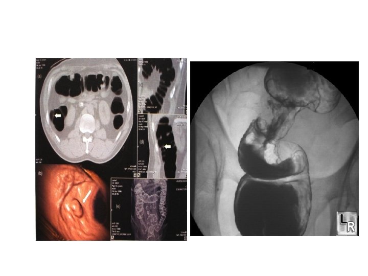
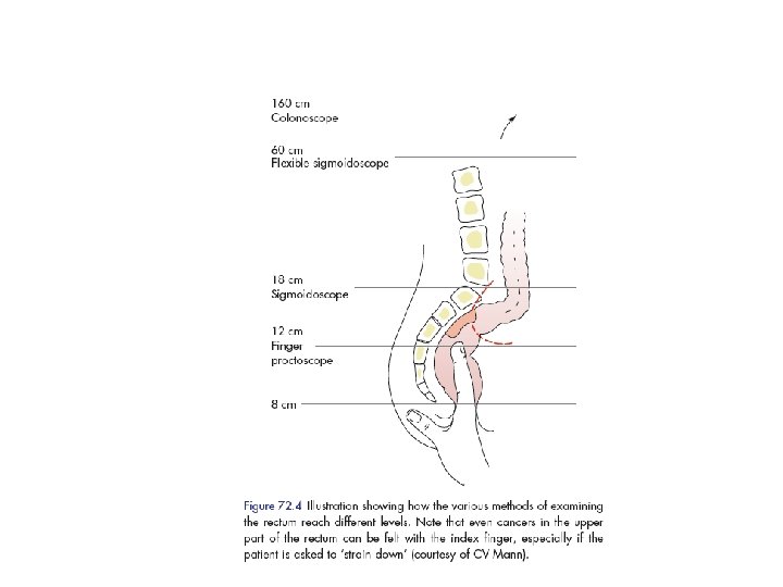
Diagnosis • • • Complete history Physical examination /DRE Routine investigations Confirmatory- Biopsy Staging workup – – – – CXR Barium enema Colonoscopy CT abdomen- pelvis Virtual colonoscopy MRI PET • Gold standard- Colonoscopy+ Biopsy • • • Others FOBT Stool cytology CEA IHC markers Molecular markersoncogenes

Screening Guidelines • Screening for asymptomatic men and women at age 50, using a menu of screening options. • Mortality rates have been declining for the past 2 decades, largely attributable to the contribution of screening to prevention and early detection.

Recommended CRC screening tests 1. Annual high-sensitivity g. FOBT or FIT, following the manufacturer's recommendations for specimen collection 2. FSIG every 5 years 3. Colonoscopy every 10 years 4. Double-contrast barium enema every 5 years 5. CT colonography every 5 years. Stool DNA testing, which also was among the recommended options in the 2008 update, is no longer commercially available for screening.

SPREAD OF CARCINOMA OF THE COLON • • Direct spread Lymphatic spread Haematogenous spread Transcoelomic spread
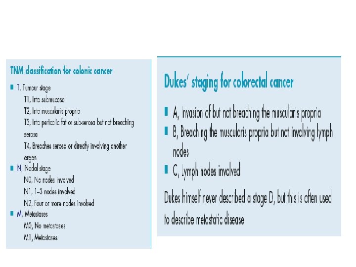

Dukes classification- Dukes A: Invasion into but not through the bowel wall. Dukes B: Invasion through the bowel wall but not involving lymph nodes. Dukes C: Involvement of lymph nodes Dukes D: Widespread metastases Modified astler coller classification- Stage A : Limited to mucosa. Stage B 1 : Extending into muscularis propria but not penetrating through it; nodes not involved. Stage B 2 : Penetrating through muscularis propria; nodes not involved Stage C 1 : Extending into muscularis propria but not penetrating through it. Nodes involved Stage C 2 : Penetrating through muscularis propria. Nodes involved Stage D: Distant metastatic spread

TNM Classification TX T 0 Tis T 1 T 2 Primary tumor cannot be assessed No evidence of primary tumor Carcinoma in situ: intraepithelial or invasion of lamina propria Tumor invades submucosa Tumor invades muscularis propria T 3 Tumor invades through the muscularis propria into pericolorectal tissues T 4 a Tumor penetrates to the surface of the visceral peritoneum T 4 b Tumor directly invades or is adherent to other organs or structures Tis T 1 T 2 T 3 T 4 Mucosa Muscularis mucosae Submucosa Muscularis propria Subserosa Serosa Extension to an adjacent organ

TNM Classification

Stage Grouping

SURGERY • SURGRY is the GOLD STANDARD and principle therapy of primary and non metastatic ca colon – Curative – Palliative – Accurate disease staging – Guides adjuvant treatment • Likelihood of cure is greater when disease is detected at early stage • AIM • To excise the primary lesion with adequate margin ~5 cm of normal bowel proximal and distal to the tumor • To reconstitute bowel continuity

• The operations described are designed to remove the primary tumour and its draining locoregional lymph nodes.

FAP Total proctocolectomy and IPAA Various designs of ileal pouchs

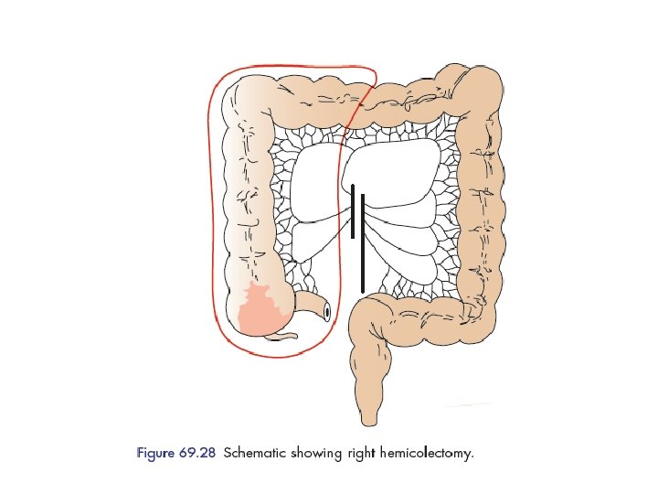
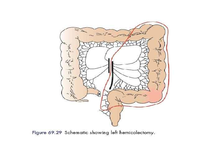
Emergency surgery • 20 % of patients with colonic cancer will present as an emergency, the majority with obstruction, but occasionally with haemorrhage or perforation. • If the lesion is right sided, it is usually possible to perform a right hemicolectomy and anastomosis in the usual manner; this can be facilitated by decompressing the bowel at the start of the operation • Perforation with substantial contamination or if the patient is unstable, it may be advisable to bring out an ileo/colostomy rather than anastomosing bowel in these circumstances. • For a left-sided lesion, the decision-making process is similar to that in diverticular disease between a Hartmann’s procedure and resection and anastomosis
 T 1 N 0 97 T 2")
Stage Mean 5 yr survival rate (%) T 1 N 0 97 T 2 N 0 90 T 3 N 0 78 T 2 N + 74 T 4 N 0 63 T 3 N + 48 T 4 N + 38

• The most important determinant of prognosis is tumour stage and, in particular, lymph node status. • Overall five-year survival for colorectal cancer is approximately 50 %. • Follow up aims to identify synchronous bowel tumours that were not picked up at original diagnosis due to emergency presentation or incomplete assessment

Chemotherapy Combinations § § § 5 Fu Leucovorin Oxaliplatin Irinotecan Bevacizumab cetuximab § FOLFOX § § FOLFIRI Leucovorin/5 FU Capecitabine Bevacizumab in combination with the above regimens.

Stage IV/ Metastatic PRIMARY SURGERY RESECTION OF LOCAL/ METS PALLIATIVE CCT ADJUVANT IMMUNOTHEARPY

Rectum • The rectum measures approximately 15 cm in length • It is divided into lower, middle and upper thirds • The blood supply consists of superior, middle and inferior rectal vessels • Although the lymphatic drainage follows the blood supply, the principal route is upwards along the superior rectal vessels to the para-aortic nodes

• Main symptoms of rectal disease – – – Bleeding per rectum Altered bowel habit Mucus discharge Tenesmus Prolapse

Rectal polyps • Either single or multiple • Adenomas are the most frequent histological type • Villous adenomas may be extensive and undergo malignant changes • All adenomas must be removed to avoid carcinomatous change • All patients must undergo colonoscopy to determine whether further polyps are present • Most polyps can be removed by endoscopic techniques, but sometimes major surgery is required

Workup • CT of the chest and abdomen to exclude distant metastases • PET scanning can be helpful in identifying metastases if imaging is otherwise equivocal • Endoluminal ultrasound, performed using a probe placed in the rectal lumen, can be used to assess the local spread of the tumour • MRI

Transrectal ultrasound –EUS • use for clinical staging. • 80 -95% accurate in tumor staging • 70 -75% accurate in mesorectal lymph node staging • Very good at demonstrating layers of rectal wall • Use is limited to lesion < 14 cm from anus, not applicable for upper rectum, for stenosing tumor • Very useful in determining extension of disease into anal canal (clinical important for planning sphincter preserving surgery) Figure. Endorectal ultrasound of a T 3 tumor of the rectum, extension through the muscularis propria, and into perirectal fat.
")
MRI Circumferential Resection Margin (CRM)

Management • Management of rectal cancer has become increasingly complex, because of the various surgical, neoadjuvant and adjuvant options available, and is best delivered in a multidisciplinary setting. • Before treatment can be planned, it is necessary to assess: – Fitness of the patient – Extent of spread

• Radical excision of the rectum, together with the mesorectum and associated lymph nodes, should be the aim in most cases. • When a tumour appears to be locally advanced , the administration of a course of neoadjuvant chemoradiotherapy over approximately 6 weeks may reduce its size and make curative surgery

• For patients who are unfit for radical surgery, who have very early tumours or who have widespread metastases, a local procedure such as transanal excision, laser destruction or interstitial radiation should be considered • Sphincter-saving operation (anterior resection) is usually possible for tumours whose lower margin is 2 cm above the anal canal.

Operative Position
")
High Anterior Resection 15 cm Low Anterior Resection Ultra-low Anterior Resection Abdominoperineal Resection (APR)

Mid-rectal Anastomosis
- Slides: 65