Collecting Duct Carcinoma of Kidney Differential Diagnosis of


 (+) E-cadherin (+) epithelial membrane antigen")


- Slides: 33
Collecting Duct Carcinoma of Kidney Differential Diagnosis of Neoplasms Involving the Renal Medulla Merce Jorda, MD, Ph. D, † and Murugesan Manoharan, MD* (Pathology Case Reviews 2006; 11: 191– 196)
Case Presentation n n A 61 -year-old woman hematuria and abdominal discomfort.
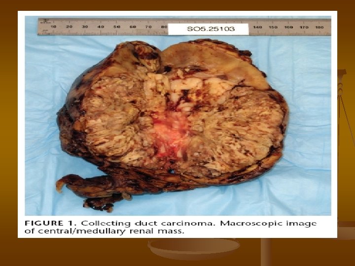
macroscopic examination n n Left kidney showed a central mass with necrosis measuring 12 cm in greatest dimension The tumor replaced the entire pyelocaliceal system, invaded the renal vein and the Gerota fascia, and metastasized to the adjacent adrenal gland
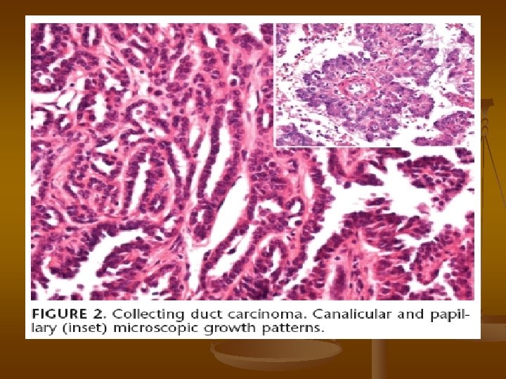
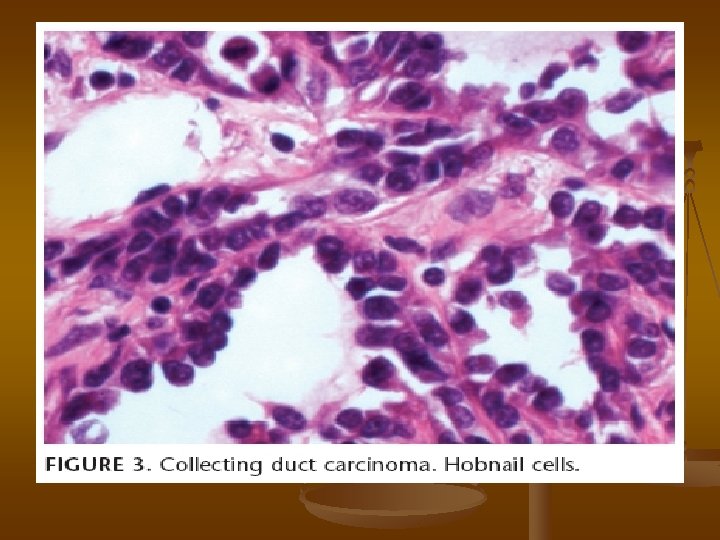
Microscopic n n n this neoplasm was characterized by a predominantly solid and canalicular pattern of growth with papillary areas Cell size ranged from small cuboidal to large, with eccentric nuclei, distinct nucleoli, and frequent mitoses. Hobnail cells were also identified
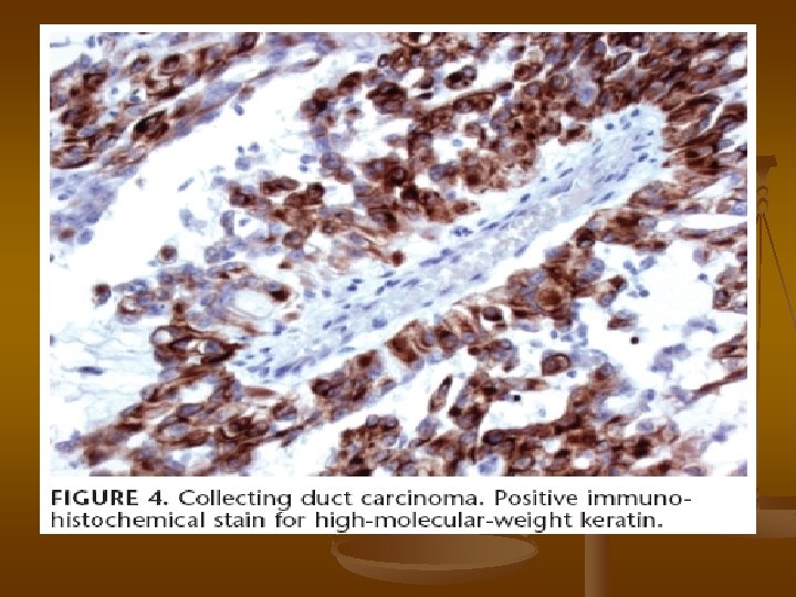
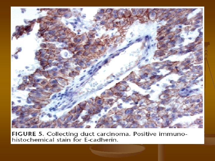
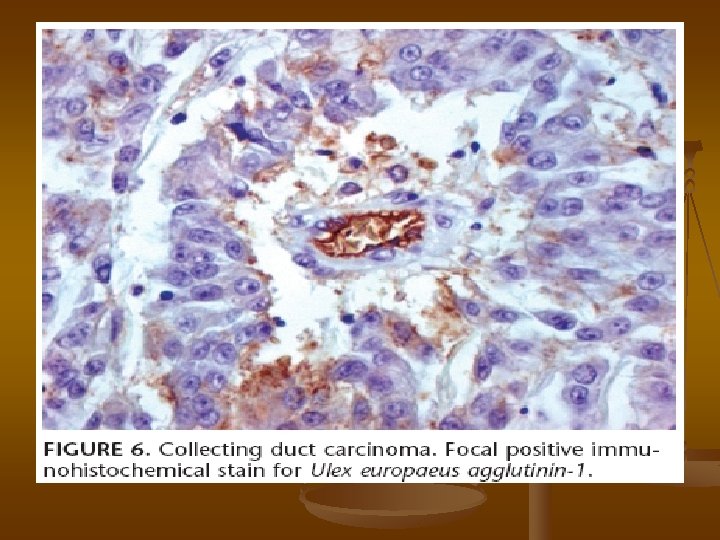
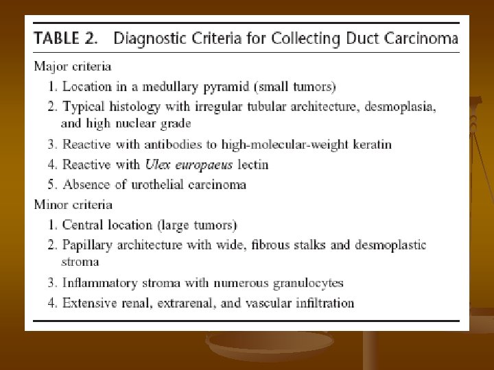
Immunohistochemical stains n n n high-molecular-weight keratin (HMWK) (+) E-cadherin (+) epithelial membrane antigen (EMA) (+) Carcinoembryonic antigen (CEA) (+) Ulex europaeus agglutinin-1 UEA-1 (+) P 63 / renal cell carcinoma antigen (RCC) / CD-10 (-)
DISCUSSION n n CDC, previously called Bellini duct carcinoma, was first described by Mancilla. Jimenez et al in 1976. CDC is a rare renal neoplasm that originates from the distal segment of the collecting duct in the renal medulla pyramids known as the collecting ducts of Bellini
DISCUSSION n n n CDC is thought to arise from the ureteral bud or mesonephros, hence sharing the same origin as urothelial carcinomas. CDC accounts for 0. 6% to 3% of all kidney carcinomas Of all renal epithelial neoplasms, CDC is the most aggressive, with early metastasis common at the time of clinical presentation
Clinical Features n n n 2: 1 male to female ratio Mean age is 55 years, wide range (13 to 83) Association with chronic dialysis Abdominal pain, flank mass, and hematuria Imaging studies are usually suggestive of RPUC (pelvic/medullary location)
Clinical Features n n n Approximately 35% to 40% of patients have metastasis at presentation Common metastatic sites include regional lymph nodes, adrenal gland, bone, lung, and liver. More than 75% of patients die with disease within 2 years of diagnosis
Clinical Features n n Conventional treatments for renal carcinoma, including radiation, chemotherapy Immunotherapy are not effective
Gross Pathology n n n CDC is located in the central region of the kidney and often involves hilar structures. It ranges in size from 2. 5 to 12 cm and is typically firm and gray-white, with infiltrating borders. It may present as multifocal, probably representing intrarenal metastasis.
Microscopic Characteristics n n n This neoplasm is characterized by tubular, tubulopapillary, or solid growth patterns with desmoplasia and inflammatory reaction. Small cyst formations may be present within folding of papillary growth. Sarcomatoid differentiation as a sign of dedifferentiation has also been described.
Microscopic Characteristics n n n Mucin production may be present Cytologically, CDC is composed of eosinophilic or basophilic high-nuclear grade cells with frequent hobnail pattern. (Papillary RCCs lack hobnail cells)
Immunohistochemistry n n n n HMWK + UEA-1 + E-Cad + CK 5/CK 6/CK 17 CD 10/15 N-Cad P 63 -
Cytogenetics n n Monosomy of chromosome 1 appears to be a constant finding in CDC. Other alterations described are monosomy of chromosomes 18 and 21, loss of the Y chromosome, and gains of chromosome 7, 12, 17, and 20.
Molecular Alterations n n CDCs lose chromosomal arm 1 q more frequently than other renal neoplasms, a feature shared with urothelial carcinomas. Loss of heterozygosity of chromosome 6 p is observed in 50% of the CDCs.
Molecular Alterations n n Loss of chromosome 3 p, frequently seen in conventional RCCs, is a rare event in CDC. Only 8% of CDC demonstrated alteration of the VHL gene (3 p 25 -26). RCCs have not included CDC due to the rarity of this neoplasm
Differential Diagnosis n n The principal differential diagnosis of CDC includes PC, RPUC, and metastatic carcinoma. Less frequently, other medullary renal neoplasms such as MC and TC may be part of the differential diagnosis
Immunohistochemistry
Papillary RCC n n a large neoplasm that may invade the renal medulla histologically characterized by the presence of fibrovascular cores with tumor cells arranged in papillary configuration.
RPUC n n n RPUC bears little morphologic resemblance to CDC occasionally extend to collecting ducts of the renal papillae immunohistochemical markers that may be useful
Medullary carcinoma n n MC is a rare neoplasm first distinguished from CDC by Davis et al in 1995 the strong association of this neoplasm with sickle cell trait is a helpful hint in the differential diagnosis.
Tubulocystic carcinoma n n n TC, also known as low-grade CDC Their immunohistochemical profile is similar to CDC Low nuclear grade and dilated tubules are key diagnostic features of TC
Metastaric carcinoma n n n Metastatic carcinomas, particularly those of gastrointestinal tract or lung origin, are part of the differential diagnosis. Immunohistochemistry for CDX-2 and TTF 1 may Be helpful Frequently multiple, well-circumscribed, and usually not associated with dysplastic changes of the collecting ducts.
CONCLUSION n n n CDC is a rare and aggressive renal neoplasm that shares biologic characteristics with carcinomas of urothelial origin. Because of origin in the renal medulla, the differential diagnosis is with other neoplasms that may involve the central area of the kidney. A correct diagnosis and distinction from other RCCs are imperative since prognosis and treatment modalities are different.