Coagulation Physiology and Acquired Coagulation Disorders Guy Young


 V VIIa IXa Ca++ X")
 VII Ca++ XI XIa TF Ca++ IX VIIIa Thrombin (IIa)")
 VII Ca++ XI XIa TF Ca++ IX VIIIa Thrombin (IIa)")
 Va")


 Thrombin (IIa) VII Ca++ XI XIa Ca++ IX")
 Thrombin")

 PAI-1")

, u. PA (not")


 Fibrinogen (I) Fibrin Assesses presence and function of fibrinogen Abnormal")


 factor II, VII, IX, X -COOH CH")

")
 • Increases VWF • Increases FVIII – Probably due to")


 • Bleeding time –")


 Consumption of prohemostatic clotting factors and")


")


 can contain warfarin-like drugs – Currently,")


 • Elevated PT – Early marker of liver disease (short")

 Cryoprecipitate (to")


 • Specific factor")


 that doesn’t")


")
")
")
")
")
")
")
")
")
- Slides: 106
Coagulation Physiology and Acquired Coagulation Disorders Guy Young, MD Children’s Hospital Los Angeles University of Southern California Keck School of Medicine
Abbreviations • Abbreviations will be explained the first time the term appears on a slide
Lecture Outline • Primary Hemostasis • Disorders of primary hemostasis • Plasma coagulation – Clotting factors – Natural inhibitors • • Fibrinolytic system Laboratory Testing Warfarin and DDAVP Acquired Hemostatic Disorders
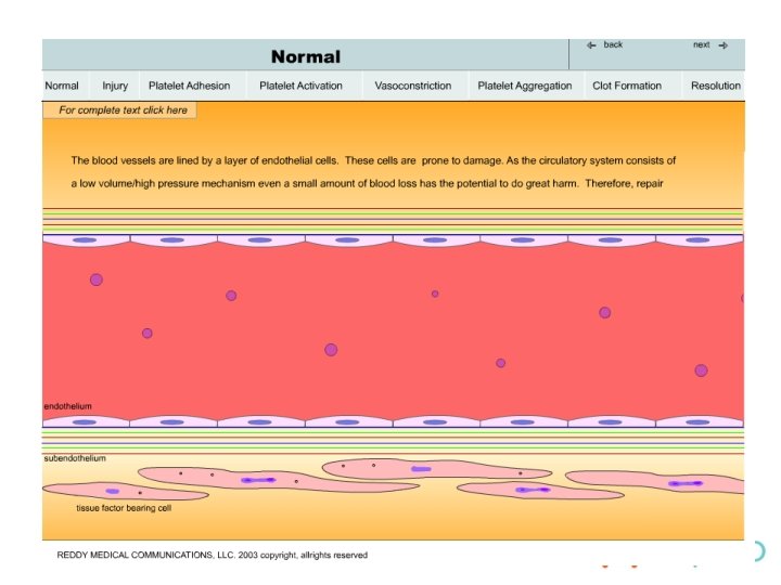
Primary Hemostasis • Subendothelium – Normal collagen – Tissue factor • Platelets – Need adequate number – Need adequate function (see platelet section for details) • von Willebrand factor – Need normal amount – Need normal function
Lecture Outline • Primary Hemostasis • Disorders of primary hemostasis • Plasma coagulation – Clotting factors – Natural inhibitors • • Fibrinolytic system Laboratory Testing Warfarin and DDAVP Acquired Hemostatic Disorders
Disorders of Primary Hemostasis • Collagen disorders – Ehlers Danlos – Osteogenesis imperfecta • Platelet disorders (covered in platelet section) • von Willebrand disease (will be covered in next presentation)
Ehler-Danlos Syndrome
Diagnosis: Physical Examination is usually sufficient. Genetic testing is available in some laboratories
Blue sclera Osteogenesis imperfecta Fractures Diagnosis: Physical Examination is usually sufficient. Genetic testing is available—there at least 4 subtypes.
Lecture Outline • Primary Hemostasis • Disorders of primary hemostasis • Plasma coagulation – Clotting factors – Natural inhibitors • • Fibrinolytic system Laboratory Testing Warfarin and DDAVP Acquired Hemostatic Disorders
Properties of Hemostatic Factors Factor Site of synthesis Levels in infancy Half-life Vitamin K- Other dependent Fibrinogen (I) Liver Normal 2 -4 d No Prothrombin (II) Liver Low 3 d Yes Factor V Liver Megakaryocytes Low 36 hr No Factor VII Liver Low 3 -6 hr Yes Factor VIII Liver Endothelial Cells Normal/ High 8 -12 hr No Factor IX Liver Low 22 hr Yes Factor X Liver Low 40 hr Yes Factor XI Liver Low 80 hr No Factor XIII Liver/Macrophage Low 10 d No Important in wound healing VWF Endothelial cells Megakaryocytes 12 hr No Stored in Weibel. Palade bodies in endothelial cells Normal/ High fibrinogen increases ESR Circulates with VWF Increased by DDAVP
Classical Coagulation Cascade K HMWK XII PK=Prekallikrein K=Kallikrein HMWK=High molecular weight kininogen TF=Tissue factor Ca++=Calcium PK XIIa VII Ca++ XI XIa TF Ca++ IX VIIIa Thrombin (IIa) V VIIa IXa Ca++ X Xa Va Prothrombin (II) Ca++ XIII Thrombin (IIa) Fibrinogen (I) Fibrin XIIIa Cross-linked fibrin
Contact Activation System HMWK K Components of the contact activation system: High molecular weight kininogen Prekallikrein Factor XII Factor XI PK XIIa Ca++ XI XIa C 3 C 1 C 3 active C 1 active Complement system Bradykinin/Kinin (Inflammation) Among the contact factors, only FXI deficiency is associated with bleeding (bleeding tendency is highly variable—some patients don’t bleed at all) Factor XIIa and Kallikrein are contact activation factors with interactions in the complement system
Physiologic Coagulation VII TF Ca++ IX VIIIa Thrombin (IIa) V VIIa IXa Ca++ X Xa Va Prothrombin (II) Ca++ XIII Thrombin (IIa) Fibrinogen (I) Fibrin XIIIa Cross-linked fibrin
Physiologic Coagulation Thrombin (IIa) VII Ca++ XI XIa TF Ca++ IX VIIIa Thrombin (IIa) V VIIa IXa Ca++ X Xa Va Prothrombin (II) Ca++ XIII Thrombin (IIa) Fibrinogen (I) Fibrin XIIIa Cross-linked fibrin
Physiologic Coagulation Thrombin (IIa) VII Ca++ XI XIa TF Ca++ IX VIIIa Thrombin (IIa) V VIIa IXa Ca++ X Xa Va Prothrombin (II) XIII Ca++ Thrombin (IIa) Fibrinogen (I) Fibrin XIIIa TAFI Thrombin (IIa) Cross-linked fibrin TAFIa
Thrombin’s procoagulant/antifibrinolytic effects on coagulation factors VIIIa XI XIa XIII V Thrombin (IIa) Va XIIIa TAFI Fibrinogen (I) Fibrin TAFIa Antifibrinolytic effect
Thrombin’s interaction with platelets and endothelial cells TM --Thrombomodulin --platelet --activated platelet Protein Ca Thrombin (IIa) TM --endothelial cell Va and VIIIa Thrombin (IIa) --fibrinogen --fibrin --inhibits
Fibrinogen structure and gene Chromosome 4 FGA Aα chain FGB Bβ chain FGG γ chain Thrombin binding site Fibrinogen Fibrinopeptide B Fibrinopeptide A Fibrin
Fibrin-platelet interaction GPIIb. IIIa Fibrin GPIIb. IIIa
Fibrinogen special features • Fibrinogen is an acute phase reactant • An elevated fibrinogen causes an elevated ESR • Tests for deficiency of fibrinogen – PT, PTT and TT (thrombin time) are prolonged when fibrinogen is <1 g/L (100 mg/d. L) – Measure functional fibrinogen (Clauss method) • Tests for dysfibrinogenemia – Thrombin time – Fibrinogen antigen to activity ratio • Tests for fibrinogen and dysfibrinogen in presence of heparin – Reptilase time (converts fibrinogen to fibrin but insensitive to presence of heparin)
Factor XIII special features • It has the longest half-life of all the clotting factors (10 days) • Screening coagulation tests (PT and PTT) are normal • Factor XIII testing – Clot solubility assay (with urea or acetic acid) • Qualitative test (normal or abnormal) • Sensitive only for very low levels (<5%) – Chromogenic assay • More sensitive • Not as readily available • Factor XIII is associated with poor wound healing – Delayed separation of umbilical stump (can also occur with leukocyte adhesion deficiency) • Common presentation is bleeding from the umbilical stump and intracranial hemorrhage
Lecture Outline • Primary Hemostasis • Disorders of primary hemostasis • Plasma coagulation – Clotting factors – Natural inhibitors • • Fibrinolytic system Laboratory Testing Warfarin and DDAVP Acquired Hemostatic Disorders
Natural Coagulation Inhibitors Inhibits ( ) Thrombin (IIa) VII Ca++ XI XIa Ca++ IX VIII V VIIa IXa Ca++ X Thrombin (IIa) Tissue factor pathway inhibitor (TFPI) TF Xa X Antithrombin Xa Va XIII Protein S Prothrombin (II) Protein Ca Ca++ Thrombin (IIa) Fibrinogen (I) Thrombin (IIa) Fibrin XIIIa Protein C Cross-linked fibrin
Endothelial cell’s antithrombotic effects PS --Protein S HS --heparan sulfate PC APC (PS) Thrombin PC—protein C TPA --tissue plasminogen activator APC—activated protein C TM --Thrombomodulin PAI-1 --plasminogen activator inhibitor type 1 Va and VIIIa AT Thrombin TM fibrinolysis PS TPA HS PAI-1
Lecture Outline • Primary Hemostasis • Disorders of primary hemostasis • Plasma coagulation – Clotting factors – Natural inhibitors • • Fibrinolytic system Laboratory Testing Warfarin and DDAVP Acquired Hemostatic Disorders
Fibrinolysis and natural inhibitors t. PA-tissue plasminogen activator u. PA-urine plasminogen activator (urokinase) PAI-1 -plasminogen activator inhibitor-1 TAFI-thrombin activatable fibrinolysis inhibitor t. PA u. PA Plasminogen PAI-1 Plasmin α 2 -antiplasmin TAFIa Fibrin clot Clot lysis Fibrin split products D-dimer Thrombin (IIa) TAFI
Tests of fibrinolytic system • Signs of increased fibrinolysis – Abnormal in DIC, venous thrombosis and liver disease • D-dimer • Fibrin degradation (split) products • Assessment of fibrinolytic proteins – Decreased in deficiency states [congenital (rare), acquired] associated with thrombosis (at least in theory) • Plasmin • t. PA • Assessment of fibrinolytic inhibitors – Decreased in deficiency states [congenital (rare)] associated with bleeding • PAI-1 • α-2 antiplasmin
Fibrinolytic and anti-fibrinolytic drugs • Fibrinolytic – t. PA (and analogs), u. PA (not available in USA) • Used for treatment of arterial and venous thrombosis • Used to treat central venous catheter lumen occlusions • Antifibrinolytic – ε-aminocaproic acid and tranexamic acid (amino acid analogs of lysine) • Inhibit fibrinolysis by blocking plasmin’s binding site for fibrin (plasmin recognizes exposed lysine residues on fibrinogen)
Mechanism of action of antifibrinolytic drugs
Lecture Outline • Primary Hemostasis • Disorders of primary hemostasis • Plasma coagulation – Clotting factors – Natural inhibitors • • Fibrinolytic system Laboratory Testing Warfarin and DDAVP Acquired Hemostatic Disorders
Factor Deficiencies and Coagulation Assays Intrinsic Pathway=a. PTT K HMWK PK Extrinsic Pathway=PT XIIa VII Ca++ XI TF VIIa XIa Ca++ IX IXa/VIIIa Common Pathway=PT and a. PTT Ca++ X Prothrombin (II) Xa/Va Ca++ Thrombin (IIa) Fibrinogen (I) Fibrin
Thrombin Time Thrombin (IIa) Fibrinogen (I) Fibrin Assesses presence and function of fibrinogen Abnormal in hypo- and afibrinogenemia Abnormal in dysfibrinogenemia
Factor Deficiencies and Coagulation Assays • a. PTT – Contact factors • PK, HWMK, FXII – Factor IX – Factor VIII – Common Pathway • • Factor X Factor V Factor II (Prothrombin) Fibrinogen • PT – Factor VII – Common Pathway • • Factor X Factor V Factor II (Prothrombin) Fibrinogen
PTT PT NORMAL PROLONGED PT PROLONGED NORMAL FXIII deficiency FVII deficiency PROLONGED NORMAL Liver disease Afibrinogenemia Vitamin K deficiency Dysfibrinogenemia Circulating anticoagulant DIC Warfarin effect Heparin effect Deficiencies of factors VIII, IX, XII, Prekallikrein, HMWK Circulating anticoagulant Heparin effect
Factor XIII and Coagulation Assays • • The a. PTT and PT reactions are complete once fibrin monomers are formed They do not take into account the cross-linking effect of FXIII Therefore deficiency of FXIII does NOT affect either of these tests Testing for FXIII deficiency requires specifically assaying for the presence of this factor – Urea clot lysis assay (qualitative only and only abnormal in severe deficiency) – Chromogenic assay (quantitative but only available in selected laboratories) XIII Prothrombin (II) Ca++ Thrombin (IIa) Fibrinogen (I) Fibrin XIIIa Cross-linked fibrin
Lecture Outline • Primary Hemostasis • Disorders of primary hemostasis • Plasma coagulation – Clotting factors – Natural inhibitors • • Fibrinolytic system Laboratory Testing Warfarin and DDAVP Acquired Hemostatic Disorders
Vitamin K metabolism and warfarin γ-carboxylated (functional) factor II, VII, IX, X -COOH CH -COOH Factor II, VIII, IX, X precursor (PIVKA) CH 2 -COOH Vitamin K γ-glutamyl carboxylase Vitamin K hydroxyquinone Vitamin K epoxide warfarin Vitamin K Epoxide reductase Vitamin K Dietary sources Vitamin K Epoxide reductase
Warfarin special features • Warfarin is monitored with the INR – The INR is a mathematical calculation of the PT ratio based on the sensitivity of the PT reagent the lab uses • The half-life of FVII (3 -6 hours) is short while the half-life of factor X is long (40 hours) – This can lead to an early rapid rise in the INR due to the drop in factor VII – This change is NOT a sign of therapeutic warfarin as that effects takes 5 -7 days when the factor X level falls
Warfarin special features • Warfarin should always be overlapped with another anticoagulant (heparin, LMWH) until the INR is therapeutic for 2 consecutive days • Protein C and S are vitamin K dependent – Some patients develop warfarin-induced skin necrosis due to the drop in protein C which happens early after warfarin initiation
Effects of DDAVP (desmopressin) • Increases VWF • Increases FVIII – Probably due to increased VWF • Increases t. PA release
Lecture Outline • Primary Hemostasis • Disorders of primary hemostasis • Plasma coagulation – Clotting factors – Natural inhibitors • • Fibrinolytic system Laboratory Testing Warfarin and DDAVP Acquired Hemostatic Disorders
Disorders of Coagulation • General – Screening tests – Artifacts in coagulation tests – Limitations as pre-operative tests – Factors that are acute phase reactants
Screening tests • PT, PTT, and TT (already covered above) • Bleeding time – Essentially a historical relic – Assesses primary hemostasis • Prolonged in disorders of primary hemostasis – Collagen disorders – Platelet function disorders/thrombocytopenia – May be abnormal in v. WD • PFA-100 – A “modern” bleeding time (see platelet section) • Platelet aggregation studies – Abnormal in platelet function disorders (see platelet section)
Disorders of Coagulation • General – Screening tests – Artifacts in coagulation tests – Limitations as pre-operative tests – Factors that are acute phase reactants
Artifacts in coagulation tests • PT, PTT, TT – Sample relies on proper ratio of plasma to citrate (9: 1) • Too little plasma leads to elevated levels – Tube not properly filled – Patient with polycythemia • Too much plasma (severe anemia—falsely normal results) – Other pre-analytic issues that could lead to a falsely elevated result • Sample not processed quickly (temperature artifact) • Difficult lab draw (sample begins to clot consuming the factors) • Heparin in sample – Use heparin neutralization procedure
Disorders of Coagulation • General – Screening tests – Artifacts in coagulation tests – Limitations as pre-operative tests – Factors that are acute phase reactants
Limitations as pre-operative tests • PT, PTT screen for factor deficiencies not bleeding – Factor XII deficiency has no bleeding but very prolonged PTT – Factor XIII deficiency has severe bleeding but normal screening labs • PT, PTT are subject to many false positives (previous slide) • PT, PTT can be truly abnormal (aside from factor deficiencies) yet still not suggest an increased risk for bleeding – Lupus anticoagulant (more on this later)
Disorders of Coagulation • General – Screening tests – Artifacts in coagulation tests – Limitations as pre-operative tests – Factors that are acute phase reactants
Factors that are acute phase reactants • Factor VIII • v. WF • Fibrinogen
Disseminated intravascular coagulation • • Pathophysiology Clinical features Laboratory abnormalities Treatment and outcome
Inciting Event Activation of coagulation (circulating tissue factor) Consumption of prohemostatic clotting factors and platelets Bleeding Consumption of anticoagulant clotting factors Thrombosis Fibrinolysis Disruption of endothelial cell anticoagulant surface
Causes of DIC • DIC does not occur as a primary illness • Typical causes in children – – – Sepsis Kasabach-Merritt syndrome (unique to neonates) Trauma (brain, burns, gunshot wounds) Malignancy (APML) Severe hemolysis Severe protein C deficiency (unique to neonates)
Purpura fulminans • Severe skin necrosis – Due to microvascular thrombosis • Acute onset
Disseminated intravascular coagulation • • Pathophysiology Clinical features Laboratory abnormalities Treatment and outcome
Clinical features • Symptoms – Patients may have no bleeding or thrombosis but still have DIC – Typical symptoms • Bleeding – Bruising, petechia, mucus membrane bleeding • Thrombosis – Purpura fulminans – Organ dysfunction • Hemolytic anemia
Disseminated intravascular coagulation • • Pathophysiology Clinical features Laboratory abnormalities Treatment and outcome
Laboratory findings • No single lab test diagnoses DIC • Common lab findings (none are required) – Prolonged PT and PTT • Any clotting factor can be reduced – Decreased fibrinogen – Thrombocytopenia – Elevated FDP, D-dimer, and fibrinopeptide A and B – Decreased protein C and S and antithrombin • Neonates are physiologically deficient in proteins C/S and antithrombin impairing their protective mechanisms for DIC
Disseminated intravascular coagulation • • Pathophysiology Clinical features Laboratory abnormalities Treatment and outcome
Treatment and outcome • Treating the underlying disease is the main therapeutic option • Management of coagulopathy – No clear consensus – Transfusion therapy • Platelet transfusions are not generally indicated (risk for increased thrombosis) • Fresh frozen plasma – Provides balanced replacement of all prohemostatic clotting factors and their inhibitors (standard risks for FFP) – No evidence it affects outcome • Cryo – Can be given for low fibrinogen (if bleeding occurs) – No clear evidence of benefit (standard risks for cryo) – Anticoagulation is not indicated (risk of bleeding)
Vitamin K deficiency • • • Pathophysiology Etiology and clinical features Laboratory findings Treatment and response Drug-induced vitamin K deficiency
Pathophysiology • Vitamin K is fat soluble • Tissue stores are limited – Can become deficient quickly • Human milk has little to no vitamin K
Vitamin K deficiency • • • Pathophysiology Etiology and clinical features Laboratory findings Treatment and response Drug-induced vitamin K deficiency
Pathophysiology Vitamin K deficiency bleeding Early Classical Late Age First 24 hours 2 -7 days 1 -6 months (even later) Risk factors Maternal use of drugs that interfere with vitamin K metabolism (warfarin, anticonvulsants) None (all newborns are prone) Disorders that interfere with vitamin K intake (cystic fibrosis, other GI fat malabosrption disorders, chronic antibiotic use, liver disease) Bleeding sites ICH, GI, umbilical stump, bruising ICH, GI, mucocutaneous Treatment Recognition of drugs that can cause this and eliminating them from maternal use Prevention with neonatal vitamin K administration Parenteral vitamin K
Vitamin K deficient newborn (did not receive prophylactic vitamin K)
Vitamin K deficiency • • • Pathophysiology Etiology and clinical features Laboratory findings Treatment and response Drug-induced vitamin K deficiency
Laboratory findings • Early and Classical – PT and PTT very prolonged (often unclottable, i. e. >200 seconds) – In less severe cases (tests do clot), PT can be as long or even longer than the PTT (absence of factor VII) – Vitamin K dependent factors (II, VII, IX, X, PC and PS severely deficient (if measured)
Vitamin K deficiency • Late – – PT is always prolonged (factor VII has the shortest half-life) PTT is variably prolonged (depends on severity) Vitamin K dependent factors deficient (if measured) Elevated PIVKA (sent only if diagnosis is in question) • PIVKA (proteins induced in vitamin K absence)
Vitamin K deficiency • • • Pathophysiology Etiology and clinical features Laboratory findings Treatment and response Drug-induced vitamin K deficiency
Treatment and response • Replace vitamin K – Different preparations • Oral/Parenteral – Phytonadione (vitamin K 1) is currently the only form of vitamin K approved by the FDA – It takes 12 -24 hours after a parenteral dose to correct the deficiency • Prothrombin complex concentrates – Plasma-derived products containing factors II, VII, IX, and X – Can be given if immediate correction is warranted (ICH) – Carry a risk for thrombosis
Vitamin K deficient newborn before and 3 days after vitamin K
Vitamin K deficiency • • • Pathophysiology Etiology and clinical features Laboratory findings Treatment and response Drug-induced vitamin K deficiency
Drug-induced vitamin K deficiency • Pesticides (rats, mice) can contain warfarin-like drugs – Currently, declining in use due to resistance and better pesticides – These drugs are more potent with much longer half-lives than human warfarin (often weeks) • Can cause prolonged severe coagulopathy if accidentally ingested • Treatment is prolonged therapy with vitamin K (until poison has been cleared)
Liver disease • Pathophysiology • Laboratory features • Treatment
Pathophysiology • Mechanisms responsible for excess bleeding – Decreased production of liver synthesized factors • Most factors are decreased (see table on clotting factors) • FVIII is elevated in liver disease – Thrombocytopenia • Various causes – Hypersplenism – Decreased thrombopoietin production – Hyperfibrinolysis • Increased t. PA release from endothelial cells
Liver disease • Pathophysiology • Laboratory features • Treatment
Laboratory features (coagulation tests) • Elevated PT – Early marker of liver disease (short half-life of FVII) • Elevated PTT – Due to decreased production of most other factors • Low fibrinogen – Varies with severity of liver failure – Fibrinogen can be high as an acute phase reactant • Thrombocytopenia • Elevated D-dimers – Liver clears D-dimers – Hyperfibrinolysis
Liver disease • Pathophysiology • Laboratory features • Treatment
Treatment • • Fresh frozen plasma (contains all the plasma clotting factors) Cryoprecipitate (to correct low fibrinogen) Platelet transfusions (for bleeding with thrombocytopenia) r. FVIIa – Useful if factor VII is particularly low • Prothrombin complex concentrates (PCCs) – Contain all the vitamin K-dependent factors (II, VII, IX, X) – Cases of thrombosis have been reported with PCC use in liver failure (ABP outline states “contraindicated in the treatment of bleeding due to liver disease”)
Blood coagulation inhibitors • Inhibitors against specific factors • Lupus-type anticoagulants
Mixing Studies Prolonged a. PTT Mixing study with normal plasma Corrects Factor Deficiency Does not correct Inhibitors Lupus anticoagulant Specific factor inhibitors
Blood coagulation inhibitors • Inhibitors against specific factors • Lupus-type anticoagulants
Specific factor inhibitors • Extremely rare in childhood (outside of hemophilia) • Specific factor inhibitors – Factor VIII—secondary to autoantibodies – Factor V—secondary to exposure to bovine thrombin • Passively acquired maternal antibodies – Factor VIII autoantibodies in mothers can be transplacentally transferred – Lupus anticoagulant that is passively transferred can lead to thrombosis in the neonate (renal vein thrombosis)
Blood coagulation inhibitors • Inhibitors against specific factors • Lupus-type anticoagulants
Lupus anticoagulant Most lupus anticoagulants are transient, asymptomatic and result from an acute viral infection (one of the most common causes of a prolonged PTT) Lupus anticoagulants can be part of the antiphospholipid syndrome and cause thrombosis (to be discussed in thrombosis section) Rarely, patients with the lupus anticoagulant also have acquired hypoprothrombinemia (due to increased clearance of prothrombin) which can result in bleeding (known as “Lupus anticoagulant hypoprothrombinemia syndrome”) Bleeding Thrombosis Lupus anticoagulant
Lupus anticoagulant laboratory testing • Screening – Prolonged PTT (or rarely PT) that doesn’t correct with mixing study • Diagnosing specific factor inhibitors – Specific factor assays (in patients with a non-correcting mixing study) • Diagnosing lupus anticoagulant – Specific clotting assays for lupus anticoagulants (often referred to as “lupus anticoagulant assay”) • Several base assays – Most common (by far) is the dilute Russell’s viper venom time (d. RVVT)
d. RVVT • 2 Steps – Screen • Run the d. RVVT on patient sample • If abnormal, proceed to confirm – Confirm • Run d. RVVT on same sample adding exogenous phospholipid • Result – Ratio of screen to confirm – Adding back phospholipid will “correct” the abnormality if the abnormality was due to an antiphospholipid antibody – Abnormal ratio is generally >1. 2 (significant if >1. 5)
Lupus anticoagulant management • Most lupus anticoagulants are transient/asymptomatic – Observation – Repeat assay in 6 -12 weeks – No specific therapy is indicated • Bleeding (lupus anticoagulant hypoprothrombinemia syndrome) – Replace prothrombin with PCC for bleeding – Observation (usually transient) • Thrombosis in antiphospholipid antibody syndrome – Will be covered in thrombosis section
Miscellaneous acquired bleeding disorders • Hemophagocytic lymphohistioctyosis syndrome – Can have severe coagulopathy with hypofibrinogenemia • Treatment with asparaginase – Asparagine is important in many coagulation proteins – Patients can have thrombosis from decrease in antithrombin, proteins C and S – Patients can have bleeding from hypofibrinogenemia • Nephrotic syndrome – Leads to loss of antithrombin in the urine (follows albumin) – Increases the risk for thrombosis • Protein losing enteropathy – Loss of proteins C/S and antithrombin – Increased risk for thrombosis
Appendix—ABP Outline and Slide Number A. Coagulation 1. Physiology of coagulation, fibrinolysis and the vessel wall a. Contact activation i. iii. iv. Know the components of the contact activation system (slide 14) Know the consequences of deficiencies in the contact activation system on coagulation assays (slides 33, 35 -36) Know the interaction between the contact activation system and the complement system (slide 14) Know the function of factor XI in the coagulation cascade (Slides 13 -14, 16) b. Factor IX i. iii. iv. v. vi. Know the age-related changes in factor IX concentrations (slide 12) Know the site of synthesis of factor IX (slide 12) Know the half-life of factor IX (slide 12) Know the role of vitamin K in the synthesis and activity of factor IX and also factors II (prothrombin), VII, and X (slide 39) Know the mechanism of activation and the function of factor IX in the coagulation cascade (slide 15 -17) Know the consequences of deficiency of factor IX on the laboratory assessment of hemostasis (slides 33, 35 -36)
Appendix—ABP Outline and Slide Number A. Coagulation 1. Physiology of coagulation, fibrinolysis and the vessel wall c. Factor VIII i. iii. iv. Know that DDAVP increases plasma factor VIII concentration (slide 42) Know the natural inhibitors of factor VIII (slides 19, 25 -26) Know the function of factor VIII in coagulation (slides 15 -17) Know the consequences of a deficiency of factor VIII on the laboratory assessment of hemostasis (slides 33, 35 -36) v. Know the normal value of factor VIII in a newborn infant (slide 12) vi. Know the half-life of factor VIII (slide 12) vii. Know that factor VIII circulates as a complex with von Willebrand factor (slide 12) d. von Willebrand factor (v. WF)—see section on v. WD except as noted below i. iii. iv. v. vi. Know the sites of synthesis, storage, and release of v. WF (slide 12) Know the platelet aggregation patterns associated with the different types of von Willebrand disease Know the laboratory methods for measuring the concentrations, structure, and function of v. WF Know the interaction between v. WF, platelets, and the vessel wall Know the consequences of a deficiency of v. WF on the laboratory assessment of hemostasis Know the factors that affect the serum concentration of v. WF Know the half-life of v. WF (slide 12)
Appendix—ABP Outline and Slide Number A. Coagulation 1. Physiology of coagulation, fibrinolysis and the vessel wall e. Factor VII and tissue factor i. iii. iv. v. Know the function of factor VII/tissue factor in coagulation (slides 15 -17) Know the consequences of deficiency of factor VII on the laboratory assessment of hemostasis (slides 33, 35 -36) Know the age-related changes in factor VII concentrations (slide 12) Know the site of synthesis of factor VII (slide 12) Know the half-life of factor VII (slide 12) f. Factor X i. ii. Know the mechanism of activation and function of factor X in coagulation (slide 15 -17) Know the consequences of a deficiency of factor X on the laboratory assessment of hemostasis (slides 33, 35 -36) iii. Know that the half-life of factor X is longer than the half-life of factor VII and know why this is important when switching anticoagulation from heparin to coumadin (slide 40) g. Factor V i. Know the mechanism of activation and the function of factor V in coagulation (slide 15 -17) ii. Know the consequences of a deficiency of factor V on the laboratory assessment of hemostasis (slides 33, 35 -36) iii. Know which inhibitors regulate factor V (slides 19, 25 -26)
Appendix—ABP Outline and Slide Number A. Coagulation 1. Physiology of coagulation, fibrinolysis and the vessel wall h. Prothrombin and thrombin i. iii. iv. v. Know the mechanism of activation of prothrombin (slides 15 -17) Know the function prothrombin and thrombin in coagulation, natural anticoagulation, and fibrinolysis (slides 18) Know the consequences of deficiency of prothrombin on the laboratory assessment of hemostasis (slides 33, 35 -36) Know the natural inhibitors of thrombin (slides 25 -26) Know the interaction of thrombin with platelets and endothelial cells (slide 19)
Appendix—ABP Outline and Slide Number A. Coagulation 1. Physiology of coagulation, fibrinolysis and the vessel wall i. Fibrinogen and fibrin a. Know the association of fibrinogen concentration and erythrocyte sedimentation rate (slide 22) b. Know the basic structure of fibrinogen and its gene control (slide 20) c. Know the function of fibrinogen and fibrin in coagulation (slides 15 -17) d. Know the consequences of fibrinogen deficiency on the laboratory assessment of hemostasis (slides 22, 33 -36) e. Know the normal value of fibrinogen in a newborn infant (slide 12) f. Know the sites of synthesis of fibrinogen (slide 12) g. Know the half-life of fibrinogen (slide 12) h. Know the interaction of fibrinogen with platelets (slide 21) i. Know the screening tests for fibrinogen deficiency and dysfibrinogenemia (slide 22)
Appendix—ABP Outline and Slide Number A. Coagulation 1. Physiology of coagulation, fibrinolysis and the vessel wall j. Factor XIII i. iii. iv. v. vi. Know the association of factor XIII with poor wound healing (slide 23) Know the consequences of a deficiency of factor XIII on the laboratory assessment of hemostasis (slide 23, 37) Know the function of factor XIII in coagulation (slides 15 -17) Know that the half-life of factor XIII (slide 12, 23) Know the sites of synthesis of factor XIII (slide 12) Know the laboratory test for factor XIII (slide 23, 37) k. Fibrinolysis i. iii. iv. v. Know the mechanism of activation of plasminogen (slide 28) Know the effects of DDAVP on the tissue plasminogen activator (slide 42) Know the natural inhibitors of plasminogen and its activators (slide 28) Know the laboratory tests which measure the fibrinolytic system (slide 29) Know the fibrinolytic and antifibrinolytic drugs and their mechanisms of action (slides 30 -31)
Appendix—ABP Outline and Slide Number A. Coagulation 1. Physiology of coagulation, fibrinolysis and the vessel wall l. Blood vessels i. iii. iv. v. vi. Know which connective tissue diseases are associated with bleeding (slide 7 -10) Know that thrombomodulin is an endothelial cell surface protein which binds to thrombin (slide 19, 26) Know the role of heparan sulfate proteoglycans on the endothelial cell surface in maintaining a non-thrombogenic surfce (slide 26) Know that endothelial cells synthesize and secrete tissue plasminogen activator (slide 26) Know that endothelial cells synthesize and secrete plasminogen activator inhibitor (slide 26) Know that endothelial cells synthesize and secrete protein S (slide 26)
Appendix—ABP Outline and Slide Number A. Coagulation 2. Disorders of coagulation (diagnosis and therapy) a. General i. ii. iv. Understand which components of the hemostatic system are measured by screening tests, e. g. prothrombin time, partial thromboplastin time, thrombin time, bleeding time, platelet function screen (PFA 100), platelet aggregation studies (slides 22, 3336, 45) Know the effects of specimen collections artifacts on coagulation tests (heparin, polycythemia, inadequate specimen) (slide 47) Understand the limitations of and the use of preoperative screening tests to rule out bleeding tendencies (slide 49) Know which coagulation factors are acute phase reactants (slide 51)
Appendix—ABP Outline and Slide Number A. Coagulation 2. Disorders of coagulation (diagnosis and therapy) b. Acquired defects i. Disseminated intravascular coagulation (DIC) a) Pathophysiology and clinical features 1) Recognize the underlying conditions associated with DIC (slide 54) 2) Recognize the pathophysiology of purpura fulminans (slides 53, 55) 3) Know that DIC does not occur as a primary illness and may occur in severely ill patients without bleeding or thrombosis (slides 54, 57) 4) Know the triggering events, e. g. endotoxin, viruses, procoagulant factors released from the tissues, toxins) that activate blood coagulation (slide 54) 5) Know the mechanism by which triggering events lead to DIC (slide 53) 6) Know which protective mechanisms against disseminated intravascular coagulation are physiologically impaired in sick neonates as compared to older infants and children (slide 59) 7) Know that the clinical features of DIC can include hemorrhage (localized or diffuse), thrombosis, hemolytic anemia, and organ dysfunction (slide 57) 8) Know the underlying diseases unique to the neonate that are associated with DIC (slide 59)
Appendix—ABP Outline and Slide Number A. Coagulation 2. Disorders of coagulation (diagnosis and therapy) b. Acquired defects i. Disseminated intravascular coagulation (DIC) b) Laboratory abnormalities 1) Know which tests to perform and how to interpret their results in patients with DIC (slide 59) 2) Know that fibrinolysis usually accompanies DIC (slide 53) 3) Know which blood coagulation factors are reduced in the plasma of patients with DIC (slide 59) 4) Know the significance of the measurement of fibrinogen, fibrin products, and degradation, e. g. D-dimer, fibrin monomer, fibrinopeptide A and B (slide 59) c) Treatment and outcome 1) Recognize the risks and benefits of the various therapeutic options for DIC (slide 61) 2) Know that control of the primary disorder is the main treatment for DIC (slide 61)
Appendix—ABP Outline and Slide Number A. Coagulation 2. Disorders of coagulation (diagnosis and therapy) b. Acquired defects ii. Vitamin K deficiency a) Pathophysiology 1) Know that vitamin K is fat soluble (slide 63) 2) Know that vitamin K tissues stores are limited and that patients can become rapidly deficient (slide 63) 3) Know that human milk contains very little vitamin K (slide 63) b) Etiology and clinical features 1) Recognize the hematologic manifestations of cystic fibrosis in an infant (slide 65) 2) Know the clinical conditions associated with vitamin K deficiency (slide 65) 3) Recognize the clinical features of classic vitamin K deficiency in the newborn period (2 -7 days of age) (slide 65) 4) Recognize the clinical features and the underlying causes of late hemorrhagic disease of the newborn, i. e. occurring between 4 -12 weeks of age (slide 65) 5) Know that vitamin K deficiency and hemorrhage may occur by 24 hours of age in infants of mothers who have taken anti-convulsant drugs or
Appendix—ABP Outline and Slide Number A. Coagulation 2. Disorders of coagulation (diagnosis and therapy) b. Acquired defects ii. Vitamin K deficiency c) Laboratory response 1) Know the laboratory features of vitamin K deficiency (altered coagulation test results and which specific coagulation proteins are affected) (slides 68 -69) d) Treatment and response 1) Know the efficacy and response of different vitamin K preparations (slide 71) 2) Know that hemorrhagic disease of the newborn can be prevented by vitamin K shortly after birth (slide 65) e) Drug-induced vitamin K deficiency 1) Know that certain long-acting (super) warfarin-containing rat poisons may cause a severe and prolonged coagulopathy following their ingestion by an infant or young child (slide 74)
Appendix—ABP Outline and Slide Number A. Coagulation 1. Disorders of coagulation (diagnosis and therapy) b. Acquired defects iii. Liver disease a) Pathophysiology 1) Know that multiple mechanisms responsible for excessive bleeding in patients with severe liver disease (slide 76) b) Laboratory features 1) Know the laboratory test abnormalities most commonly seen inpatients with severe liver disease and clinical bleeding (slide 78) c) Treatment 1) Know the treatment options of various therapeutic modalities in patients who are bleeding from liver disease (slide 80) 2) Know why the use of prothrombin complex concentrates is contraindicated in the treatment of bleeding due to liver disease (slide 80)
Appendix—ABP Outline and Slide Number A. Coagulation 1. Disorders of coagulation (diagnosis and therapy) b. Acquired defects iv. Blood coagulation inhibitors a) Heparins and heparin-like substances (see thrombosis section) b) Inhibitors against specific factors 1) Recognize that inhibitors against specific blood coagulation factors in a patient without hemophilia are rare during childhood (slide 84) 2) Know that passively acquired maternal coagulation inhibitors can adversely affect the newborn infant (slide 84) 3) Know how to differentiate inhibitor from a factor deficiency using conventional coagulation screening tests (slide 82)
Appendix—ABP Outline and Slide Number A. Coagulation 1. Disorders of coagulation (diagnosis and therapy) b. Acquired defects iv. Blood coagulation inhibitors a) Lupus-type anticoagulants 1) Know what hemosatic defects associated with lupus-type anticoagulants may either cause bleeding or thrombosis (slide 86) 2) Know that screening and specific laboratory tests to detect the lupustype anticoagulant, including the 1: 1 mixing study (slides 82, 87) 3) Know that the lupus-type anticoagulant is often transiently observed in healthy children following a viral infection and is one of the most common causes of a prolonged PTT during childhood (slide 86) 4) Know that the management of children and adolescent patients with a lupus-type anticoagulant (slide 89)
Appendix—ABP Outline and Slide Number A. Coagulation 1. Disorders of coagulation (diagnosis and therapy) b. Acquired defects v. Miscellaneous acquired bleeding disorders a) Know what children with hemophagocytic syndromes may have a severe coagulopathy characterized by marked hypofibrinogenemia (slide 90) b) Know the mechanism by which L-Asparaginase effects the coagulation system and the clinical consequences (slide 90) c) Know the coagulation patterns in patients with the nephrotic syndrome and the clinical consequences (slide 90) d) Know the coagulation abnormalities in protein-losing enteropathy and their clinical consequences (slide 90)