
Coagulation Bruno Sopko Content Biochemistry of haemocoagulation Laboratory
Coagulation Bruno Sopko
Content � Biochemistry of haemocoagulation � Laboratory test
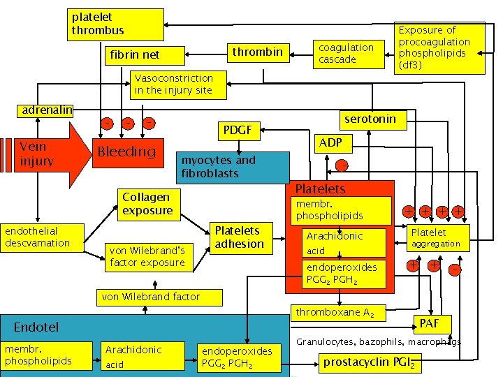
platelet thrombus thrombin fibrin net coagulation cascade Vasoconstriction in the injury site adrenalin Vein injury - - - Bleeding PDGF myocytes and fibroblasts Collagen exposure endothelial descvamation von Wilebrand’s factor exposure Platelets adhesion Exposure of procoagulation phospholipids (df 3) serotonin ADP - Platelets membr. phospholipids Arachidonic acid endoperoxides PGG 2 PGH 2 + + Platelet aggregation + + - von Wilebrand factor thromboxane A 2 Endotel membr. phospholipids Arachidonic acid endoperoxides PGG 2 PGH 2 PAF Granulocytes, bazophils, macrophágs prostacyclin PGI 2
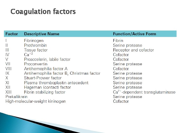
Coagulation factors
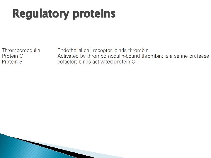
Regulatory proteins
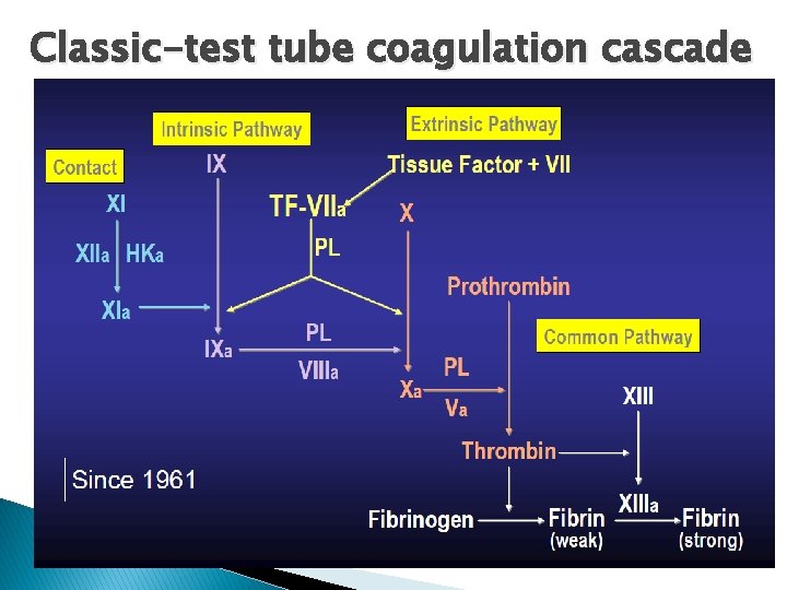
Classic-test tube coagulation cascade
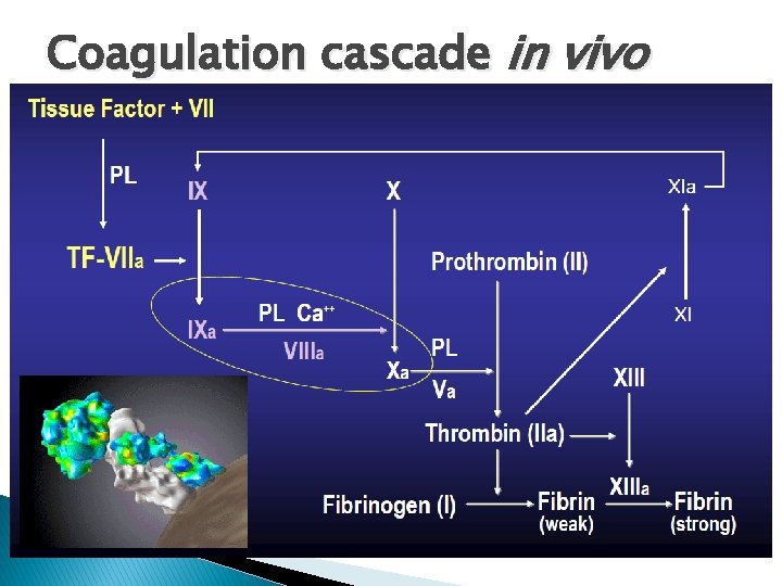
Coagulation cascade in vivo

Formation of fibrin clot
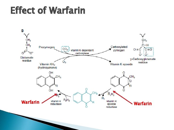
Effect of Warfarin

Thrombin roles
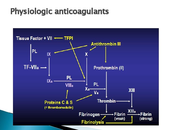
Physiologic anticoagulants
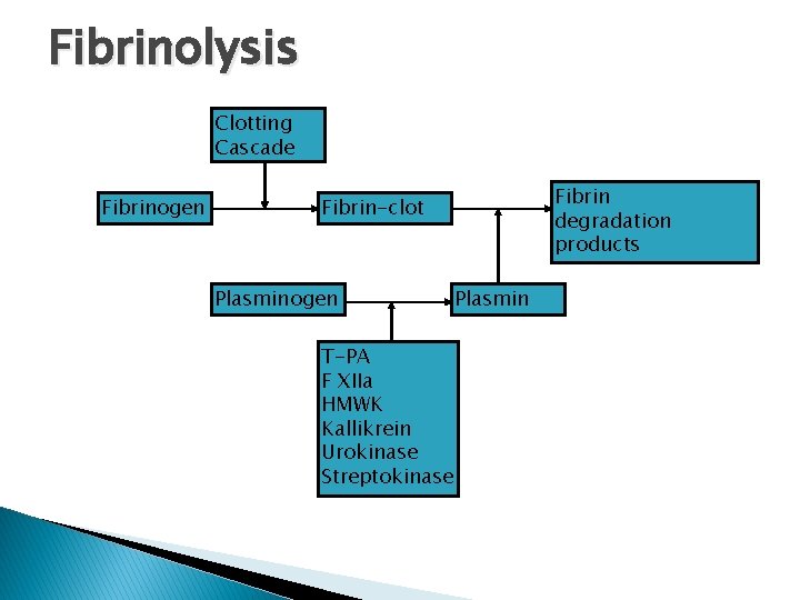
Fibrinolysis Clotting Cascade Fibrinogen Fibrin degradation products Fibrin-clot Plasminogen T-PA F XIIa HMWK Kallikrein Urokinase Streptokinase Plasmin
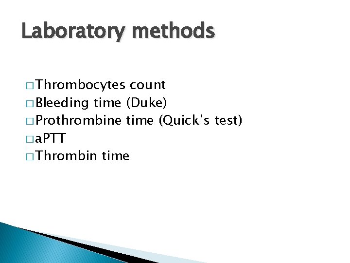
Laboratory methods � Thrombocytes count � Bleeding time (Duke) � Prothrombine time (Quick’s test) � a. PTT � Thrombin time
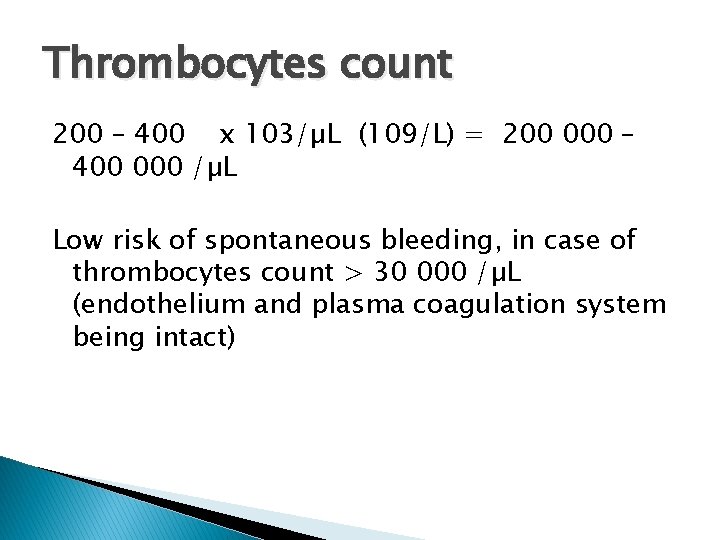
Thrombocytes count 200 – 400 x 103/µL (109/L) = 200 000 – 400 000 /µL Low risk of spontaneous bleeding, in case of thrombocytes count > 30 000 /µL (endothelium and plasma coagulation system being intact)
Bleeding time � � Ivy method: is the traditional format for this test. In the Ivy method, a blood pressure cuff is placed on the upper arm and inflated. A lancet or scalpel blade is used to make a stab wound on the underside of the forearm. The time from when the stab wound is made until all bleeding has stopped is measured and is called the bleeding time. Every 30 seconds, filter paper or a paper towel is used to draw off the blood. The test is finished when bleeding has stopped completely. Template method: a template is placed over the area to be stabbed and two incisions are made in the forearm using the template as a location guide. Duke method: a nick is made in an ear lobe or a fingertip is pricked to cause bleeding. A normal bleeding time for the Ivy method is less than five minutes from the time of the stab, 3 minutes for Duke method
Protrombin Time (Quick’s test) � Prothrombin Time (PT) ◦ Plasma + Calcium + Tissue Thromboplastin TF + VIIa → Xa + V → IIa → CLOT � PT ◦ ◦ ◦ only elevated Factor VII deficiency Congenital (very rare) Acquired (Vit K deficiency, liver disease) Factor VII inhibitor Rarely in pts with modest decreases of factor V or X
a. PTT � Activated Partial Thromboplastin Time (a. PTT) ◦ Plasma + Calcium + Kaolin + Phospholipids Contact → XIa → IXa + VIII →Xa + Va →IIa →CLOT � a. PTT ◦ ◦ only elevated Factor XI, IX, or VIII deficiency Factor XI, IX, or VIII specific factor inhibitor Heparin contamination Antiphospholipid antibodies
Both PT-INR and a. PTT are elevated � Factor(s) X, V, or II deficiency � Factor(s) X, V, or II inhibitor � Improper anticoagulation ratio (Hct >60 or <15) � High doses of heparin (↑ a. PTT > ↑ PT-INR) � Large Warfarin effect ((↑ PT-INR > ↑ a. PTT) � Low fibrinogen (<80 mg/dl)
Thrombin Clotting Time (TCT) � Add thrombin to patient’s plasma ◦ This should directly clot fibrinogen � Elevated ◦ ◦ ◦ in Heparin use DIC Dysfibrinogenemia Low fibrinogen levels High fibrinogen levels Uremia
Marks´ Basic Medical Biochemistry, A Clinical Approach, third edition, 2009 (M. Lieberman, A. D. Marks) Color Atlas of Biochemistry (J. Koolman, K. H. Roehm) Stanislav Matoušek: Patofyziologie koagulace, Thomas A. Whitehill: Coagulation Made Simple
- Slides: 20