Co Vi D19 EPIDEMIC BASICS OF MECHANICAL VENTILATION

Co. Vi. D-19 EPIDEMIC: BASICS OF MECHANICAL VENTILATION Dr Suhail Sarwar Siddiqui MD, DM, EDIC Assistant Professor Department of Critical Care Medicine King George’s Medical University (KGMU) Lucknow

Contents • Sensitization • Background of MV • Indications • Classification • Basic design and concepts: ventilator design and modes • Initial ventilator setting • Troubleshooting • Monitoring during MV • Complications • Weaning from MV

Sensitization • Co. Vid -19: Pandemic/national disaster • All routine services closure • Gearing up to fight the pandemic • All the forces unite • Know your armamentarium • Protect yourself

Background of MV
 Municipal Hospital, Copehagen")
Background of MV Bjorn Ibsen (1915 -2007) Municipal Hospital, Copehagen

Indications of Mechanical Ventilation • Poor Oxygenation • Raised CO 2 level • Increased Work of breathing

Classifications of MV Non Invasive Ventilation
")
Basic design and concepts: modes • Controlled/ /Mandatory breaths: • Volume Control Ventilation (VCV) • Pressure Control Ventilation (PCV) • Supported breaths: • Pressure support ventilation (PSV) • Mixed breaths: • Synchronized Intermittent Mandatory Ventilation (SIMV) • Assisted modes

Basic design and concepts • All ventilators do only one thing: Push in air in inspiration • Compressor +Two valves • Inspiration ØI. valve =Open ØE. valve = Closed • Insp Pause = both valves closed • Expiration Ø I. valve = Closed ØE. valve = Open • Exp Pause (PEEP) = both valves closed
 Trigger Cycle Time")
Phase variables of a Breath Limit Inspiration Time Pressure Flow (L/min) Trigger Cycle Time (sec) Trigger Expiration Cycle Time/Volume Pressure Flow Courtesy: Prof J V Divatia
 Patient Machine Time (sec) Courtesy: Prof J V Divatia")
Triggering Pressure (cm. H 20) Patient Machine Time (sec) Courtesy: Prof J V Divatia

Inspiration - Limit • If one or more inspiratory variables rises no higher than some preset value • However inspiration is not terminated because the limit is reached • It remains at that level till changeover to expiration occurs

Inspiratory Plateau Pressure Volume Controlled Ventilation P Plateau by End inspiratory hold maneuver Paw (cm H 2 O) PIP Exhalation Valve Opens Pplateau Inspiratory Pause (Palveolar) Expiration Time (sec) Begin Inspiration Inspiratory Hold Begin Expiration Courtesy: Prof J V Divatia
 Beginning of")
Inspiration Peak inspiratory flow rate PIFR Inspiration Inspiratory valve closes Flow (L/min) Beginning of expiration exhalation valve opens Inspiratory Time TI Expiratory Time TE Time (sec) Insp Pause Beginning of inspiration exhalation valve closes Expiration Courtesy: Prof J V Divatia

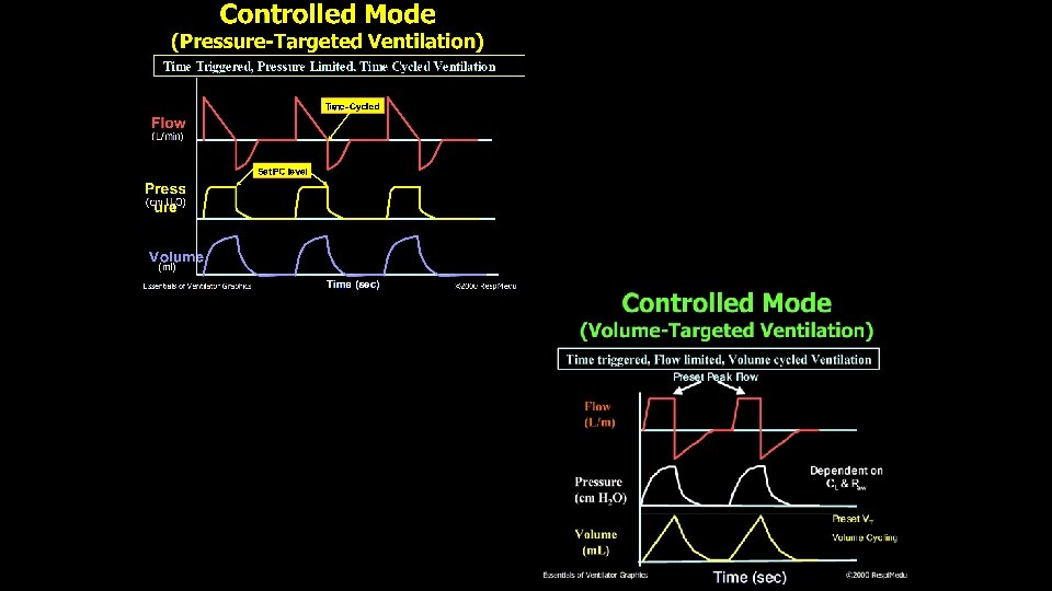
Volume vs. Pressure Control Volume Preset Pressure Preset • Set parameter is the tidal volume; airway pressure is variable • Set parameter is airway pressure; tidal volume delivered is variable • Constant tidal volume in the face of changing lung characteristics • Tidal volume varies with changes in lung characteristics • Patient-ventilator asynchrony due to fixed flow rate • Flow will vary according to patient's demands • No leak compensation • Compensates for leaks

CMV • All breaths are mandatory • Preset frequency, inspiratory time/Inspiratory flow • Pressure or volume control • Trigger: Time • Limit: Volume, flow or pressure • Cycle: Time, volume

Like CMV • Each additional assisted breath at prefixed tidal volume or pressure. Trigger: ventilator or patient *Limit: Flow / volume or Pressure *Cycling: volume or time Assist Control

SIMV • Machine triggered breaths with spontaneous breaths allowed in between • SIMV • Synchronised mandatory breaths with spontaneous breaths allowed in between • Ventilator creates a time window around the scheduled delivery of mandatory breath • If a patient effort is detected, it synchronises the machine breath with the patient’s inspiration • If no patient effort is detected, it delivers a breath at the scheduled time
 Pressure (cm H 2 O) Volume (ml) Time (sec) Spontaneous")
SIMV Mode Flow (L/min) Pressure (cm H 2 O) Volume (ml) Time (sec) Spontaneous Breath Courtesy: Prof J V Divatia

Pressure Support • Completely spontaneous mode in which patient triggers each breath • On inspiration patient exposed to a preset pressure • Inspiration is terminated when the flow rate reaches a minimum level or % of peak flow ØTrigger: Patient ØLimit: Pressure ØCycling: flow
 Pressure (cm H 2 O) Set PS level Rise Time Volume")
PSV Flow (L/m) Pressure (cm H 2 O) Set PS level Rise Time Volume (m. L) Time (sec) Courtesy: Prof J V Divatia

Initiating Mechanical Ventilation • Check ventilator assembly: power connection, circuit connection, HME filter catheter mount, gas connections • Switch on ventilator, check on test lung • Begin Preoxygenation (aerosol precaution) • Watch for Hypotension • Infuse Fluids • Start Mechanical Ventilation • A/C: TV=350 -450, RR=20 -30, FIO 2= 60 -100% • Peak Flow rate 40 -60 l/min • PEEP=5 -10, I: E=1: 2, Sens= -0. 8 - 2

Predicted Body Weight, TV and Minute Ventilation • The predicted body weight calculated as • Males: 50+0. 91(height cm -152. 4); • Female: 45. 5+0. 91( height cm -152. 4). • Calculate predicted body weight (PBW) • Males = 50 + 2. 3 [height (inches) - 60] • Females = 45. 5 + 2. 3 [height (inches) -60] TV= 6 ml/kg PBW MVm= 4 XBSA MVf= 3. 5 XBSA MV= 100 ml/kg of PBW

GOALS • OXYGENATION GOAL: Pa. O 255 -80 mm. Hg or Sp. O 2 88 -95% PLATEAU PRESSURE GOAL: ≤ 30 cm H 2 O p. H GOAL: 7. 30 -7. 45 Acidosis Management: (p. H < 7. 30) If p. H 7. 15 -7. 30: Increase RR until p. H > 7. 30 or Pa. CO 2 < 25 (Maximum set RR = 35). If p. H < 7. 15: Increase RR to 35. If p. H remains < 7. 15, TV may be increased in 1 ml/kg steps until p. H > 7. 15 (Pplat target of 30 may be exceeded). (Max TV =8, Min TV=4 ml/kg) • Alkalosis Management: (p. H > 7. 45) Decrease vent rate if possible. • • •

Other settings • I: E=1: 2, Vt: 500 ml, RR= 20/min • Inspiratory flow rate=? • Total cycle time (TCT)= 60 sec/RR=60 sec/20= 3 • Required I: E= 1: 2= hence 1+2=3, divide 3 seconds into 3 parts. • So Ti=1 sec, Te=2 sec. • So to deliver 500 ml in 1 seconds inspiratory flow should be 0. 5 L/sec. Which is equal to 30 litre/min.

Initiating Mechanical Ventilation • Confirm bilateral chest expansions • Start Sedation +/- Relaxants • Insert Central Line, Foleys Catheter & NG Tube • Check X Ray & ABG in 10 -20 Min. • Start Anti Gastritis and DVT Prophylaxis & Eye Care

Monitoring on MV • Heart rate • Sp. O 2 • Respiratory rate • Pattern of respiration and signs of respiratory distress • Blood pressure • ETCO 2 • Expired tidal volume: equal to set tidal volume (leaks) • Peak pressure, Plateau pressure (< 30 mm Hg) TREND IS YOUR FRIEND

Types of Waveforms • Scalars and Loops: • Scalars: Plot pressure, volume, or flow against time. Time is the x-axis • Loops: Plot pressure or flow against volume. (P/V or F/V). There is no time component.

Types of Scalars

Types of Loops P-V Loop F-V Loop

Ventilator alarms and troubleshooting Alarms Priority Causes Steps Electrical power/ gas delivery/ Battery Highest Disconnection of power supply or oxygen source Connect the power or Oxygen source Low airway pressure High Disconnection or volume leak, in appropriate alarm setting Connect ventilator, check alarm setting High airway pressure High Obstruction to flow, pneumothorax and other compliance problem (fluid overload/ abdominal hypertension) Check for Ppeak and Pplat (diff <3 -5) if both high: Compliance problem (pneumothorax etc) If Ppeak and Pplat (diff>3 -5) : Resistance problem (tube/circuit block etc): suction/change tube, bronchodilator Low Expired Vt High ETT cuff leak, Circuit leak, leak from Check/change ETT, Circuit, HME filter, leak from ICD filter, ICD High RR High PEEP Moderate Blocked tube/bronchoconstriction Suction/bonchodilation

Management of other problems • Hypoxemia: check for disconnection, Increase Fio 2, Call for help, check for tube block, fluid overload, check air entry, patient ventilator asynchrony DOPE: D: Disconnection/ Dislodging O: Obstruction, P: Pneumothorax E: Equipment failure There may exist multiple problems

Criteria for consideration for Weaning/discontinuation • Underlying disease stable or improving • Pa. O 2 / Fi. O 2 > 200 • PEEP < 5 -8 cm. H 2 O • Fi. O 2 < 0. 5 • Reliable respiratory drive • Stable CVS • Minimal pressors or inotropes • Absence of myocardial ischemia • Capable of initiating inspiratory effort

Underlying condition has Resolved or improved and there is no other condition mandating MV Daily screening of RS function Not Ready MV and Daily screening Weaning Ready SBT T-piece or PSV 30 min is enough Tolerated Not tolerated Gradual Withdrawal Esteban. AJRCCM 1997; 156: 45 Once-daily T-piece PSV Esteban. AJRCCM 1999; 159: 51 Ely. NEJM 1996; 335: 1864
 • B: Brain dysfunction")
Difficult weaning • A: Airway and lung dysfunction (resistance/compliance problem) • B: Brain dysfunction (delirium, depression) • C: Cardiac dysfunction (Cardiomyopathy) • D: Diaphragm/ respiratory muscle • E: Endocrine and metabolic (hypokalemia, hypomagenesimia, hypophosphatemia, malnutrition, hypothyroidism, met. Acidosis, fever) Heunks et al Critical Care; 2010

Complications of MV • Airway management related complications • Hypotension • Pneumothorax/Subcutaneous emphysema • Ventilator induced lung injury • Ventilator associated pneumonia

CARRY HOME MESSAGE • Co. Vi. D-19 patients usually present to ICU with ARDS. • For ventilating these patients Tidal volume (TV) should be calculated by 6 ml/kg PBW. (TVmax=8 ml/kg, TVmin=4 ml/kg) • Ventilating a patient with ARDS: Low TV, High RR, High PEEP and Plateau pressure < 30 cm H 2 O. • One should be aware of monitoring and troubleshooting of mechanical ventilator. • Patient should be initiated on Controlled or assist control mode of MV and once recovers can be weaned using spontaneous breathing trial. • In patients with difficult weaning ascertain and correct the cause.

THANK YOU
- Slides: 39