CNS Scintigraphy 1 Planar 2 D imaging 2
 imaging 2. SPECT (3 D) imaging 3. CSF")


 study ie cerebral perfusion mapping § Tc 99 m HMPAO is")

 is divided into")


- Slides: 16
CNS Scintigraphy 1. Planar (2 D) imaging 2. SPECT (3 D) imaging 3. CSF Flow studies
Radiopharmaceuticals: § A. Traces that do not cross intact BBB: § i. e localizes in the lesion. Tc 99 m DTPA, Tc 99 m Glucoheptonate, Tc 99 m DMSA, and free Tc 99 m § B. Tracers that cross intact BBB: i. e used mainly with SPECT to reveal regional cerebral blood flow both visually and quantitatively. § Tc 99 m HMPAO, Tc 99 m ECD and I 123 iodoamphetamine.
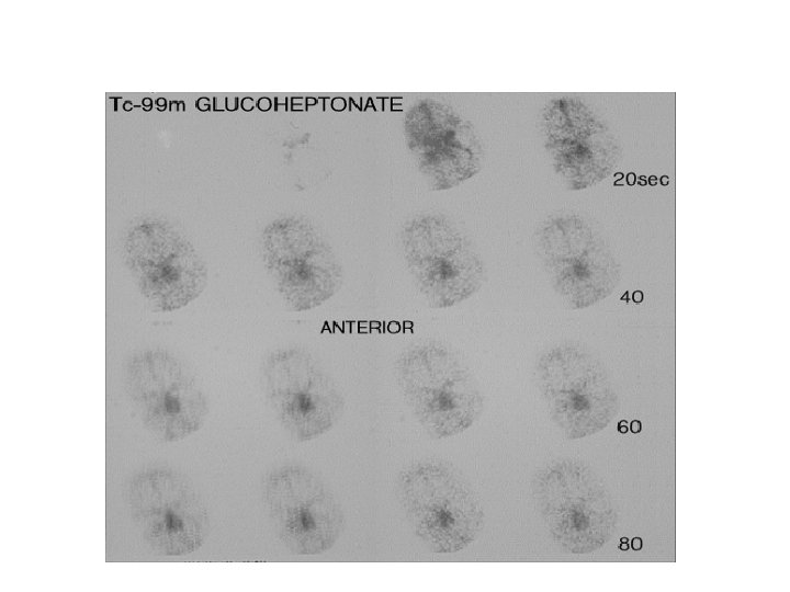
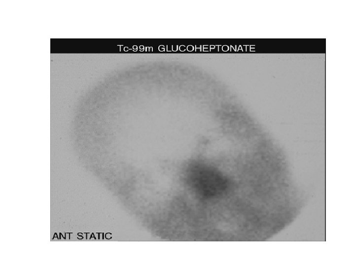
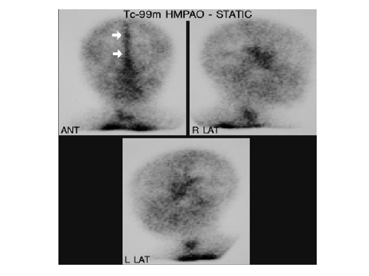
Technique of Planar brain study: § Patient preparation § Dose 15 -20 m. Ci of Tc 99 m DTPA or Glucoheptonate IVI. § Dynamic imaging: flow images at a rate of 1 image/3 sec for 60 sec. usually ant views are taken. § Immediate static images to evaluate blood pool abnormalities. § Delayed static images at 90 -120 min after clearance of background activity to detect breakdown of BBB. Views: ant, post, lateral, -+vertex images
: Normal planar scan § Dynamic images reveal visualization of both carotids, circle of Willis and later the venous sinuses. Time/activity curves are generated after drawing ROI to obtain global cerebral perfusion of either side of brain (amplitude, peak timing and time seq). No focal foci in brain. § Static images: the brain is outlined by a radioactive rim representing passively flowing tracers into scalp and skull. Lesions appear as foci of active hot tracer uptake. Venous sinuses are seen with clarity. The transverse and sigmoid sinuses represent the upper boundary of cerebellum.
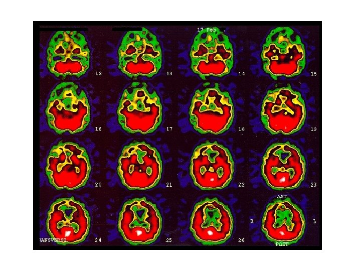
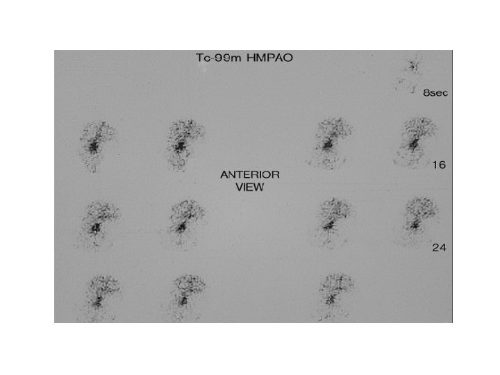
SPECT (3 D) study ie cerebral perfusion mapping § Tc 99 m HMPAO is a chemical micosphere that crosses BBB irreversibly to give a snapshot of brain activity over a short time, peak activity within 1 min after ivi. § Iodoamphetamine cross BBB and reversibly redistributes to become sequestered and slowly released from lungs ie represent integration of all brain activity from time of ini until the study is complete. It is cyclotron produced (expensive). § Tc 99 m ethyl cysteine dimer (ECD), adv. Can localize in areas of luxury perfusion, but more expensive than HMPAO. §
Technique of brain SPECT § Prior to exam: CP, previous CT or MRI, medications, supine , indirect lightening. § Dose: 15 -30 m. Ci. § To acess vasodilatory reserve, acetazoleamide (Dimox ) giver 20 min prior to injection of tracer. As it is a diuretic, patient is asked to void before the study CI: complicated migraine, sulpha allergy, within 3 d of acute stroke. § Images after 60 -90 min, dual head camera rotating 360 o around patient. Reconstructed trans-axial, coronal, and sagittal views are taken. §
Normal SPECT brain scan § The mid trans-axial scans (6 images) is divided into 3 parts: frontal, temoro-parietal and parieto-occipital on either side of cerebral hemisphere. +3 lower trans-axial cerebellar sections are compared as regards visual tracer intensity (color) and quantitatively by count intensity. § Cerebellar counts and tracer intensity is the reference region? Usually spared in most diseases. § Rt: Lt regional count should be within 1 -+5% , >5% is abnormal. § Coronal views are useful in temporal lobes lesions. § normally, gray and white matter are easily recognised, higher counts at BG, thalami, visual cortex, and cerebellum.
: Indications § 1. pre-surgical ictal identification of seizure foci. § 2. pre-surgical ballon dilatation. § 3. Psychiatric disorders eg. dementia. § 4. to document brain death.
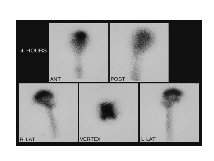
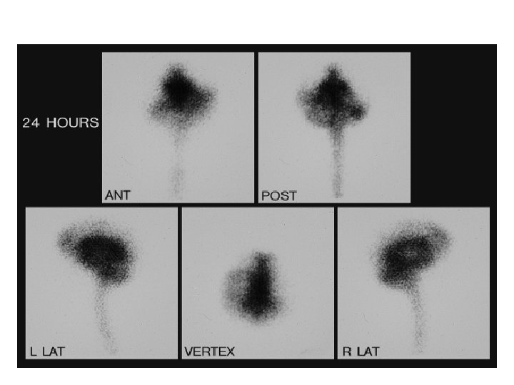
CNS fluid studies ((cisternogram § CSF is formed in choroid plexus as an ultrafiltrate of plasma. It flows from ventricles through foramina of ventricle and ascends over convexities of brain to be absorbed by the arachnoid villa. Technique: intrathecal injection of 0. 5 m. Ci of indium 111 -DTPA. Initial images are taken to ensure proper injection. The radiopharmaceutical ascend to basilar cisterns in 4 hr and flows over convexities in 24 hr. Normal scans: Images of basilar cisterns are obtained at 4 hr to 6 hr. At 24 hr images show ascent of tracer over convexities with activity in interhemispheric fissure with relative clearance of cisterns. Otherwise delayed images are obtained at 48 hr and 72 hr. CSF shunt and reservoir studies, injection is done via the reservoir, then dynamic images are obtained for 10 min in supine position. If tracer drains from the reservoir, the abd or chest images may be obtained. To detect CSF leakage ( rhinorrhea/ otorrhea ie intermittent leakage) films are obtained 1, 3 and 24 hr after administration, +-48 hr with lat and ant, respectively. Cotton pledgets may be placed in site of expected leak and radioactive count is done using well counter and compared with simultaneous serum essay that should not exceed 1. 5 times. used to diagnose normal pressure hydrocephalus. 4 th § § §