CNS Central Nervous System CNS Brain and Spinal

CNS Central Nervous System

CNS §Brain and Spinal Cord

CNS: PROTECTION §Turn to page 220 -221

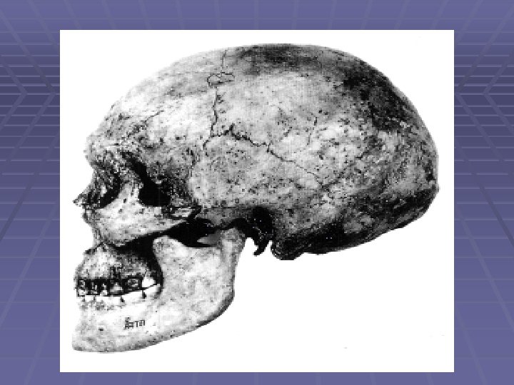
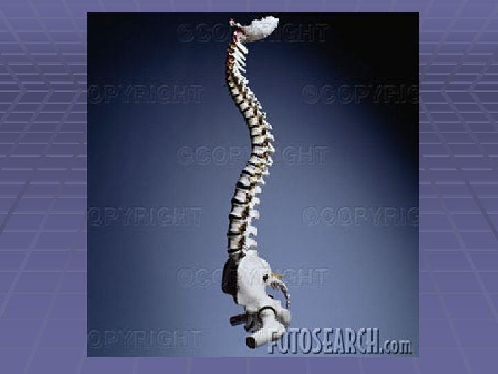
CNS: PROTECTION §BONE: §Cranium = brain §Vertebrate = spine

CNS: PROTECTION §Meninges: Fiborus tissue §Color code a, b, b 1, c
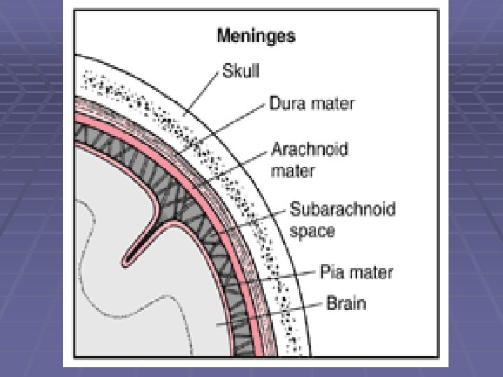

CNS: PROTECTION §Dura Mater: Toughest connective tissue §Right under cranium

CNS: PROTECTION §Arachnoid: Spider-web covering under dura mater.
")
CNS: PROTECTION §Subarachnoid: Pockets of the arachnoid that contain cerebral spinal fluid (CSF)

CNS: PROTECTION §Pia Mater: Delicate, contains many blood vessels.

CNS: PROTECTION §CSF: Circulates subarachnoid space §Cushions, and nourishes. §Surrounds entire CNS

Ohio State University: Neurology: College of Medicine. § CSF from the lumbar region contains 15 to 45 mg/dl protein (lower in childen) and 50 -80 mg/dl glucose (two-thirds of blood glucose). Protein concentration in cisternal and ventricular CSF is lower. Normal CSF contains 0 -5 mononuclear cells. The CSF pressure, measured at lumbar puncture (LP), is 100 -180 mm of H 2 O (8 -15 mm Hg) with the patient lying on the side and 200 -300 mm with the patient sitting up.

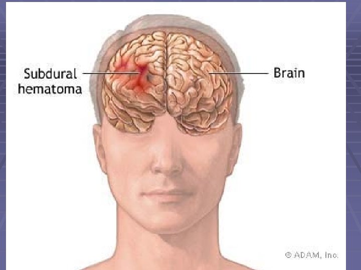
Problems § Increased protein: In bacterial meningitis, CSF protein may rise to 500 mg/dl. A more moderate increase (150 -200 mg/dl) occurs in inflammatory diseases of meninges (meningitis, encephalitis), intracranial tumors, subarachnoid hemorrhage, and cerebral infarction. A more severe increase occurs in the Guillain-Barré syndrome and acoustic and spinal schwannoma.
 of the CSF following subarachnoid hemorrhage is due to")
Problem § Xanthochromia (blonde color) of the CSF following subarachnoid hemorrhage is due to oxyhemoglobin which appears in 4 to 6 hours and bilirubin which appears in two days. Xanthochromia may also be seen with hemorrhagic infarcts, brain tumors, and jaundice.
")
§ § Normal Clear as water Abnormal findings Faint yellow, orange or pink (Xanthochromia) § CSF Protein >100 mg/dl § Red Blood Cell lysis § Red Blood Cell >100, 000/mm 3 (Subarachnoid Hemorrhage) § Cloudy or turbid § CSF Leukocytes > 200 wbc/mm 3 § Red Blood Cells > 400 per mm 3 § Brown or Dark CSF § Metastatic Melanoma (meningeal Melanomatosis) § Jaundice (Hyperbilirubinemia) § Green CSF § Hyperbilirubinemia § Purulent cerebrospinal fluid
 intervertebral discs, (2) vertebral bodies, (3) dura, (4) epidural space, (5) spinal")
§ (1) intervertebral discs, (2) vertebral bodies, (3) dura, (4) epidural space, (5) spinal cord, and (6) subdural space

BRAIN
 §Adult = 3")
BRAIN §Folded to increase surface area § 35 billion neurons (98%) §Adult = 3 lbs
 at rest needs as much oxygen as 61 lbs of")
Brain §Brain (3 lbs) at rest needs as much oxygen as 61 lbs of skeletal muscle.

Brain §Turn to page 224

BRAIN §Gyrus: Peaks of the folds, ridges. §Sulcus: furrow or groove between gyrus

Brain § Ventricles: CSF circulate in four major canals. (Travels through brain and into spine) Continuous. § Blue on page 228

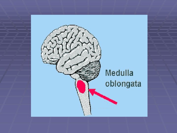
BRAINSTEM

BRAINSTEM §Lower brain §Unconscious part

BRAINSTEM § COLOR CODE: § Med. Oblongata = k § Pons = f § Midbrain = a § Reticular formation = g

MEDULLA OBLONGATA §Breathing §Heart rate §Reflex center

PONS §Connects cerebellum to cerebrum §Breathing

Midbrain §Like a hook §Diencephalon to cerebrum §Eye reflexes
 §Inactive so are")
Reticular formation §Fibers in the middle of brainstem (connects to RAS) §Inactive so are you! Consciousness.

Diencephalon

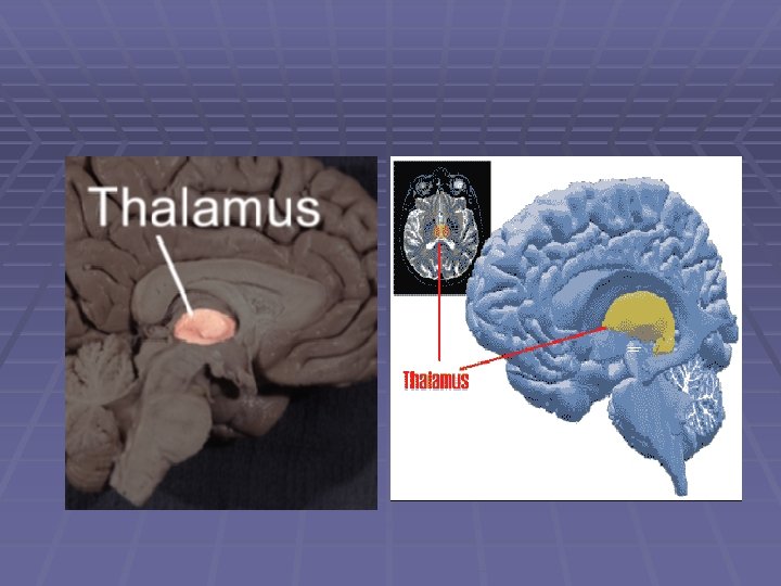
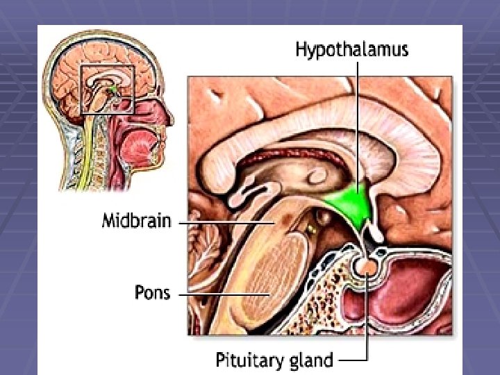
COLOR §Color code the Diencephalon to the right. §Thalamus = a §Hypothalamus = b

Diencephalon §On top of brainstem

Thalamus §Relay station for sensory headed to the cerebrum. §Filters out messages.

RAS §Reticular Activation System §Deals with arousal and consciousness.
 §Emotions: Rage, pleasure, pain, thirst, hunger")
HYPOTHALAMUS §Maintains homeostasis (temp) §Emotions: Rage, pleasure, pain, thirst, hunger

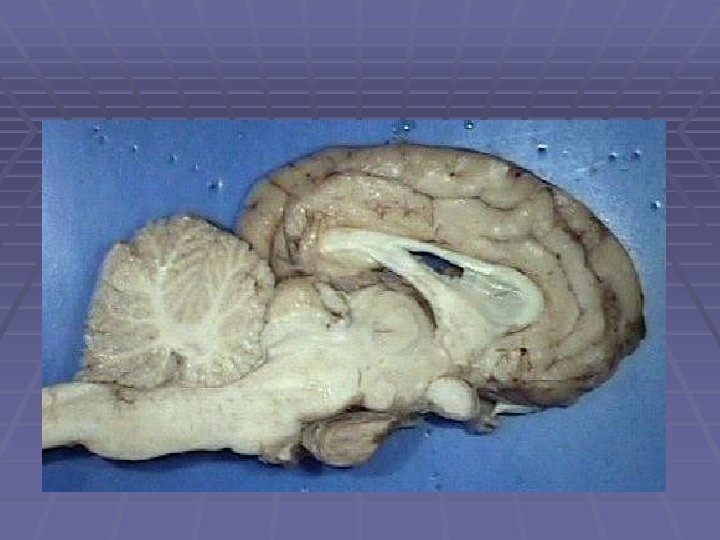
COLOR CODE §On middle picture page 10 §Cerebellum = h §Arbor Vitae = i
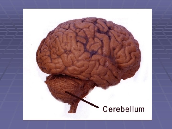

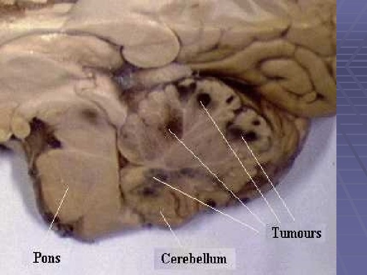
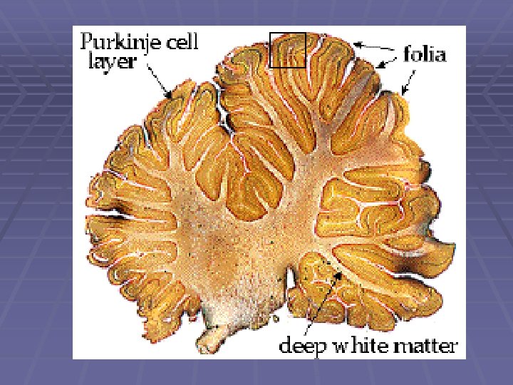
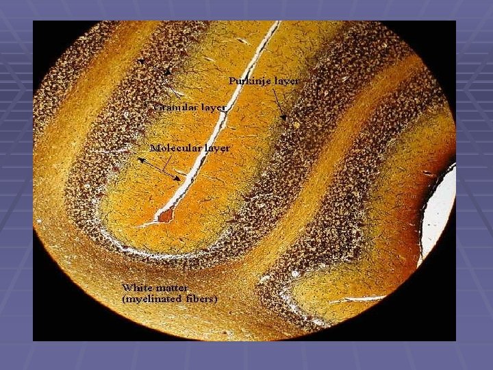
CEREBELLUM §Controls muscle balance and coordination. §Lower, posterior part of brain.

Arbor Vitae §White “tree-shaped” structure inside cerebellum.

Cerebrum §Color code §Frontal = a, a 1, a 2 §Parietal = b, b 1 §Temporal = c, c 1 §Occipital = d, d 1

Cerebrum §Outer layer, largest region, called cortex §About 1 inch thick § 4 lobes

Frontal lobe §Higher level thinking §Personality, speech center.

Parietal Lobe §Sensory for touch and pain

Temporal lobe §Sensory for hearing and smell
 Sensory for vision")
Occipital lobe §(back) Sensory for vision

§Brain time

Limbic system §Area around center of brain. §Not too much known about area.
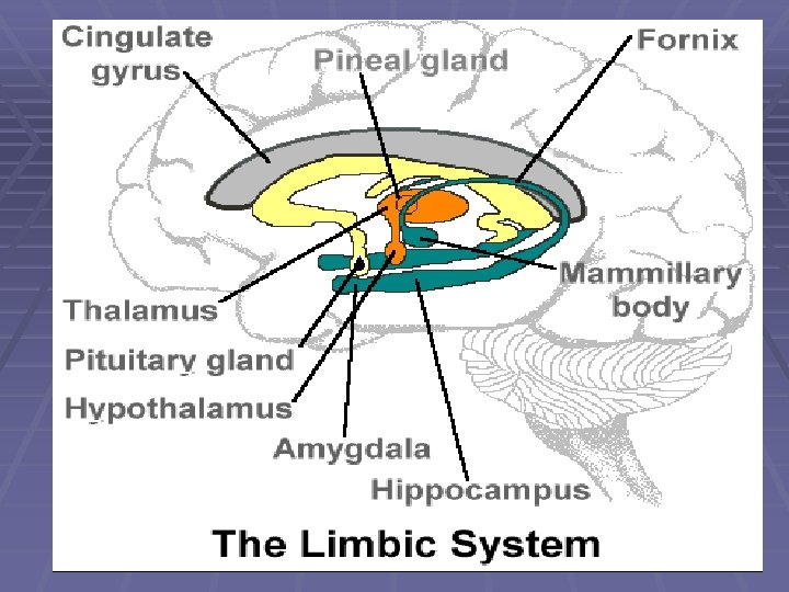

Limbic system §Emotion: Emotional states, fear, rage and sexual arousal.
 (learning)")
Limbic system §Memory and learning: §Long term memory and retrieval (hippocampus) (learning)

Lateralization §Brain divided laterally into 2 hemispheres. §Connected by corpus callosum

Lateralization §RIGHT music, art, creative §Left math, verbal
- Slides: 65