CLONING VECTORS Dr S Rubanraj Dept of Mathematics

CLONING VECTORS Dr. S. Rubanraj Dept of Mathematics St. Joseph’s College Trichy

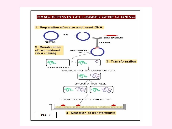
Vectors Vector is an agent that can carry a DNA fragment into a host cell in which it is capable of replication. If it is used only for reproducing the DNA fragment, it is called a cloning vector. If it is used for expression of foreign gene, it is called an expression vector. Vectors are ship for carrying the target DNA into a host cell. Cloning vector – used for obtaining millions of copies of cloned DNA segment. Used for creating genomic library or preparing the probes or genetic engineering experiments or other basic studies. Expression vector – allows expression of cloned gene, to give the product (protein). This can be achieved through the use of promoters and expression cassettes and regulatory genes. Used for transformation to generate trangenic plants, animals or microbes where cloned gene expresses to give the product.
 It should be autonomously replicating i. e. it")
Properties of a good vector: (1) It should be autonomously replicating i. e. it should have ori region. (2) It should contain at least one selectable marker e. g. gene for antibiotic resistance (tet. R for tetracycline resistance). (3) It should have unique restriction enzyme site (only one site for one RE) for different REs (preferably in one of the marker genes) to insert foreign DNA. (4) It should be preferably small in size for easy handling. (5) It should have relaxed control of replication so that multiple copies can be obtained. (6) It should contain specific control systems like promoters, terminators, ribosome binding sites etc so that the cloned DNA should express properly.

TYPES Vectors are of different types depending on the host. These are as follows: 1. Bacterial vectors 2. Yeast vectors 3. Plant vectors 4. Animal vectors

Bacterial vectors E. coli is the most commonly used bacterium for gene cloning though other bacteria such as Bacillus are also used. Vectors for cloning in these bacteria are described below: Vectors for cloning in E. coli A number of vectors are used for cloning in E. coli. Theses are categorized as plasmids, phages, cosmids, phagemids and bacterial artificial chromosomes.

Bacterial plasmid

PLASMIDS Plasmids are classified 1. by their ability to be transferred to other bacteria Conjugative The sexual transfer of plasmids to another bacterium through a pilus. those plasmids possess the 25 genes required for transfer. Non-conjugative plasmids don’t initiate conjugation. They can only be transferred with the help of conjugative plasmids. mobilisable An intermediate class of plasmids are mobilisable, and carry only a subset of the genes required for transfer. These plasmids can 'parasitise' another plasmid, transferring at high frequency in the presence of a conjugative plasmid
 plasmids, They are capable")
2. by function 1. 2. 3. 4. 5. 6. Fertility-(F) plasmids, They are capable of conjugation (they contains the genes for the pili). Resistance-(R) plasmids, contain gene (s) that can build resistance against one or several antibiotics or poisons. Col-plasmids, contain genes coding for colicines, proteins that can kill other bacteria. Degradative plasmids, able to digest unusual substances, e. g. , toluene or salicylic acid. Virulence plasmids, turn a bacterium into a pathogen. addiction system. These plasmids produce both a long-lived poison and a short-lived antidote. Daughter cells that retain a copy of the plasmid survive, while a daughter cell that fails to inherit the plasmid dies or suffers a reduced growth-rate because of the lingering poison from the parent cell.

Conjugative plasmids The sexual transfer of plasmids to another bacterium through a pilus. Those plasmids, F plasmids, possess the 25 genes required for transfer. Plasmid forms: 1. covalently closed circles –both strands of DNA intact 2. open circles-only one of the two strand is intact 3. Linear. 4. Supercoiled circles.

Plasmid vectors v. Plasmids are autonomously replicating circular, double stranded DNA molecules found in bacteria. v They have their own origin of replication (ori region), and can replicate independently of the host chromosome. v The size of plasmids ranges from a few kb to 200 kb. Plasmid vectors are often used for cloning DNA segments of small size (upto 10 kilobases). v. Single copy plasmid - maintained as single copy per cell v. Multicopy plasmid - maintained as 10 -20 copies per cell v. Plasmids under relaxed control of replication – over 1000 copies per cell – used as cloning vectors. v. In each bacterial cell about 20 -25 plasmids are maintained under normal growth condition. Some of the commonly used plasmid vectors are described below:

p. BR 322 v. The first plasmid vector that has been constructed artificially is p. BR 322. v It is named after the scientists Bolivar and Rodriguiz who constructed it in 1977. v It is 4362 bp in size and most widely used cloning vector. v It has an origin of replication derived from a colicinresistance plasmid (Col. E 1). v. This origin allows a fairly high copy number, about 100 copies of the plasmid per cell. v Plasmid p. BR 322 carries two selectable markers viz. genes for resistance to ampicillin (Apr) and tetracycline (Tcr ).
 unique RE sites are present within these genes for insertion")
§Several (over 40 enzymes) unique RE sites are present within these genes for insertion of foreign DNA (Fig 1). §Eco. RIV, Bam. HI, Sph. I, Sal. I, Xma. III, and Nru. I are present within the gene coding for tetracycline resistance, two sites (Hind. III and Cla. I) within the promoter of the tetracycline resistance gene and the three sites (Pst. I, Pvu. I and Sca. I) within the β lactamase gene that provide resistance to ampicillin §When a foreign DNA segment is inserted in any of these genes, the antibiotic resistance by that particular gene is lost. This is called insertional inactivation. §For instance, insertion of a restriction fragment in the Sal. I site of the Tcr gene inactivates that gene. §One can still select for Apr colonies, and then screen to see which ones have lost Tcr.

Figure p. BR 322 The map shows the positions of the ampicillin-resistance gene (amp R), the tetracycline-resistance gene (tet R), the origin of replication (ori) and the recognition sequences for seven restriction endonucleases.

The plasmid p. BR 322 is one of the most commonly used E. coli cloning vectors. p. BR 322 is 4361 bp in length and contains: (1) the replicon rep responsible for the replication of plasmid (source – plasmid p. MB 1); (2) rop gene coding for the Rop protein, which promotes conversion of the unstable RNA I – RNA II complex to a stable complex and serves to decrease copy number (source – plasmid p. MB 1); (3) bla gene, coding for beta-lactamase that confers resistance to ampicillin (source – transposon Tn 3); (4) tet gene, encoding tetracycline resistance protein (source – plasmid p. SC 101).

Figure 4. 18 Recombinant selection with p. BR 322
 have been")
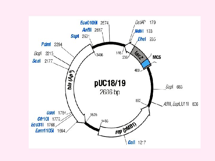
p. UC v. A series of small plasmids (about 2. 7 kb) have been developed (by Messings and co-workers in 1983) at the University of California and hence the name p. UC e. g. p. UC 7, 8, 9, 12, 13, 18 and 19 etc. v. These are high copy number plasmids that carry an ampicillin resistance gene and an origin of replication, both from p. BR 322. v They also have a multiple cloning site (MCS) – a sequence of DNA that carries unique sites for many REs. v. The MCS contains a portion of lac. Z gene that codes for the enzyme β-galactosidase. v. When such plasmids are introduced into E. coli, the colonies are blue on plates containing X-gal (5 -bromo-4 -chloro-3 -indolyl-β-dgalactopyranoside, the substrate for β- galactosidase) and IPTG (isopropyl thiogalactoside, an inducer).

v. Recombinants and non-recombinants can therefore be distinguished simply by plating the transformed cells onto agar containing ampicillin and X-gal. v. All colonies that grow on this medium are made up of transformed cells because only transformants are ampicillin resistant. v. Blue colonies contain cells with functional β-galactosidase enzymes and hence with undisrupted lac. Z′ genes these colonies are therefore non-recombinants. v. The white colonies comprise cells without β-galactosidase activity and hence with disrupted lac. Z′ genes; these are recombinants. v. Thus cells containing recombinant plasmids form white (not blue) colonies.

Recombinant selection with p. UC 8
 Ori 2) A dominant selectable Marker 3) Cleavage sites for cloning")
Must have: 1) Ori 2) A dominant selectable Marker 3) Cleavage sites for cloning 4) (high copy no. ) The plasmid cloning vector p. UC 19. This plasmid has an origin of replication (ori), an amp. R selectable marker, and a polylinker located within part of the galactosidase gene lac. Z+.

Phage vectors v. Bacteriophages or phages are viruses that specifically infect bacteria. v The phage particle attaches to the outer surface of bacterium and injects its DNA into the cell. v The phage DNA is then replicated inside the host and its genes are expressed to make phage capsid proteins and new phage particles are assembled and released from the bacterium. v. Phage vectors can accommodate more DNA (upto 25 kb) than plasmids and are often used for preparation of genomic libraries. v They also have higher transformation efficiency as compared to plasmids.

v. The main reason for seeking a different type of vector was the inability of plasmids such as p. BR 322 and p. UC 8 to handle DNA fragments greater than about 10 kb in size, larger inserts undergoing rearrangements or interfering with the plasmid replication system in such a way that the recombinant DNA molecules become lost from the host cells. v The first attempts to develop vectors able to handle larger fragments of DNA centered on bacteriophage λ. v. Two bacteriophages namely, Lambda (λ) and M 13 have been commonly used for construction of vectors for cloning in E. coli. v. The phage can have two modes of life cycles i. e. lytic and lysogenic. v. During lytic cycle, it replicates independently in the host cell and produces a large number of phage particles which are released by lysis of the host. Alternatively, it can take up lysogenic growth, meaning that it integrates its DNA into the bacterial chromosome and multiplies along with it.
 The")
Map of the λ chromosome of wild type. (N, cro, c. I genes) The genes are not essential for phage growth and can be deleted or replaced without seriously impairing the infectious growth cycle

The lysogenic infection cycle of bacteriophage λ The special feature of the lysogenic cycle is the insertion of the phage genome into the bacterium's chromosomal DNA, where it can remain quiescent for many generations.
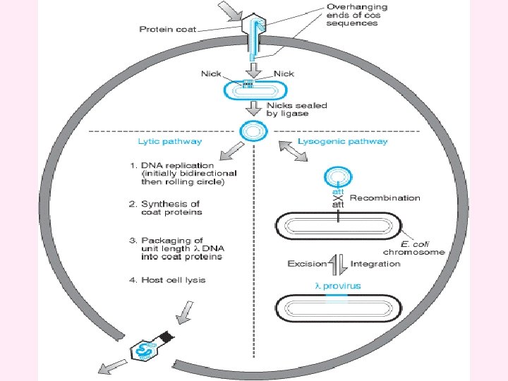

Three temporal stages of λ transcription occurs in the lytic cycle : vearly gene transcription establishes the lytic cycle (in competition with lysogeny) - early transcription proceeds from promoters PL and PR, stop at termination sites t. L and t. R 1 - transcription is subject to repression by the product of the c. I gene. vmiddle gene products replicate and recombine the DNA – It is directed by N gene product - PL and PR transcripts extend into genes such as red, O and P necessary for the middle stage. vlate gene products are packaged into mature phage particles - The cro product accumulate and prevents transcription from PL and PR. Q product is responsible for middle-to-late switch and the Q product specifically anti-terminates the short PR ´ transcript - many mature phage particles are produced.
 phage vectors v. Lambda is a temperate bacteriophage with a genome size")
Lambda (λ) phage vectors v. Lambda is a temperate bacteriophage with a genome size of 48. 5 kb. Its entire DNA sequence is known. v The lambda genome is a linear, double-stranded molecule with single-stranded, complementary ends. These ends can hybridize with each other (and do so when the DNA is within an infected cell) and are thus termed cohesive (cos) sites. v. The λ genome is 48. 5 kb, of which some 15 kb or so is ‘optional’ in that it contains genes that are only needed for integration of the phage DNA into the E. coli chromosome. These segments can therefore be deleted without impairing the ability of the phage to infect bacteria and direct synthesis of new λ particles by the lytic cycle.

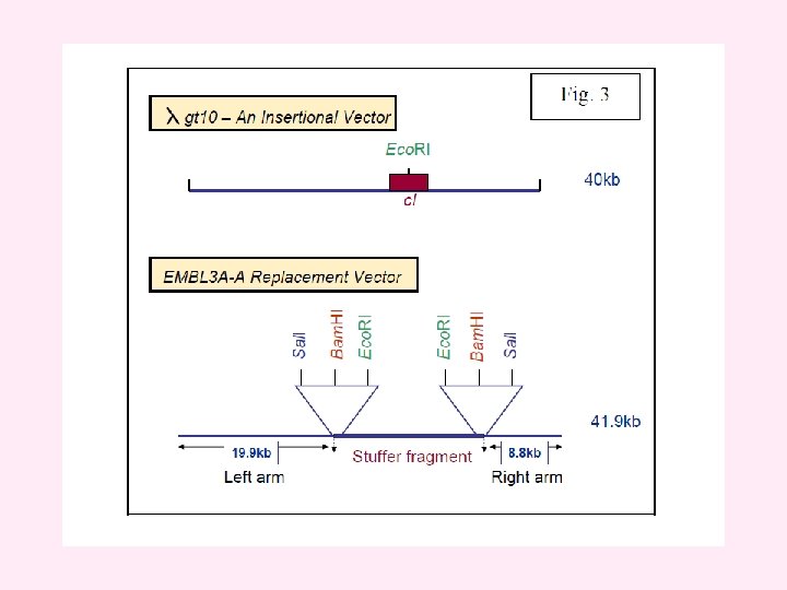
Two types of vector have been developed: v. Insertion vectors, in which part or all of the optional DNA has been removed and a unique restriction site introduced at some position within the trimmed down genome; v. Replacement vectors, in which the optional DNA is contained within a stuffer fragment, flanked by a pair of restriction sites, that is replaced when the DNA to be cloned is ligated into the vector. v. The λ genome is linear, but the two natural ends of the molecule have 12 -nucleotide single-stranded overhangs, called cos sites, which have complementary sequences and so can base-pair to one another.

v. Insertional vectors have one unique restriction site for a particular restriction enzyme and can accommodate 6 -7 kb DNA. v. Examples of insertional vectors are λgt 10, λgt 11 and λZAP II. v On the other hand, replacement vectors have two cleavage sites for a restriction enzyme and can accommodate up to 25 kb DNA. v. When vector is cut with a restriction endonuclease, a stuffer fragment is removed and replaced with a foreign DNA. v. Some examples of replacement vectors are EMBL 3, EMBL 3 A, EMBL 4, λDASH, λFIX, GEM 11 and GEM 12.
 In the λ genome, the genes are")
Cloning vectors based on bacteriophage λ (A) In the λ genome, the genes are arranged into functional groups. For example, the region marked as ‘protein coat’ comprises 21 genes coding for proteins that are either components of the phage capsid or are required for capsid assembly, and ‘cell lysis’ comprises four genes involved in lysis of the bacterium at the end of the lytic phase of the infection cycle. The regions of the genome that can be deleted without impairing the ability of the phage to follow the lytic cycle are indicated in green. (B) The differences between a λ insertion vector and a λ replacement vector.

v. Bacteriophage lambda can be reconstituted in a test tube by simply mixing phage DNA with a mixture of phage proteins, an infective viral particle with the DNA inside the phage head can be produced. This process is called in vitro packaging. v. There is a strict size requirement for the piece of DNA that goes into the phage head. That is, it should not be more than 52 kb and less than 38 kb. v. This feature allows only the recombinants to be packaged inside the phage head. v In addition, some lambda phage vectors have a stuffer fragment that carries the β-galactosidase gene. v. When it is removed or when foreign DNA is cloned within the gene, β-galactosidase activity may be abolished. The accompanying loss of activity may be used to select recombinant clones.

Cloning with a λ insertion vector The linear form of the vector is shown at the top of the diagram. Treatment with the appropriate restriction endonuclease produces the left and right arms, both of which have one blunt end and one end with the 12 nucleotide overhang of the cos site. The DNA to be cloned is blunt ended and so is inserted between the two arms during the ligation step. These arms also ligate to one another via their cos sites, forming a concatamer. Some parts of the concatamer comprise left arm-insert DNA-right arm and, assuming this combination is 37– 52 kb in length, will be enclosed inside the capsid by the in vitro packaging mix. Parts of the concatamer made up of left arm ligated directly to right arm, without new DNA, are too short to be packaged.

Bacteriophage infection is visualized as a plaque on a lawn of bacteria

DNA cloning with single stranded DNA vectors v. These coliphages are developed as cloning vectors v. This phage particles have dimensions 900 nm × 9 nm contain a single-stranded circular DNA molecule (6407 (M 13) or 6408 (fd) nucleotides long). vthey have many advantages over other vectors v. M 13, f 1 and fd are filamentous coliphages containing a circular single-stranded DNA molecule.

The biology of the filamentous coliphages v. The phages only infect enteric bacteria harbouring F pili vthe end of the F pilus is the adsorption site. vinfected cells continue to grow and divide, and extrude virus particles. v. Up to 100 phage particles may be released into the medium per cell per generation v. Replication of phage DNA does not result in host -cell lysis. v. The process of phage transfect and release – v 1. The single-stranded phage DNA enters the cell v 2. the single-stranded phage DNA is released.

The single-stranded phage DNA enters the cell §the virus discard the capsid proteins §the viral DNA passes into the cell §converted to a doublestranded replicative form. §the decapsidation and replication are tightly coupled. The RF multiplies rapidly inside the cell until about 100 RF molecules.

§a viral-encoded protein accumulated. §The protein binds to the viral strand prevents synthesis of the complementary strand. §Replication of the RF becomes asymmetric §only viral single strands are synthesized.

Why use single-stranded vectors? l. Sequencing by dideoxy method required singlestranded DNA loligonucleotide-directed mutagenesis required singlestranded DNA. lcertain probe preparation required single-stranded DNA. l. In summary filamentous phages possess all the advantages of plasmids and producing particles containing single-stranded DNA in an easily obtainable form.

M 13 Phage vectors v. M 13 is a filamentous bacteriophage of E. coli and contains a single stranded circular DNA of 7. 2 kb. v A series of vectors (M 13 mp series) have been developed from this phage. v. These vectors have a polylinker with unique restriction enzyme sites in lac Z gene that complements host (e. g. JM 103 or JM 104). v Screening of recombinants is done based on formation of blue/white plaques. v M 13 vectors are used for obtaining sufficient quantity of DNA for sequencing by Sanger's dideoxy chain termination method.
 Plasmid")
Cloning vectors and their insert capacities Vector system Host cell Insert capacity (kb) Plasmid E. coli 0. 1 -10 Bacteriophage l E. coli 10 -20 Cosmid E. coli 35 -45 Bacteriophage P 1 E. coli 80 -100 BAC (bacterial artificial E. coli chromosome) 50 -300 P 1 bacteriophagederived AC E. coli 100 -300 YAC Yeast 100 -2, 000 Human AC Cultured human cells >2, 000

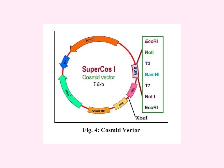
A typical cosmid p. JB 8 is 5. 4 kb in size and carries the ampicillin-resistance gene (amp R), a segment of λ DNA containing the cos site, and an Escherichia coli origin of replication (ori).

cosmid vector The cosmid vector is a combination of the plasmid and bacteriophage lambda. It is small (5 -7 kb) circular DNA containing an origin for DNA replication (ori), selectable markers and restriction sites from plasmid plus a sequence from lambda needed for packaging the DNA (cos site). Cosmids may be used to clone large DNA molecules of up to 45 kb. They also have high transformation efficiency. Some examples of cosmid vectors include p. JB, PWE and Super. Cos series.

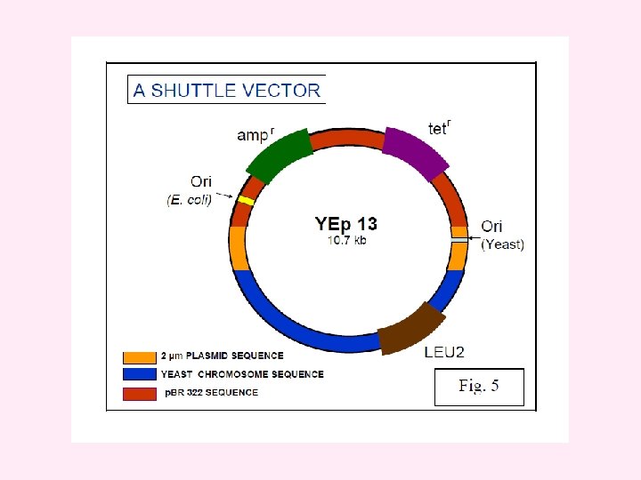
Vectors for cloning in yeast The discovery of a 2μm plasmid in most strains of Saccharomyces cerevisiae led to the development of cloning vectors in yeast. The 2μm plasmid is 6 kb in size. It is present in 50 -100 copies per cell. A number of shuttle vectors based on 2μm plasmid and bacterial plasmids have been constructed which can replicate either in E. coli or yeast. Yeast plasmid vectors are of four types, yeast episomal plasmids (YEps), yeast integrative plasmids (YIps) yeast replicative plasmids (YRps) and yeast centromeric plasmids (Ycps). In addition to plasmid vectors, yeast artificial chromosomes (YACs) are also used as vectors for cloning large pieces of DNA.
 Yeast episomal plasmids (YEps) These are derived from 2μm plasmid. Some YEps contain")
i) Yeast episomal plasmids (YEps) These are derived from 2μm plasmid. Some YEps contain the entire 2μm plasmid; others include just the 2μm origin of replication. An example of latter type is YEp 13. It is a shuttle vector and can be replicated both in E. coli and yeast. It contains 2μm origin of replication, yeast gene leu 2 as selectable marker and entire sequence of p. BR 322. The leu 2 gene codes for an enzyme involved in biosynthesis of amino acid leucine. YEps may replicate autonomously or integrate in one of the yeast chromosomes by homologous recombination. They have high transformation frequency of 10, 000 to 100, 000 transformants/ μg DNA.
 Yeast integrative plasmids (YIps) These are basically bacterial plasmids carrying a yeast gene.")
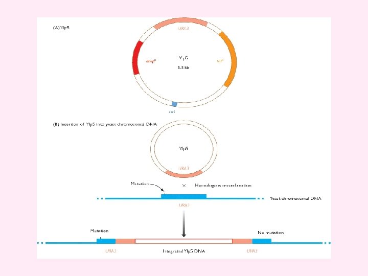
ii) Yeast integrative plasmids (YIps) These are basically bacterial plasmids carrying a yeast gene. YIp 5 is an example of yeast integrative plasmid. It has ura 3 gene inserted in p. BR 322. The gene ura 3 codes for an enzyme involved in biosynthesis of pyrimidine nucleotides and acts as selectable marker. The plasmid cannot replicate autonomously as it lacks 2μm origin of replication and survives by integrating in yeast chromosomal DNA. They have very low transformation frequency, less than 100 transformants/ μg DNA.
 Yeast replicative plasmids (YRps) They carry a part of chromosomal DNA with an")
iii) Yeast replicative plasmids (YRps) They carry a part of chromosomal DNA with an origin of replication and one or two selectable markers and are capable of independent replication. They have transformation frequency between 1000 and 10, 000 transformants/ μg DNA. iv) Yeast centromeric plasmids (YCps) These are shuttle vectors that behave as small chromosomes and replicate only once during each cell divison. They contain i) origin of replication called ARS sequence , ii) CEN sequence (for proper segregation of chromosomes) and iii) a selectable marker such as leu 2 from yeast and sequences from bacterial plasmid having ori region and selectable marker (Apr). They are stably maintained at one copy per cell.
 Yeast Artificial Chromosomes (YACs) YACs are artificial chromosomes that replicate in yeast cells.")
v) Yeast Artificial Chromosomes (YACs) YACs are artificial chromosomes that replicate in yeast cells. Main features of these vectors are: 1. Autonomously replicating sequence (ARS) necessary for the replication in yeast cells (Fig 6). 2. Telomeres (TEL), which are ends of chromosomes involved in the replication and stability of chromosomes. 3. A yeast centromere (CEN), required for proper segregation of chromosomes 4. Selectable markers that allow the easy isolation of yeast cells that have taken up the artificial chromosome. 5. Unique RE sites. YACs are capable of carrying a large DNA fragment (up to 3000 kb), but their transformation efficiency is very low.
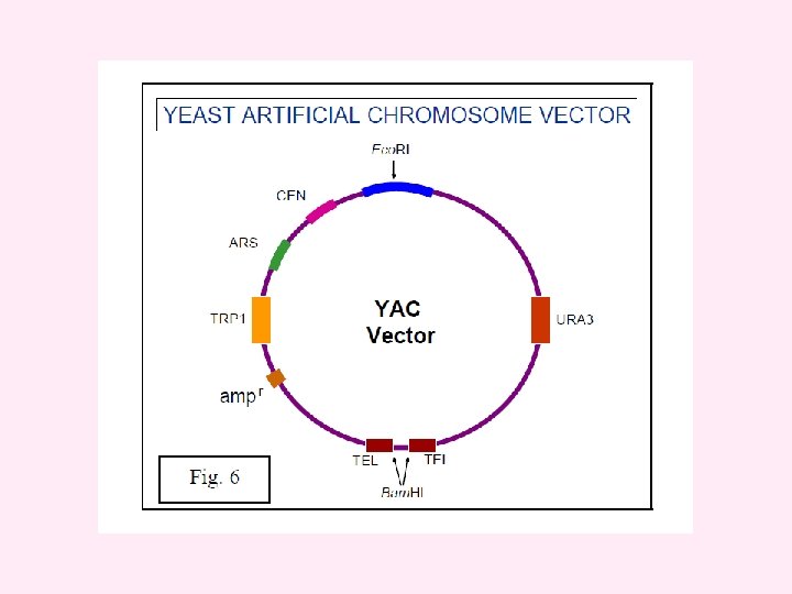

Table 4. 4 Sizes of human genomic libraries prepared in different types of cloning vector * Calculated from the equation: where N is the number of clones required, P is the probability that any given segment of the genome is present in the library, a is the average size of the DNA fragments inserted into the vector, and b is the size of the genome. Number of clones* Type of vector Insert size (kb) P = 95% P = 99% λ replacement 18 532 500 820 000 Cosmid, fosmid 40 240 000 370 000 P 1 125 77 000 118 000 BAC, PAC 300 32 000 50 000 YAC 600 16 000 24 500 Mega-YAC 1400 6850 10 500
 The cloning vector p. YAC 3.")
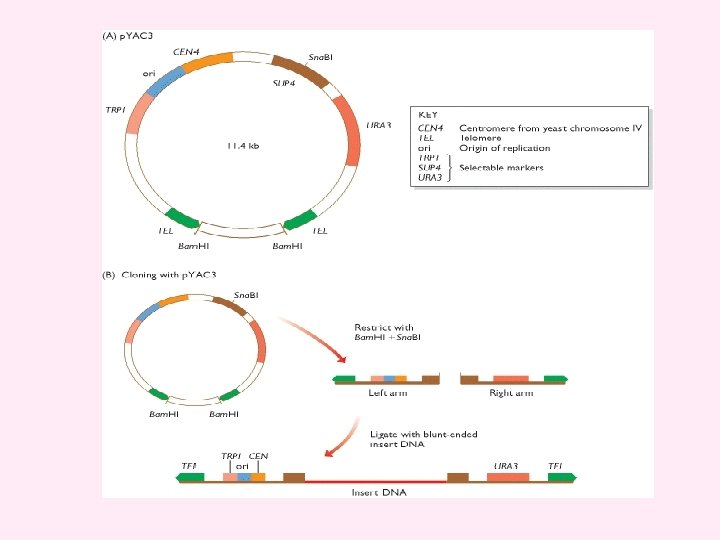
Figure 4. 25 Working with a YAC (A) The cloning vector p. YAC 3. (B) To clone with p. YAC 3, the circular vector is digested with Bam. HI and Sna. BI. Bam. HI restriction removes the stuffer fragment held between the two telomeres in the circular molecule. Sna. BI cuts within the SUP 4 gene and provides the site into which new DNA will be inserted. Ligation of the two vector arms with new DNA produces the structure shown at the bottom. This structure carries functional copies of the TRP 1 and URA 3 selectable markers. The host strain has inactivated copies of these genes, which means that it requires tryptophan and uracil as nutrients. After transformation, cells are plated onto a minimal medium, lacking tryptophan and uracil. Only cells that contain the vector, and so can synthesize tryptophan and uracil, are able to survive on this medium and produce colonies. Note that if a vector comprises two right arms, or two left arms, then it will not give rise to colonies because the transformed cells will still require one of the nutrients. The presence of insert DNA in the cloned vector molecules is checked by testing for inactivation of SUP 4. This is done by a color test: on the appropriate medium, colonies containing recombinant vectors (i. e. with an insert) are white; non-recombinants (vector but no insert) are red.
 YIp 5, a typical yeast integrative")
Figure 4. 26 Cloning with a YIp (A) YIp 5, a typical yeast integrative plasmid. The plasmid contains the ampicillin-resistance gene (amp R), the tetracycline-resistance gene (tet R), the yeast gene URA 3, and an Escherichia coli origin of replication (ori). The presence of the E. coli ori means that recombinant YIp 5 molecules can be constructed in E. coli before their transfer into yeast cells. YIp 5 is therefore a shuttle vector - it can be shuttled between two species. (B) YIp 5 has no origin of replication that can function inside yeast cells, but can survive if it integrates into the yeast chromosomal DNA by homologous recombination between the plasmid and chromosomal copies of the URA 3 gene. The chromosomal gene carries a small mutation which means that it is non-functional and the host cells are ura 3. One of the pair of URA 3 genes that are formed after integration of the plasmid DNA is mutated, but the other is not. Recombinant cells are therefore ura + and can be selected by plating on to minimal medium, which does not contain uracil.

Figure 4. 27 The plant cloning vector p. BIN 19 carries the lac. Z′ gene (see Figure 4. 19), the kanamycin-resistance gene (kan R), an Escherichia coli origin of replication (ori), and the two boundary sequences from the T-DNA region of the Ti plasmid. These two boundary sequences recombine with plant chromosomal DNA, inserting the segment of DNA between them into the plant DNA. The orientation of the boundary sequences in p. BIN 19 means that the lac. Z′ and kan R genes, as well as any new DNA ligated into the restriction sites within lac. Z′, are transferred to the plant DNA. Recombinant plant cells are selected by plating onto kanamycin agar, and then regenerated into whole plants. Note that p. BIN 19 is another example of a shuttle vector, recombinant molecules being constructed in E. coli, using the lac. Z′ selection system, before transfer to Agrobacterium tumefaciens and thence to the plant. For more details, see Brown (2001). From: Chapter 4, Studying DNA
 DNA cloning, and")
1. Draw diagrams that outline the events that occur during (a) DNA cloning, and (b) PCR. What are the limitations of each of these two techniques? 3. 4. 5. 6. 7. 8. 9. 10. 11. 12. 13. 2. List the types of enzyme used in recombinant DNA research. Distinguish between the two types of exonuclease activity that can be possessed by a DNA polymerase, and explain how these activities influence the potential applications of individual DNA polymerases in recombinant DNA research. Using examples, describe the various types of end produced after digestion of DNA with a restriction endonuclease. How are agarose gel electrophoresis and Southern hybridization used to examine the results of a restriction digest? Explain why the efficiency of blunt-end ligation is less than that of sticky-end ligation. What steps can be taken to improve the efficiency of blunt-end ligation? Draw diagrams of (a) p. BR 322, and (b) p. UC 8. Explain how the differences between these two vectors influence the ways in which they are used to clone DNA fragments. Distinguish between the lytic and lysogenic infection cycles for a bacteriophage. Write a short description of the way in which a bacteriophage λ vector is used to clone DNA. How does a cosmid differ from a standard λ vector? Draw a diagram showing a typical YAC. Indicate the key features and explain how a YAC is used to clone DNA. What problems might arise when a YAC is used to clone a large fragment of DNA? To what extent can these problems be solved by the use of other types of high-capacity cloning vector? How is DNA cloned in organisms other than Escherichia coli? Describe how a PCR is carried out, paying particular attention to the role of the primers and the temperatures used during thermal cycling.
 • Single stranded RNA genome •")
Retroviruses (including Lentivirus, HIV and MMLV based vectors) • Single stranded RNA genome • Lipid membrane enveloped • Host range determined by envelope proteins Retrovirus ss. RNA Genome Reverse Transcription into ds. DNA Random integration into host genome Host DNA Host Cell
: Necessary for integration")
The Retroviral Genome LTR gag pol env Long Terminal Repeat (LTR): Necessary for integration into host genome (Psi): packaging signal gag: Packages viral genome into viral particles pol: viral polymerase necessary for viral replication env: viral envelope proteins, necessary for entry into host cells, dictate host range
 The viral vector is “gutted”")
Design of Replication Incompetent Lentiviral Vectors (3 rd Generation) The viral vector is “gutted” as much as possible to create room for the insert gene and to divide the viral genome into cis- and trans- acting regions Promoter and Insert Gene LTR∆ gag Transfer Vector Regulatory Signals (LTR and ), promoter and Insert Gene pol Packaging Vector Structural & Packaging Genes (may already be present in packaging line) env LTR ∆ Modified LTR To impede the Virus from Performing more Than one round Of reverse Transcription Envelope Vector Viral envelope protein alters host range of the viral vector “self inactivating”

Principles of Retroviral Vector Design It is possible to make replication-competent retroviral vectors by adding sequences to existing viruses, but a more common design involves the replacement of retroviral sequences to create replication-defective vectors. In addition, the amount of foreign DNA that can be accommodated in replicationcompetent vectors is much smaller than can be accommodated in replication-defective vectors. Expression of retroviral proteins in most of the naturally occurring oncogenic retroviruses is driven by a single promoter in the 5′long terminal repeat (LTR), and the expression of multiple viral coding regions is achieved by alternative splicing. However, vector design is not limited to the use of the single retroviral promoter with alternative splicing. Other strategies include the use of multiple promoters, insertion of genes in the reverse orientation, and the use of internal ribosome entry sites (IRESs).

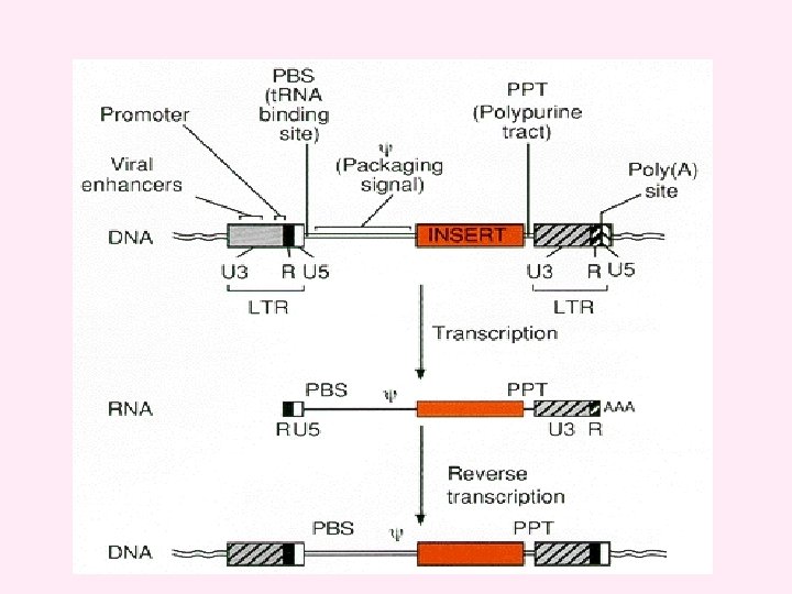
v. Efficient gene transduction and integration depend on the inclusion in the retroviral vector of a number of cis-acting viral elements. These include v (1) a promoter and polyadenylation signal in the viral genome; v (2) a viral packaging signal (ψ or E) to direct incorporation of vector RNA into virions; v(3) signals required for reverse transcription, including a transfer RNAbinding site (PBS) and polypurine tract (PPT) for initiation of first- and second-strand DNA synthesis, and a repeated (R) region at both ends of the viral RNA required for transfer of DNA synthesis between templates; v (4) short, partially inverted repeats located at the termini of the viral LTRs required for integration. v. An important general consideration in the design of retroviral vectors is the effect of viral replication on vector structure. After one round of viral replication, the U 3 regions in both LTRs are derived from the U 3 region originally present in the 3′LTR in the plasmid form of the vector, and both U 5 regions are derived from the U 5 region originally present in the 5′LTR in the plasmid. v Ordinarily, R sequences should arise primarily from the 5′plasmid LTR, but they may also include 3′plasmid LTR sequences.

Packaging Recombinant Lentiviral Particles The three plasmids containing the viral genome components are transfected into the packaging line to create the infectious viral particles. Multiple plasmids are used so multiple recombination events would be required to reconstitute a replication competent virus. www. sigma. com/RNAI

Viral Psuedotyping: A Double Edged Sword Tropism: The ability of a virus to infect a particular type of host cell Psuedotyping: Altering the viral envelope protein to alter tropism, thus allowing the virus to infect cells it originally could not Tropism Host Range Viral Envelope Protein Receptor for Viral Envelope Ecotropic Mouse / Rat Gap 70 m. CAT-1 Amphotropic / Dualtropic Mammals 4070 A / 10 A 1 Ram-1 / GALV Pantropic All Animals VSV-G Phosphotidyl serine Phosphotidyl inositol GM 3 ganglioside Special care should be used when working with pantropic or amphotropic viruses which can infect humans!

Replication Deficient Viral Vectors: Genetically Engineered So The Viral Infection Cannot Spread • The viral DNA does not contain the viral genes needed to make more viruses. Viral DNA Gene of Interest Target Cell Virus Target Cell Infected With Viral DNA Containing The Gene of Interest Cell’s DNA No New Viral Particles are Created Infection dose not spread

Rescue of Replication Deficient Viruses by superinfection with Wild Viruses Viral DNA Gene of Interest Target Cell Virus Wild Virus Cell’s DNA Complementation: The genome from the wild virus provides the missing proteins needed for the viral vector to replicate. The superinfected cell functions similarly to a packaging line.

Rescue of Replication Deficient Viruses by superinfection with Wild Viruses Viral DNA Gene of Interest Target Cell Virus Wild Virus Cell’s DNA Recombination: The genome from the wild virus randomly recombines with the viral vector, providing sufficient genetic material for the viral vector to replicate. The resulting rescued virus may possess pieces of the original insert gene. The viral genome is impossible to predict due to random recombination. The virus may exhibit altered virulence.

Risks Associated with Retroviruses: Insertional Mutagenesis Viral DNA Gene of Interest Virus Target Cell Host Cell DNA Oncogene Proto-Oncogene Random integration of viral genome may disrupt endogenous host genes. Of special concern Is disruption of proto-oncogenes, which can lead to increased cancer risk.

Risks Associated with Retroviral Vectors: Viral Transduction Viral DNA Gene of Interest Target Cell Virus Target Cell Infected With Viral DNA Containing The Gene of Interest Cell’s DNA Individuals infected with the viral vector may express the insert gene at the site of infection.
- Slides: 72