
Clinical Use of Cardiac Biomarkers Name DVM DACVIMCardiology
Clinical Use of Cardiac Biomarkers **Name**, DVM, DACVIM-Cardiology **Date**
Cardiac Biomarkers: Outline • Cardiac Biomarkers – Cardiac Troponins (c. Tn. I) – Natriuretic Peptides (BNP and ANP) • Physiology • Assays • Clinical Uses
What are biomarkers? • Characteristic objectively measured and evaluated as an indicator of normal biologic processes, pathogenic processes, or pharmacologic responses to a therapeutic intervention.
Common Cardiac Biomarkers • • • Blood Sample Measurements Blood Pressure ECG Radiographs Echocardiogram
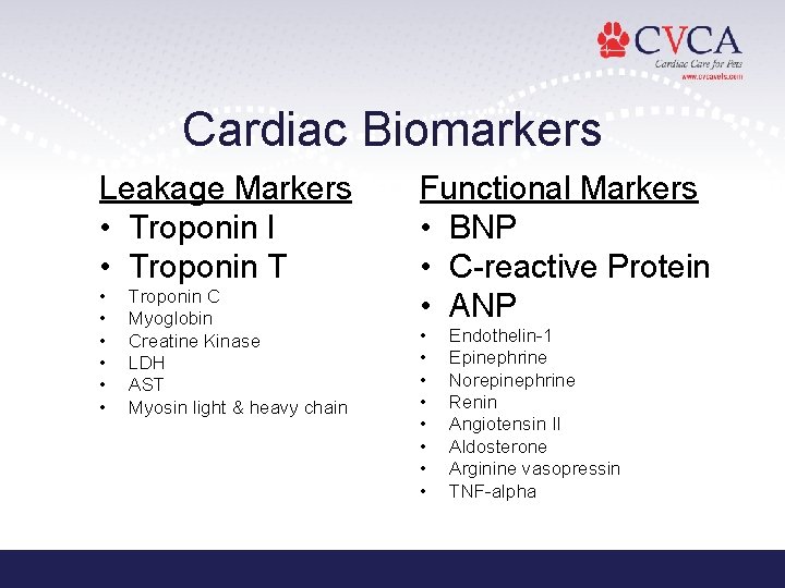
Cardiac Biomarkers Leakage Markers • Troponin I • Troponin T • • • Troponin C Myoglobin Creatine Kinase LDH AST Myosin light & heavy chain Functional Markers • BNP • C-reactive Protein • ANP • • Endothelin-1 Epinephrine Norepinephrine Renin Angiotensin II Aldosterone Arginine vasopressin TNF-alpha
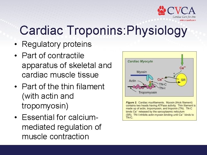
Cardiac Troponins: Physiology • Regulatory proteins • Part of contractile apparatus of skeletal and cardiac muscle tissue • Part of the thin filament (with actin and tropomyosin) • Essential for calciummediated regulation of muscle contraction
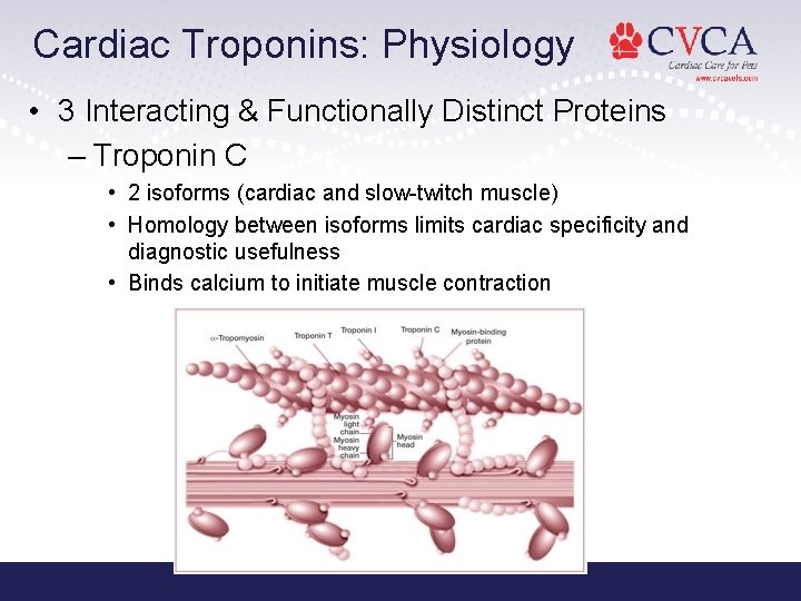
Cardiac Troponins: Physiology • 3 Interacting & Functionally Distinct Proteins – Troponin C • 2 isoforms (cardiac and slow-twitch muscle) • Homology between isoforms limits cardiac specificity and diagnostic usefulness • Binds calcium to initiate muscle contraction
Cardiac Troponins: Physiology • Troponin T – Multiple isoforms in skeletal tissue – 4 isoforms in human cardiac tissue (1 adult, 3 fetal tissue) – Fetal isoforms may be re-expressed during heart failure or in damaged skeletal muscle – Attaches the troponin complex to tropomyosin and actin • Troponin I – 3 isoforms (2 skeletal, 1 cardiac) – Not expressed in fetal tissue or after damage/regeneration in adult tissue – Inhibits actomyosin ATPase, preventing the structural interaction of myosin with actin-binding sites • Calcium binds c. Tn. C c. Tn. I displaced tropomyosin conformational change (no longer interferes with myosin/actin) muscle contraction occurs
Cardiac Troponins: Physiology • Proteins exists in 2 populations within the cells – Structurally bound within filaments of contractile apparatus – Free in cytosol (2 -4% c. Tn. I, 6 -8% c. Tn. T) • Leakage Markers – Damage to cardiac myocytes results in loss of membrane integrity release of c. Tn
Cardiac Troponins: Physiology • Release Kinetics – Acute cardiac injury release of cytosolic pool early rise blood levels slow release of bound protein sustained blood elevations – T 1/2 in circulation = 2 hours • Humans with acute MI: levels rise 4 -12 hr after infarction, peak 12 -48 hr; remain elevated 7 -10 d (c. Tn. I) and 10 -14 d (c. Tn. T) • Dog MI model: peak 10 -16 hr (more rapid development of necrosis? ) • Mechanism of elimination unknown
Cardiac Troponin I • Unique to heart – Healthy animals may have low, but detectable levels • Time to initial elevation = 3 -8 hours • Time to peak elevation = 12 -24 hours • Time to return to baseline = 5 -10 days
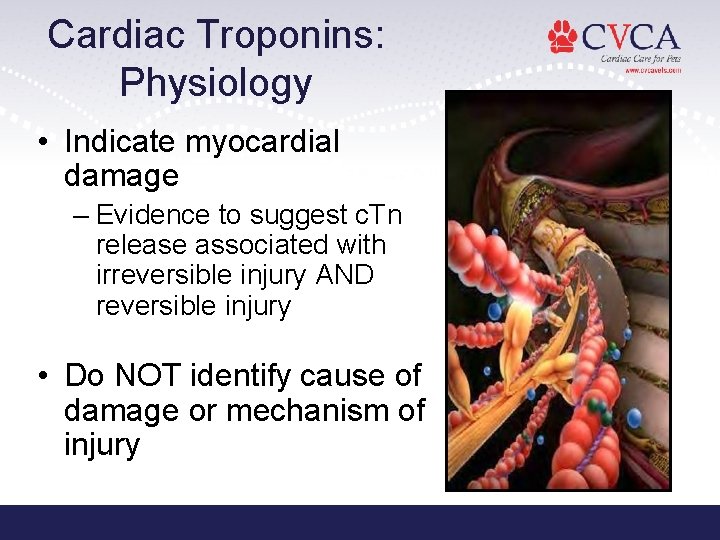
Cardiac Troponins: Physiology • Indicate myocardial damage – Evidence to suggest c. Tn release associated with irreversible injury AND reversible injury • Do NOT identify cause of damage or mechanism of injury
Cardiac Troponins: Assays • Levels determined with ELISA – Using antibodies specific for c. Tn I and T • c. Tn. I – No gold standard assay – Can be modified or complexed to other proteins (ex. c. Tn. C) – Assay antibodies may have differing specificity for each circulating form – Can not compare results from different assays • False Positives – Rheumatoid factor, excess fibrin, hemolysis, lipemia, ALP, etc. • c. Tn. I more sensitive than c. Tn. T
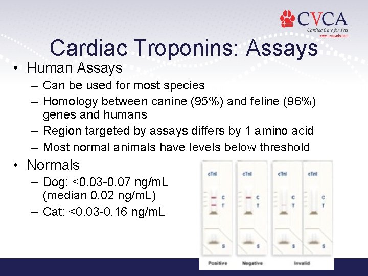
Cardiac Troponins: Assays • Human Assays – Can be used for most species – Homology between canine (95%) and feline (96%) genes and humans – Region targeted by assays differs by 1 amino acid – Most normal animals have levels below threshold • Normals – Dog: <0. 03 -0. 07 ng/m. L (median 0. 02 ng/m. L) – Cat: <0. 03 -0. 16 ng/m. L
Cardiac Troponins: Prognosis in Humans • Magnitude of troponin level elevation correlates with risk of future cardiac events or death • Aids in indentification of patients with greater disease severity who may benefit from more aggressive therapy • Size of infarcted area may be predicted based on c. Tn. I or c. Tn. T levels at 72 hr
Cardiac Troponins: Causes of Elevation • Breed – Greyhound, Boxer • Direct Trauma – Secondary to mechanical injury – Ex. cardiac sx, catheterization procedures, myocardial contusions from blunt trauma, electrical injuries (cardioversion, defibrillation)
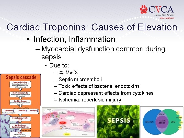
Cardiac Troponins: Causes of Elevation • Infection, Inflammation – Myocardial dysfunction common during sepsis • Due to: – – – Mv. O 2 Septic microemboli Toxic effects of bacterial endotoxins Cardiac depressant effects from cytokines Ischemia, reperfusion injury
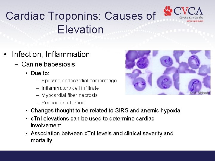
Cardiac Troponins: Causes of Elevation • Infection, Inflammation – Canine babesiosis • Due to: – – Epi- and endocardial hemorrhage Inflammatory cell infiltrate Myocardial fiber necrosis Pericardial effusion • Changes thought to be related to SIRS and anemic hypoxia • c. Tn. I elevations can be used to determine cardiac involvement • Association between c. Tn. I levels and clinical severity and mortality
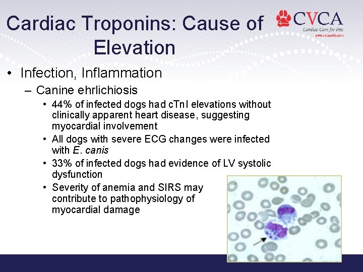
Cardiac Troponins: Cause of Elevation • Infection, Inflammation – Canine ehrlichiosis • 44% of infected dogs had c. Tn. I elevations without clinically apparent heart disease, suggesting myocardial involvement • All dogs with severe ECG changes were infected with E. canis • 33% of infected dogs had evidence of LV systolic dysfunction • Severity of anemia and SIRS may contribute to pathophysiology of myocardial damage
Cardiac Troponins: Causes of Elevation • Primary Heart Disease – HCM • Due to: – Intramural coronary artery disease areas of ischemia – Myocardial hypertrophy with cellular necrosis • Levels with moderate-severe disease • Se 85%/Sp 97% for differentiating HCM from normal • HCM + CHF had significantly high levels – Use to differentiate cardiac vs. non-cardiac cause of dyspnea in cats? – Mitral Valve Disease – Subaortic Stenosis – 3 rd Degree AVB • Due to: – Fibrosis or inflammation – Hypoxic damage (secondary to perfusion)
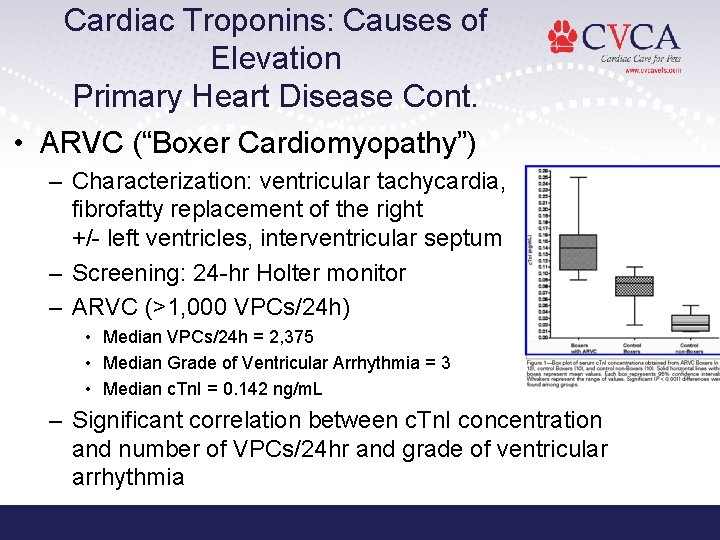
Cardiac Troponins: Causes of Elevation Primary Heart Disease Cont. • ARVC (“Boxer Cardiomyopathy”) – Characterization: ventricular tachycardia, fibrofatty replacement of the right +/- left ventricles, interventricular septum – Screening: 24 -hr Holter monitor – ARVC (>1, 000 VPCs/24 h) • Median VPCs/24 h = 2, 375 • Median Grade of Ventricular Arrhythmia = 3 • Median c. Tn. I = 0. 142 ng/m. L – Significant correlation between c. Tn. I concentration and number of VPCs/24 hr and grade of ventricular arrhythmia
Cardiac Troponins: Causes of Elevation • Heart Failure – Elevated c. Tn. I and c. Tn. T due to LV dysfunction – Levels often return to normal after successful treatment of acute episode – Class IV CHF due to MVD • 6/15 (40%) had detectable c. Tn. I (median 0. 24 ng/m. L) • 1/15 (7%) had detectable c. Tn. T (0. 02 ng/m. L) • No statistical difference in survival to discharge between dogs with detectable and non-detectable c. Tn levels • Dogs with detectable c. Tn levels had overall shorter survival times (MST 67. 5 d, compared to 390 d)
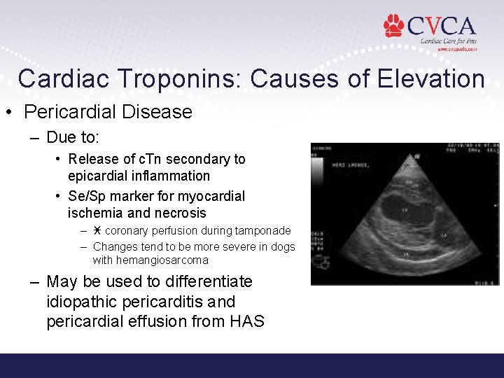
Cardiac Troponins: Causes of Elevation • Pericardial Disease – Due to: • Release of c. Tn secondary to epicardial inflammation • Se/Sp marker for myocardial ischemia and necrosis – coronary perfusion during tamponade – Changes tend to be more severe in dogs with hemangiosarcoma – May be used to differentiate idiopathic pericarditis and pericardial effusion from HAS
Cardiac Troponins: Causes of Elevation • Pericardial Disease – differentiation of HSA vs HBT vs idiopathic vs other • Comparison of plasma c. Tn. I among dogs w/ cardiac HSA, non-cardiac HSA, other neoplasms, PCE of non-HSA origin (JAVMA 2010; 237(7): 806 -811) – c. Tn. I >0. 25 ng/m. L - used to ID PCE due to cardiac HSA with 81% Se and 100% Sp • Utility of c. Tn. I in differentiating b/w underlying etiologies of PCE in K 9 patient (ACVIM Forum 2011 Abstract) – HSA vs HBT vs Idiopathic – c. Tn. I >0. 78 ng/m. L • 67% Se/95% Sp for HSA – Not reliable to DDx HBT vs. Idiopathic
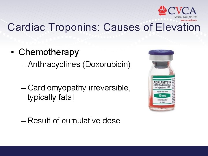
Cardiac Troponins: Causes of Elevation • Chemotherapy – Anthracyclines (Doxorubicin) – Cardiomyopathy irreversible, typically fatal – Result of cumulative dose
Cardiac Troponins: Causes of Elevation • Exercise • PTE and Pulmonary Hypertension – Due to: • pulmonary arterial resistance RV pressure myocardial perfusion and O 2 supply • PTE associated with RV infarction – Patients with elevated c. Tn have worse prognosis
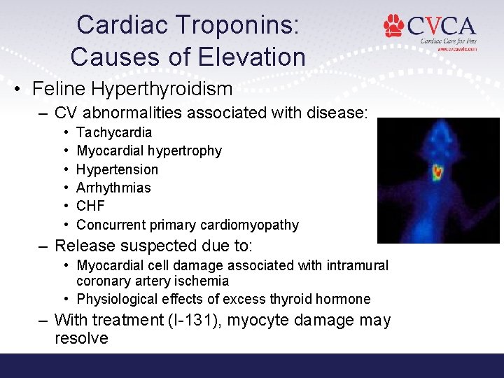
Cardiac Troponins: Causes of Elevation • Feline Hyperthyroidism – CV abnormalities associated with disease: • • • Tachycardia Myocardial hypertrophy Hypertension Arrhythmias CHF Concurrent primary cardiomyopathy – Release suspected due to: • Myocardial cell damage associated with intramural coronary artery ischemia • Physiological effects of excess thyroid hormone – With treatment (I-131), myocyte damage may resolve
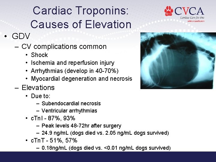
Cardiac Troponins: Causes of Elevation • GDV – CV complications common • • Shock Ischemia and reperfusion injury Arrhythmias (develop in 40 -70%) Myocardial degeneration and necrosis – Elevations • Due to: – Subendocardial necrosis – Ventricular arrhythmias • c. Tn. I - 87%, 93% – Peak levels 48 -72 hr after surgery – 24. 9 ng/m. L (dogs died vs. 2. 05 ng/m. L dogs survived) • c. Tn. T - 51%, 57% – 0. 18 ng/m. L (dogs died vs. <0. 01 ng/m. L dogs survived)
Cardiac Troponins: Causes of Elevation • Renal Failure – Chronic elevations in 50% patients • c. Tn. T more commonly associated (82%, c. Tn. I 6%) – Proposed Mechanisms: • Silent myocardial necrosis or ischemia • Ventricular hypertrophy • Inpaired renal clearance – Indicator of mortality in humans with renal disease or on hemodialysis
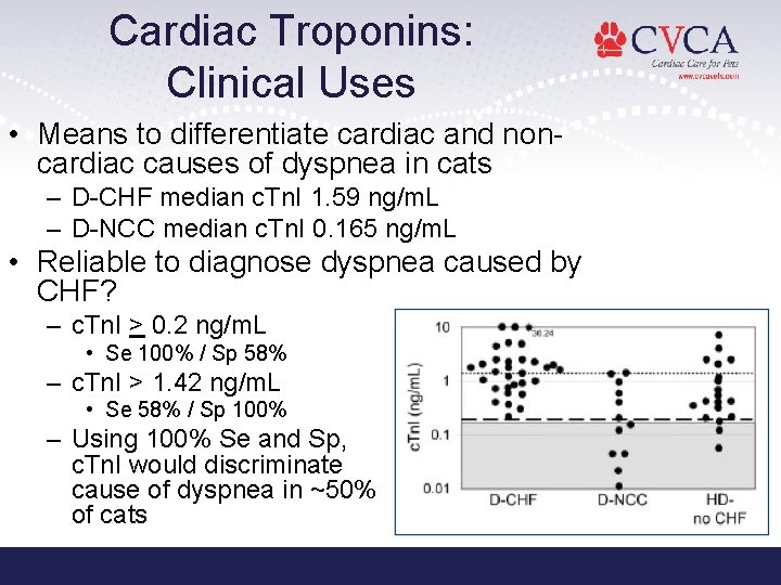
Cardiac Troponins: Clinical Uses • Means to differentiate cardiac and noncardiac causes of dyspnea in cats – D-CHF median c. Tn. I 1. 59 ng/m. L – D-NCC median c. Tn. I 0. 165 ng/m. L • Reliable to diagnose dyspnea caused by CHF? – c. Tn. I > 0. 2 ng/m. L • Se 100% / Sp 58% – c. Tn. I > 1. 42 ng/m. L • Se 58% / Sp 100% – Using 100% Se and Sp, c. Tn. I would discriminate cause of dyspnea in ~50% of cats
Cardiac Troponins: Clinical Uses • Detection of occult DCM • 3 Phases of Disease – 1: Dogs with genetic predisposition to disease w/o detectable morphologic or ECG signs – 2: Dogs with detectable abnormalities on ECG or ECHO that do not have overt clinical signs – 3: Dogs with ECG and ECHO abnormalities as well as clinical signs
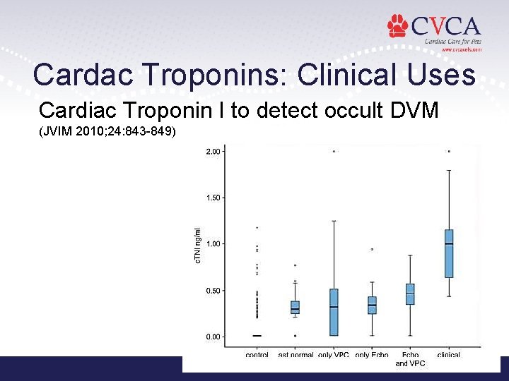
Cardac Troponins: Clinical Uses Cardiac Troponin I to detect occult DVM (JVIM 2010; 24: 843 -849)
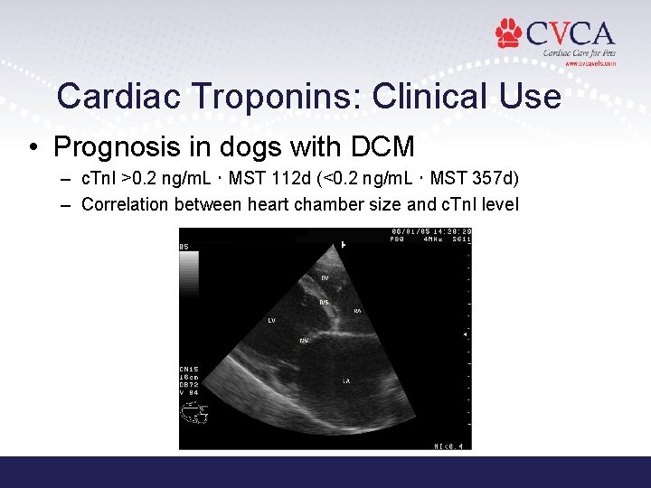
Cardiac Troponins: Clinical Use • Prognosis in dogs with DCM – c. Tn. I >0. 2 ng/m. L MST 112 d (<0. 2 ng/m. L MST 357 d) – Correlation between heart chamber size and c. Tn. I level
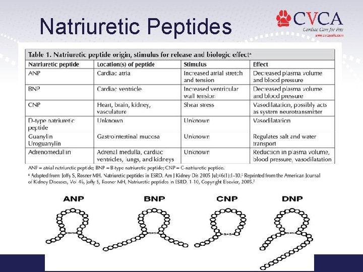
Natriuretic Peptides
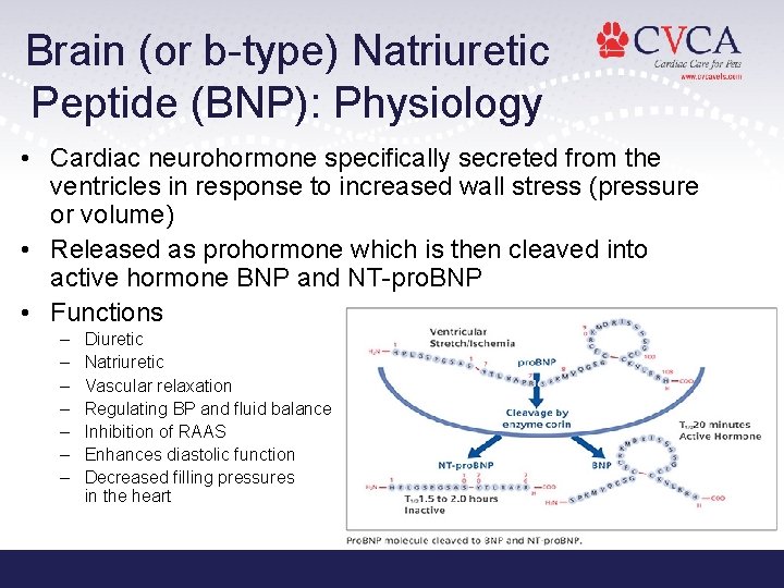
Brain (or b-type) Natriuretic Peptide (BNP): Physiology • Cardiac neurohormone specifically secreted from the ventricles in response to increased wall stress (pressure or volume) • Released as prohormone which is then cleaved into active hormone BNP and NT-pro. BNP • Functions – – – – Diuretic Natriuretic Vascular relaxation Regulating BP and fluid balance Inhibition of RAAS Enhances diastolic function Decreased filling pressures in the heart
BNP Release Kinetics • Not stored • Regulation of synthesis and excretion occurs at the level of gene expression (transcription) • BNP requires synthesis – Production is rapidly up-regulated when cardiomyocytes are stimulated • Secretion requires longer term stimulus and is regulated by myocardial wall stretch and wall tension/stretch and hypoxia – Although its production is up-regulated in situations of volume overload, the diuretic and natriuretic properties of BNP in (patho)physiological concentrations are still insufficient to retain euvolemia
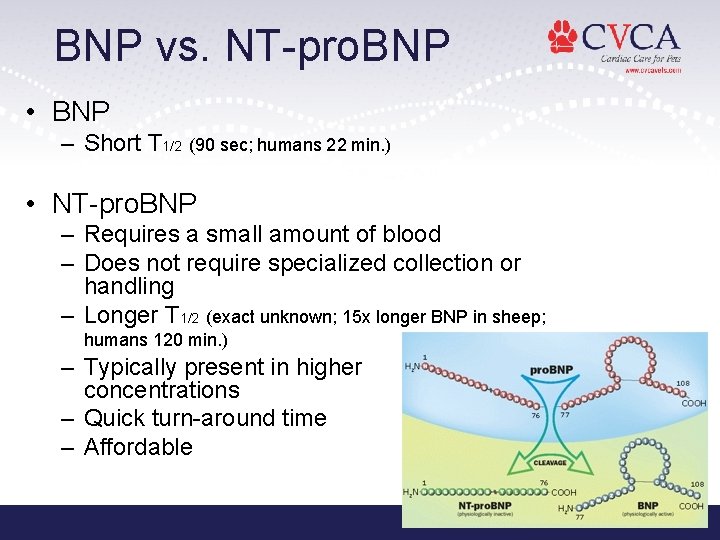
BNP vs. NT-pro. BNP • BNP – Short T 1/2 (90 sec; humans 22 min. ) • NT-pro. BNP – Requires a small amount of blood – Does not require specialized collection or handling – Longer T 1/2 (exact unknown; 15 x longer BNP in sheep; humans 120 min. ) – Typically present in higher concentrations – Quick turn-around time – Affordable
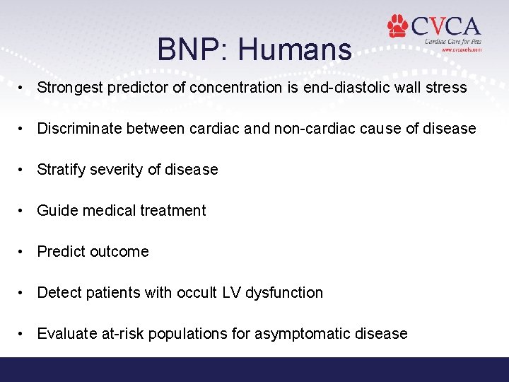
BNP: Humans • Strongest predictor of concentration is end-diastolic wall stress • Discriminate between cardiac and non-cardiac cause of disease • Stratify severity of disease • Guide medical treatment • Predict outcome • Detect patients with occult LV dysfunction • Evaluate at-risk populations for asymptomatic disease
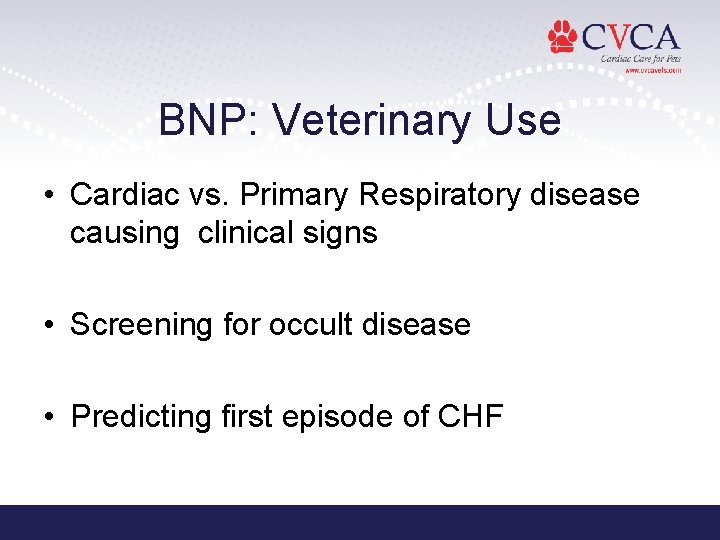
BNP: Veterinary Use • Cardiac vs. Primary Respiratory disease causing clinical signs • Screening for occult disease • Predicting first episode of CHF
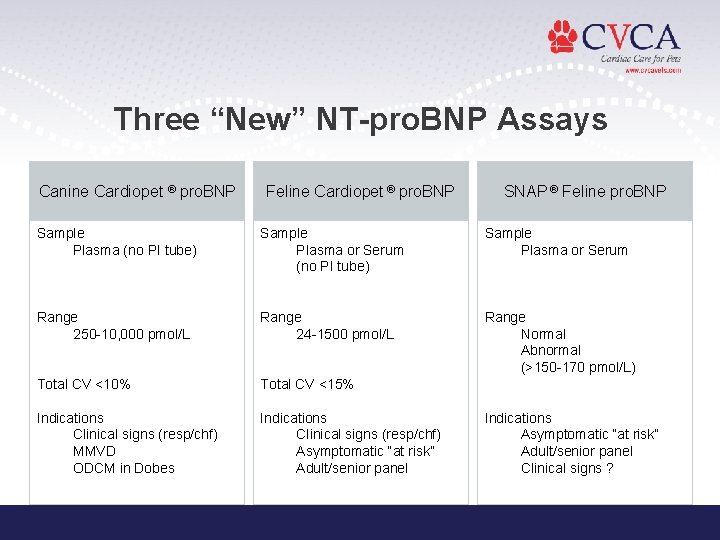
Three “New” NT-pro. BNP Assays Canine Cardiopet ® pro. BNP Feline Cardiopet ® pro. BNP SNAP ® Feline pro. BNP Sample Plasma (no PI tube) Sample Plasma or Serum Range 250 -10, 000 pmol/L Range 24 -1500 pmol/L Range Normal Abnormal (>150 -170 pmol/L) Total CV <10% Total CV <15% Indications Clinical signs (resp/chf) MMVD ODCM in Dobes Indications Clinical signs (resp/chf) Asymptomatic “at risk” Adult/senior panel Indications Asymptomatic “at risk” Adult/senior panel Clinical signs ?
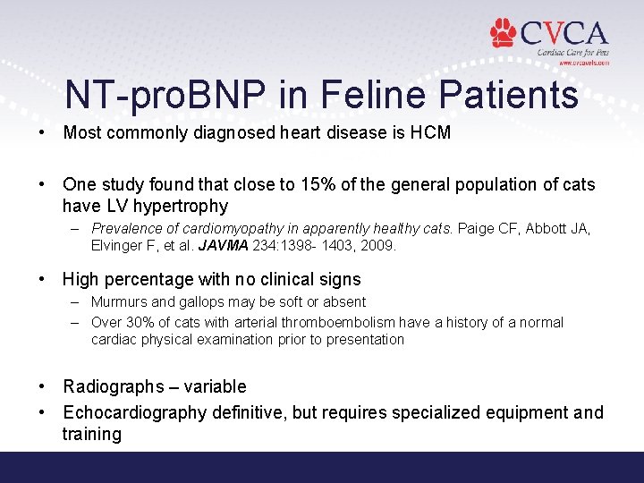
NT-pro. BNP in Feline Patients • Most commonly diagnosed heart disease is HCM • One study found that close to 15% of the general population of cats have LV hypertrophy – Prevalence of cardiomyopathy in apparently healthy cats. Paige CF, Abbott JA, Elvinger F, et al. JAVMA 234: 1398 - 1403, 2009. • High percentage with no clinical signs – Murmurs and gallops may be soft or absent – Over 30% of cats with arterial thromboembolism have a history of a normal cardiac physical examination prior to presentation • Radiographs – variable • Echocardiography definitive, but requires specialized equipment and training
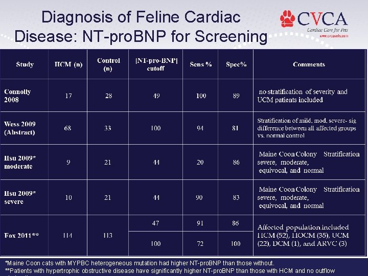
Diagnosis of Feline Cardiac Disease: NT-pro. BNP for Screening *Maine Coon cats with MYPBC heterogeneous mutation had higher NT-pro. BNP than those without. **Patients with hypertrophic obstructive disease have significantly higher NT-pro. BNP than those with HCM and no outflow
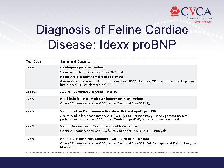
Diagnosis of Feline Cardiac Disease: Idexx pro. BNP
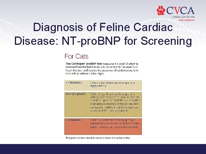
Diagnosis of Feline Cardiac Disease: NT-pro. BNP for Screening
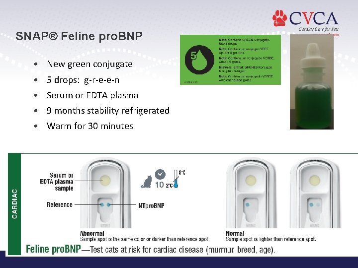
SNAP® Feline pro. BNP • New green conjugate • 5 drops: g-r-e-e-n • Serum or EDTA plasma • 9 months stability refrigerated • Warm for 30 minutes

Pricing, Packaging and Stability 9 month stability with refrigeration List Price 5/box $11. 95/test 10/box $9. 95/test
SNAP Performance Demonstrates Good Correlation with Cardiopet® pro. BNP Abnormal Normal SSDx cut-off (spots look equal) Cardiopet pro. BNP (pmol/L)
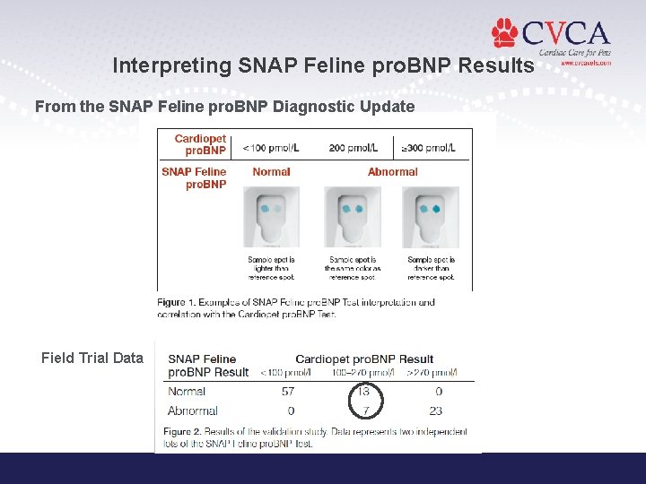
Interpreting SNAP Feline pro. BNP Results From the SNAP Feline pro. BNP Diagnostic Update Field Trial Data
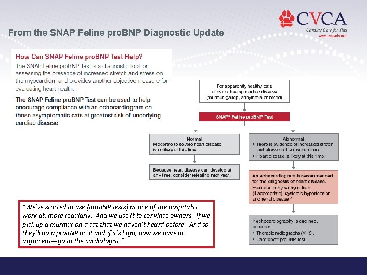
From the SNAP Feline pro. BNP Diagnostic Update “We’ve started to use [pro. BNP tests] at one of the hospitals I work at, more regularly. And we use it to convince owners. If we pick up a murmur on a cat that we haven’t heard before. And so they’ll do a pro. BNP on it and if it’s high, now we have an argument—go to the cardiologist. ”
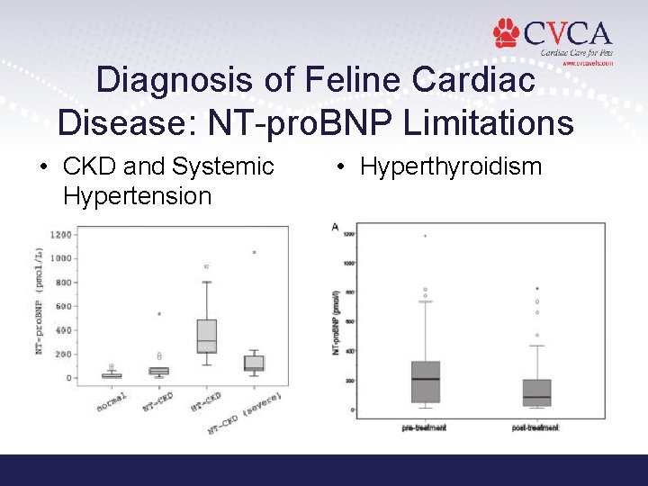
Diagnosis of Feline Cardiac Disease: NT-pro. BNP Limitations • CKD and Systemic Hypertension • Hyperthyroidism
Summary of NT-pro. BNP in Feline Patients • Prevalence of cardiac disease in the general population of feline patients is estimated to be about 15% • With a prevalence of 15% and Sens 90%/Spec 85% – PPV is 53% and NPV is 93% • A fair number of false positives • <10% chance of a false negative • Echocardiography is the gold standard for the diagnosis of feline myocardial disease
NT-pro. BNP in K 9 Patients • The most common heart disease in dogs is degenerative mitral valve disease – Accounts for 75% of acquired cardiac disease in dogs – Estimates that over 60% of older small breeds of dogs are affected many will show signs of congestive heart failure – Physical examination findings of a murmur make screening relatively easy – Long asymptomatic phase • Occult disease is more common with dilated cardiomyopathy – Doberman Pinscher incidence of 45 -62%
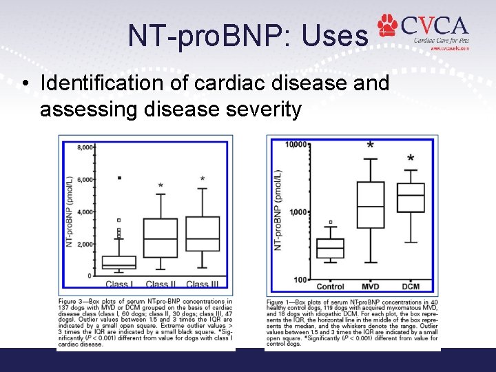
NT-pro. BNP: Uses • Identification of cardiac disease and assessing disease severity
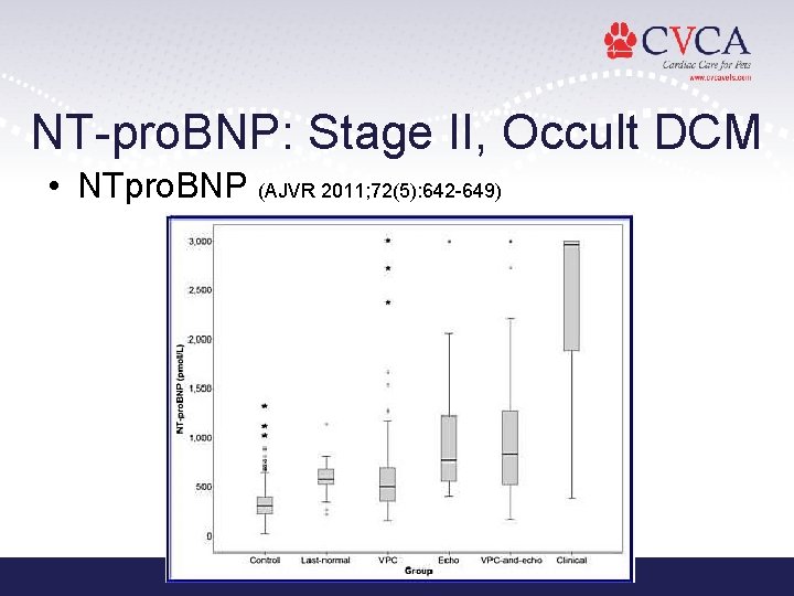
NT-pro. BNP: Stage II, Occult DCM • NTpro. BNP (AJVR 2011; 72(5): 642 -649)
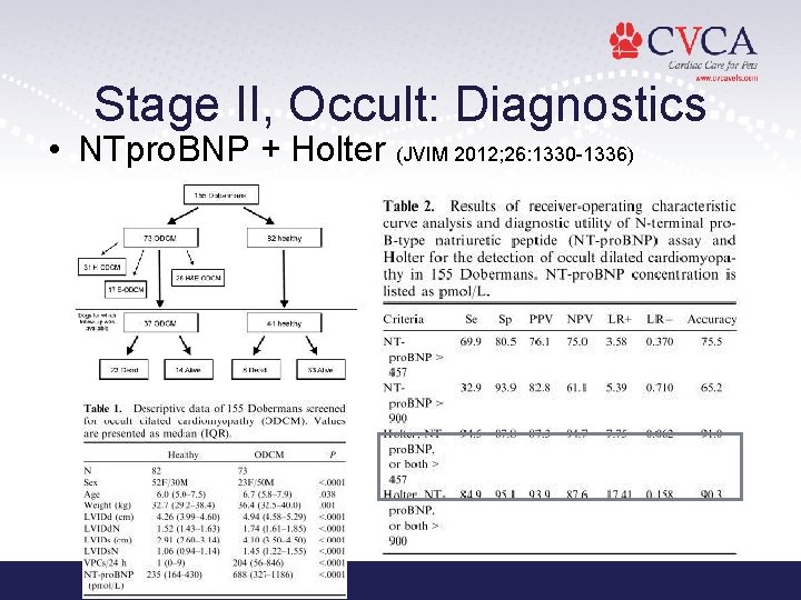
Stage II, Occult: Diagnostics • NTpro. BNP + Holter (JVIM 2012; 26: 1330 -1336)
Summary of NT-pro. BNP in Dogs • Helps differentiate between respiratory symptoms secondary to heart vs. pulmonary disease • Concentrations correlate with the severity of disease • May be predictive of impending congestive heart failure and used in combination with clinical status may predict outcome • No studies yet evaluating relation to therapy and response to treatment
CHF Challenges • Timely and accurate diagnosis can be challenging • Hx and PE findings not specific • TXR - considered to be high yield diagnostic test, can be difficult to attain and interpret • Diuretics and HF medication can be detrimental to a mis-diagnosed patient
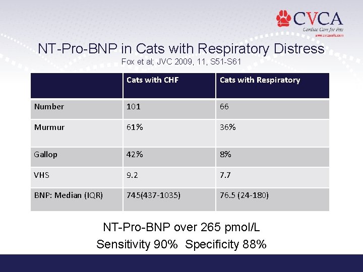
NT-Pro-BNP in Cats with Respiratory Distress Fox et al; JVC 2009, 11, S 51 -S 61 Cats with CHF Cats with Respiratory Number 101 66 Murmur 61% 36% Gallop 42% 8% VHS 9. 2 7. 7 BNP: Median (IQR) 745(437 -1035) 76. 5 (24 -180) NT-Pro-BNP over 265 pmol/L Sensitivity 90% Specificity 88%
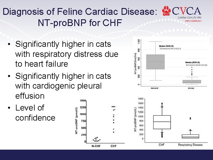
Diagnosis of Feline Cardiac Disease: NT-pro. BNP for CHF • Significantly higher in cats with respiratory distress due to heart failure • Significantly higher in cats with cardiogenic pleural effusion • Level of confidence
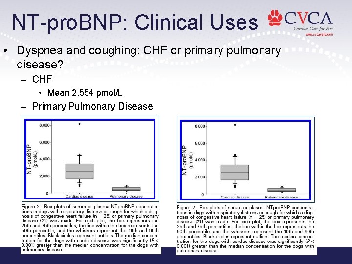
NT-pro. BNP: Clinical Uses • Dyspnea and coughing: CHF or primary pulmonary disease? – CHF • Mean 2, 554 pmol/L – Primary Pulmonary Disease • Mean 357 pmol/L
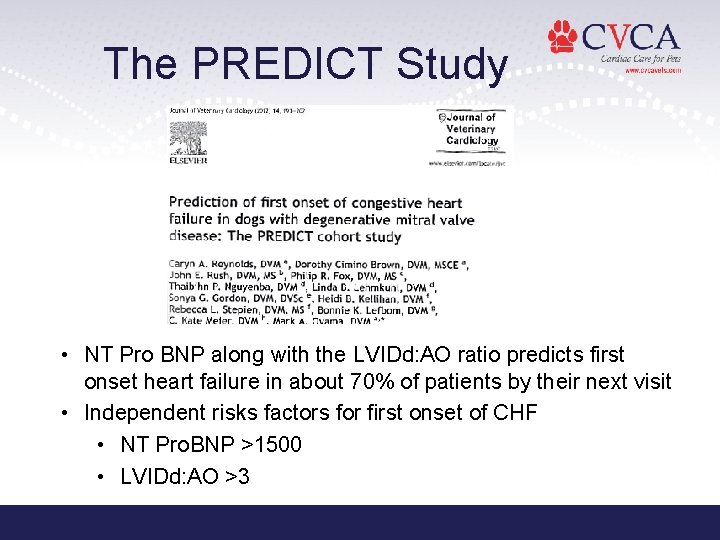
The PREDICT Study • NT Pro BNP along with the LVIDd: AO ratio predicts first onset heart failure in about 70% of patients by their next visit • Independent risks factors for first onset of CHF • NT Pro. BNP >1500 • LVIDd: AO >3
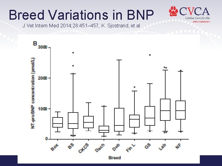
Breed Variations in BNP J Vet Intern Med 2014; 28: 451– 457, K. Sjostrand, et al
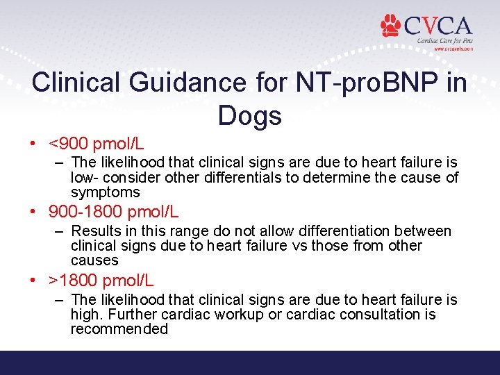
Clinical Guidance for NT-pro. BNP in Dogs • <900 pmol/L – The likelihood that clinical signs are due to heart failure is low- consider other differentials to determine the cause of symptoms • 900 -1800 pmol/L – Results in this range do not allow differentiation between clinical signs due to heart failure vs those from other causes • >1800 pmol/L – The likelihood that clinical signs are due to heart failure is high. Further cardiac workup or cardiac consultation is recommended
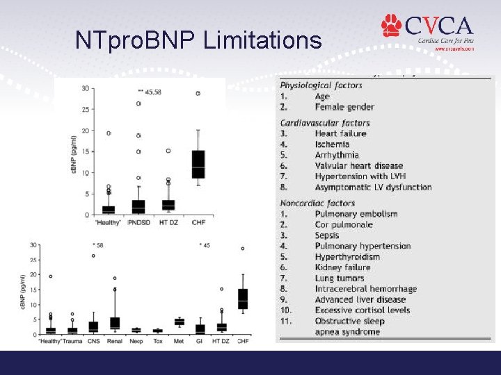
NTpro. BNP Limitations
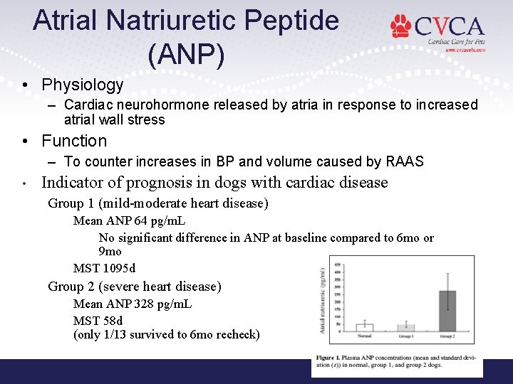
Atrial Natriuretic Peptide (ANP) • Physiology – Cardiac neurohormone released by atria in response to increased atrial wall stress • Function – To counter increases in BP and volume caused by RAAS • Indicator of prognosis in dogs with cardiac disease Group 1 (mild-moderate heart disease) Mean ANP 64 pg/m. L No significant difference in ANP at baseline compared to 6 mo or 9 mo MST 1095 d Group 2 (severe heart disease) Mean ANP 328 pg/m. L MST 58 d (only 1/13 survived to 6 mo recheck)

Comments / Questions Contact Information: info@cvcavets. com www. cvcavets. com
- Slides: 66