CLINICAL PRESENTATION OF GN DIAGNOSIS History Physical exam


. Symptoms: edema, hematuria, proteinuria, etc.")

 in nephrotic syndrome. The white line grew during a transient")


 �Diffuse: process that involve all glomeruli, >50%")


- Slides: 27
CLINICAL PRESENTATION OF GN
DIAGNOSIS • History. • Physical exam. • Investigations: Ø Labs. Ø Imaging. Ø Renal biopsy.
History • • • Onset ( sudden or gradual). Symptoms: edema, hematuria, proteinuria, etc. Family history. Drug history. Past history of infections or malignancy.
PE
Muehrcke lines (bands) in nephrotic syndrome. The white line grew during a transient period of hypoalbuminemia caused by the nephrotic syndrome.
Xanthelasmas in nephrotic syndrome.
LAB work up • antinuclear and anti DNA antibodies for lupus, • cryoglobulins and rheumatoid factor suggesting cryoglobulinemia. • anti–glomerular basement membrane (anti GBM) antibodies for Goodpasture disease, anti neutrophil cytoplasmic autoantibody (ANCA) for vasculitis, • antistreptolysin O titer or streptozyme test for poststreptococcal glomerulonephritis
• Hepatitis profile. • ASO titer. • Serum and urine electrophoresis( MM, LCD, HCD) • Measurement of systemic complement pathway activation by testing for serum C 3, C 4, and CH 50 (50% hemolyzing dose of complement) is often helpful in limiting the differential diagnosis.
Imaging • • You need to make sure you have both kidneys. Size : normally 10 13 cm in adults. No anatomical or structural abnormalities. Presence of hydronephrosis.
Hypocomplementemia in glomerular disease DDX • Classical pathway activation ( low C 3, low C 4, low CH 50): Lupus nephritis (especially Class IV), Cryoglobulinemia, Membranoproliferative GN type 1. • Alternative pathwayactivation(C 3 ↓, C 4 normal, CH 50 L): Poststreptococcal GN GN associated with other infection* (e. g. , endocarditis, shunt nephritis), HUS, Atheroembolic renal disease.
Renal biopsy • Aid in diagnosis. • Help to expect prognosis. • WHEN TO DO IT? ? this is debatable sometimes between nephrologist.
Indications For diagnosis of ? primary GN. Unexplained AKI. First presentation of CKD in young adult. When kidney disease is part of systemic illness and the kidney is the easiest to access for Dx. • Evaluation of RPGN. • •
NOT indicated • Isolated glomerular hematuria. • Isolated non-nephrotic proteinuria. • Nephrotic syndrome: when one of the following is present: Ø DM Ø history and presence of extrarenal involvement( eg: amyloidosis) Ø Children under the age of six years with the acute onset of nephrotic syndrome. Ø Malignancy, massive obesity.
Terminology �Glomerulonephritis: inflammation of the kidney (glomerulos) �Diffuse: process that involve all glomeruli, >50% �Focal: involvement of some glomeruli NOT all, <50% �Global: if the whole glomerular tuft is involved, �Segmental: only part of the tuft involved
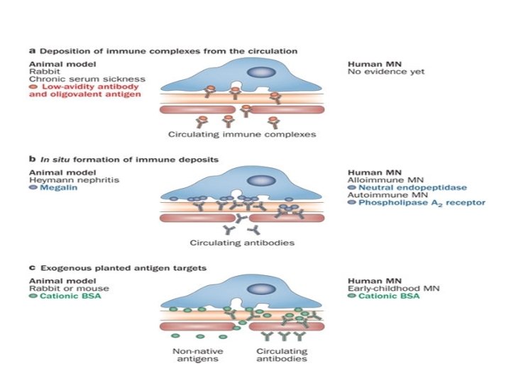
Mechanisms of glomerular inflammation � Both humoral and cell mediated immune mechanisms play a part in the pathogenesis of glomerular inflammation � Two basic mechanisms of antibody mediated glomerular injury have been identified: – antibodies can bind either to a structural component of the glomerulus or to material that is not intrinsic to the glomerulus
Mechanism. . cont’d – circulating antigen–antibody complexes form, escape clearance by the reticuloendothelial system, and are deposited in the glomerulus • activation of cell mediated immunity can also induce glomerular injury
Mechanism. . cont’d
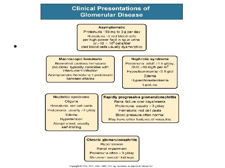
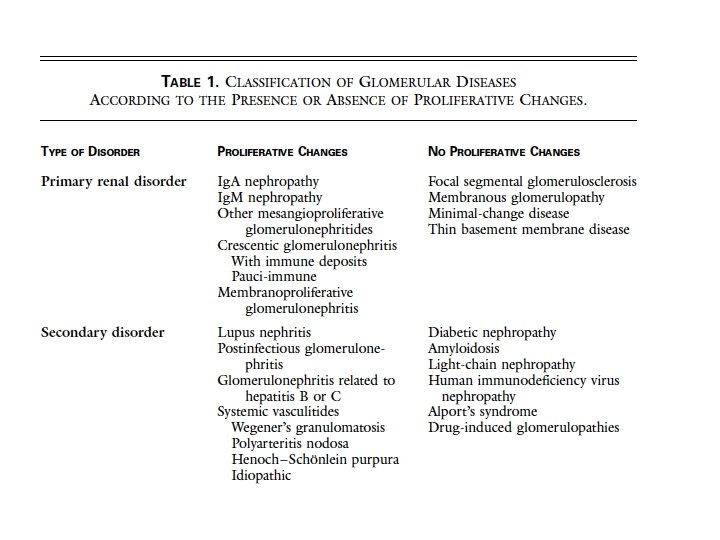
Classification Schemes �Clinical – Primary and Secondary – Nephrotic and Nephritic – Acute and Chronic
Pathologic Classification • Non Proliferative • Proliferative
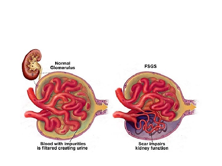
GN Pathologic Classification �Non Proliferative – Minimal Change GN – Focal Segmental Glomerulosclerosis – Membranous GN �Proliferative – Focal proliferative – Membranoproliferative – Diffuse Proliferative – Crescentic (RPGN)
Nephrotic Syndrome • • Proteinuria > 3. 5 g/d Edema Hyperlipidemia/oval fat bodies Hypoalbuminemia
Complications of nephrotic syndrome �Increased risk of atherosclerosis �Elevated levels of – Total and low density lipoprotein cholesterol – Lipoprotein (a) �Low or normal high density lipoprotein cholesterol �It is related to the hypoproteinemia and low serum oncotic pressure of nephrotic syndrome, which then leads to reactive hepatic protein synthesis, including of lipoproteins
Complications of nephrotic syndrome…cont’d • Increase risk of thrombosis DVT, PE… due to urinary anti thrombin III loss • Increase risk of infections due to urinary Ig loss.