Clinical Practice Guidelines Benign liver tumours Dr gavidel

Clinical Practice Guidelines Benign liver tumours

• Dr gavidel journal club govaresh

Overview Background Characteristics of common benign liver lesions Basic management of a ‘liver nodule’ The benign tumour MDT

Background • • Heterogenous group of liver lesions Frequently found incidentally – due to widespread imaging use Often have a benign course Some are of greater clinical relevance than others • CPG provides an aid for the practical diagnosis and management of the more common benign tumours: * – – Hepatic haemangiomas Focal nodular hyperplasia (FNH) Hepatocellular adenoma (HCA) Patients with multiple lesions *Nodular regenerative hyperplasia, although its histology is ‘benign’, has a clinical course and management distinct from other benign lesions considered in this guideline and is not reviewed here EASL CPG benign liver tumours. J Hepatol 2016; 65: 386– 98

Characteristics of common benign liver lesions Haemangioma FNH HCA Common ~5%* Less common 0. 03% Rare ≤ 0. 004% 30– 50 years 20– 40 years All ages F>M F~M F >> M US Hyperechoic Varied CT Centripetal enhancement Central scar Varied MRI Centripetal enhancement Hyperintense T 2 -w Central scar Varied Calcification Yes No No Rupture Rare No Yes Estimated prevalence Age Gender *Estimated prevalence in imaging series; has been reported to be as high as 20% in autopsy series Bahirwani R, Reddy KR. Aliment Pharmacol Ther 2008; 28: 953– 65; EASL CPG benign liver tumours. J Hepatol 2016; 65: 386– 98

Basic management of a ‘liver nodule’ Examination and baseline investigations • Associated symptoms: – – • Abdominal pain Weight loss Hepatomegaly Abnormal liver function tests Medical history – – – Conditions associated with liver lesions (e. g. cancer, anorexia, asthenia) History of foreign travel or dysentery Medication history, particularly OCPs • Exclude primary tumour distant to liver • Risk factors – – – History of/current viral hepatitis/cirrhosis History of transfusion, tattoos, IV drug abuse Family history of liver disease/tumours Alcohol excess, smoking Features of metabolic syndrome (obesity, T 2 DM, HTN, CV disease) Drug history (methotrexate, tamoxifen, androgens) Following examination and baseline investigations Contrast-enhanced imaging (CEUS, CT, MRI) for tumour characterization • • • Imaging and baseline investigations should be sufficient to diagnose benign liver tumours In cases of significant doubt, a biopsy or resection may be appropriate Invasive procedures should only be pursued after consideration by an experienced MDT EASL CPG benign liver tumours. J Hepatol 2016; 65: 386– 98

The benign liver tumour MDT Relevant expertise and experience Hepatobiliary surgeon Skills to manage benign liver lesions EASL CPG benign liver tumours. J Hepatol 2016; 65: 386– 98 Hepatologist Benign liver tumour MDT Pathologist Specific and relevant training Diagnostic and interventional radiologists Skills to manage complications of diagnostic or therapeutic interventions

Guidelines Key recommendations
 Hepatocellular adenoma (HCA)")
Topics 1. 2. 3. 4. Hepatic haemangiomas Focal nodular hyperplasia (FNH) Hepatocellular adenoma (HCA) Patients with multiple lesions EASL CPG PBC. J Hepatol 2017; 67: 145– 72 Click on a topic to skip to that section

Hepatic haemangiomas Epidemiology/clinical characteristics Key diagnostic recommendations Key management recommendations

Hepatic haemangiomas: epidemiology/clinical characteristics • Most common primary liver tumours – Prevalence on imaging series: ~5%1 – Prevalence on autopsy series: up to 20%2, 3 – Most common in women aged 30– 50 years 3 • Female to male ratio ranges from 1. 2– 6: 1 • Can occur in all age groups • Rarely of clinical significance – Often solitary and small (<4 cm), although can reach 20 cm in diameter 2, 3 – Most patients are asymptomatic even with large haemangiomas 2, 3 – Larger tumours (>10 cm) may be symptomatic – associated with pain and features of KMS (inflammatory reaction syndrome and coagulopathy)4, 5 1. Horta G, et al. Rev Med Chil 2015; 143: 197– 202; 2. Bahirwani R, Reddy KR. Aliment Pharmacol Ther 2008; 28: 953– 65; 3. Gandolfi L, et al. Gut 1991; 32: 677– 80; 4. Hall GW. Br J Haematol 2001; 112: 851– 62; 5. O’Rafferty C, et al. Br J Haematol 2015; 171: 38– 51; EASL CPG benign liver tumours. J Hepatol 2016; 65: 386– 98

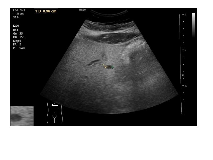
Hepatic haemangiomas: key diagnostic recommendations • Classic appearance on US is a homogenous hyperechoic mass Recommendations Grade of evidence Grade of recommendation In patients with a normal/healthy liver, a hyperechoic lesion is very likely to be a liver haemangioma US is sufficient for diagnosis in cases of typical radiology (homogeneous hyperechoic, sharp margin, posterior enhancement, absence of halo sign) in lesions <3 cm Contrast enhanced imaging (CEUS, CT or MRI) is required in oncology patients and patients with underlying liver disease Diagnosis by contrast-enhanced imaging is based on a typical vascular profile, characterized by peripheral and globular enhancement on arterial phase followed by a central enhancement on delayed phases MRI provides additional findings: e. g lesion signal on T 1 -, T 2 weighted sequences; diffusion imaging EASL CPG benign liver tumours. J Hepatol 2016; 65: 386– 98 II-2 1

Hepatic haemangiomas: imaging Typical haemangioma adjacent to FNH MRI T 2 Strongly hyperintense T 1 Hypointense CEUS EASL CPG benign liver tumours. J Hepatol 2016; 65: 386– 98 CE CE CE Lesion shows peripheral and discontinuous enhancement followed by complete fill-in on delayed-phase imaging

Hepatic haemangiomas: key management recommendations • Haemangiomas are mostly asymptomatic incidental discoveries – – May change in size during long-term follow-up No relationship between size and complications Little relationship between symptoms and characteristics Benefit of surgery debatable Recommendations Grade of evidence Grade of recommendation Due to its benign course, imaging follow-up is not required for typical haemangioma Pregnancy and OCPs are not contraindicated Conservative management is appropriate for typical cases Refer to benign liver tumour MDT in the presence of KMS, growing lesions or lesions that are symptomatic by compression EASL CPG benign liver tumours. J Hepatol 2016; 65: 386– 98 II-2 1 III 2 II-2 1 III 1

Focal nodular hyperplasia Epidemiology/clinical characteristics Diagnosis and imaging Key recommendations Management algorithm

FNH: epidemiology/clinical characteristics • Epidemiology – Clinically relevant prevalence: 0. 03% (autopsy series: 0. 4– 3%)1, 2 – Up to 90% of patients are female – Average at presentation: 35– 50 years • Clinical characteristics – – Most cases are solitary and <5 cm; multiple FNH in 20– 30% of cases 3, 4 Hyperplastic hepatocellular lesions resulting from arterial malformation Size is stable over time in most cases 5 Most cases are asymptomatic and complications are extremely rare 5 • Genetics – Upregulation of ECM genes associated with TGF- signaling 6 – Overexpression of Wnt/ -catenin target genes, e. g. GLUL 6 1. Rubin RA, Mitchell DG. Med Clin North Am 1996; 80: 907– 28; 2. Marrero JA, et al. Am J Gastroenterol 2014; 109: 1328 -47; 3. Nguyen BN, et al. Am J Surg Pathol 1999; 23: 1441– 54; 4. Vilgrain V, et al. Radiology 2003; 229: 75– 9; 5. D’Halluin V, et al. Gastroenterol Clin Biol 2001; 25: 1008– 10; 6. Rebouissou S, et al. J Hepatol 2008; 49: 61– 71; EASL CPG benign liver tumours. J Hepatol 2016; 65: 386– 98

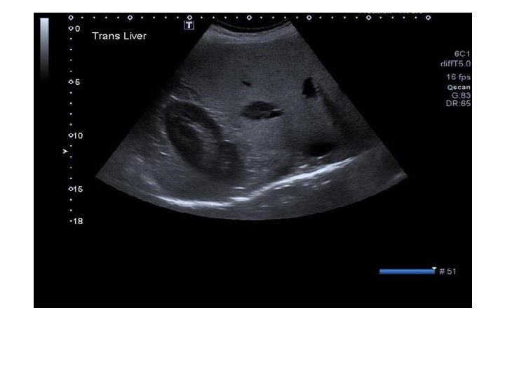
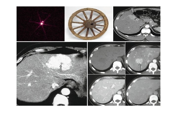
FNH: imaging • Diagnosis is based on a combination of five imaging features: 1. 2. 3. 4. 5. Lesion homogeneity, excluding the central scar Slight difference from adjacent liver tissue on pre-contrast US, CT and MRI (A & B) Strong, homogeneous enhancement on arterial phase CEUS, CT or MRI with a central vascular supply (C); becomes isointense to liver tissue on portal venous and delayed phases (D) Central scar best seen on MRI Lack of capsule with often lobulated contours T 2 - and T 1 -weighted images A B Lesion barely visible EASL CPG benign liver tumours. J Hepatol 2016; 65: 386– 98 Contrast-enhanced images C Lesion easily visible D
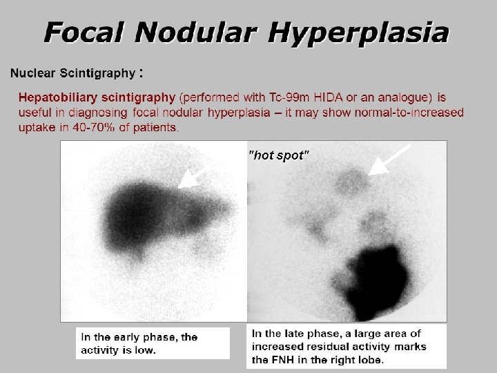

FNH: key diagnostic recommendations • MRI sensitivity – Lesion >3 cm – very good – Lesion <3 cm – second imaging modality advised, such as CEUS • Refer to a specialist centre if in doubt with two imaging modalities Recommendations Grade of evidence Grade of recommendation CEUS, CT, MRI: nearly 100% specificity with a combination of typical imaging features II-2 1 MRI has the highest diagnostic performance overall Highest diagnostic accuracy by CEUS is achieved in FNH <3 cm II-2 1 EASL CPG benign liver tumours. J Hepatol 2016; 65: 386– 98

FNH: key management recommendations • In the absence of symptoms a conservative management approach is recommended • No indication for discontinuing OCPs • Follow-up during pregnancy is not necessary Recommendations Grade of evidence Grade of recommendation For a typical FNH lesion, follow-up is not necessary unless there is underlying vascular liver disease Treatment is not recommended If imaging is atypical, or the patient is symptomatic, refer to a benign liver tumour MDT EASL CPG benign liver tumours. J Hepatol 2016; 65: 386– 98 III 2 II-3 2 III 1
* Diagnosis FNH certain <3 cm")
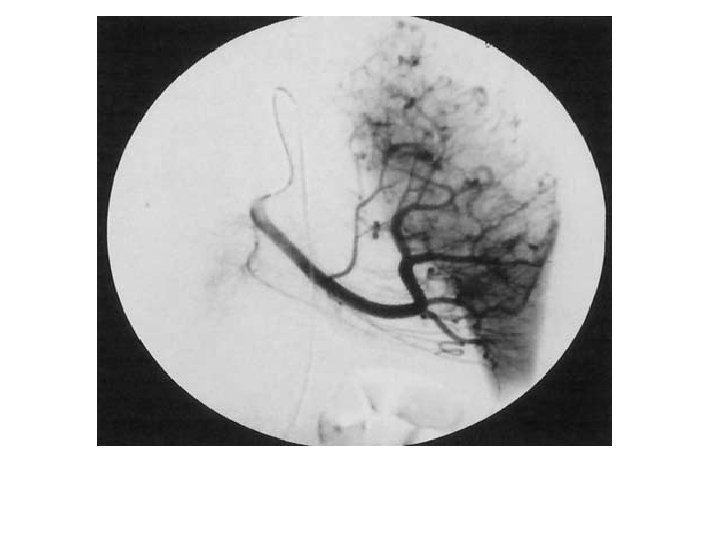
FNH: management algorithm Suspected FNH Contrast-enhanced imaging (preferably MRI)* Diagnosis FNH certain <3 cm CEUS Diagnosis uncertain Discharge, no follow-up needed *Imaging modalities may include US, CE-CT and CE-MRI EASL CPG benign liver tumours. J Hepatol 2016; 65: 386– 98 Diagnosis FNH doubtful Map-like pattern of GS is specific to FNH >3 cm Biopsy Confirmed FNH GS immunohistochemical staining is useful in difficult cases

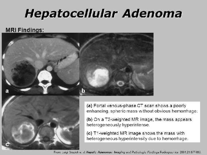
Hepatocellular adenoma Epidemiology/clinical characteristics Molecular classification Key recommendations Management algorithm

HCA: epidemiology/clinical characteristics • Epidemiology 1– 3 – Reported prevalence: 0. 001– 0. 004% – ~10 x less common than FNH – Most common in women (10: 1 female to male), especially aged 35– 40 years • Potential role of sex hormones – 30– 40 -fold increase in incidence with long-term OCP use 4 – Incidence among males is associated with androgenic steroids 5, 6 • Recent increase in prevalence associated with rising obesity and metabolic syndrome 7– 9 • Significant risk of haemorrhage and malignant transformation – Especially with lesions ≥ 5 cm HCAs need to be followed more closely than other benign tumours 1. Bonder A, Afdhal N. Clin Liver Dis 2012; 16: 271– 83; 2. Karhunen PJ. J Clin Pathol 1986; 39: 183– 8; 3. Cherqui D, et al. Gastroenterol Clin Biol 1997; 21: 929– 35; 4. Giannitrapani L, et al. Ann NY Acad Sci 2006; 1089: 228– 36; 5. Socas L, et al. Br J Sports Med 2005; 39: e 27; 6. Nakao A, et al. J Gastroenterol 2000; 35: 557– 62; 7. Bunchorntavakul C, et al. Aliment Pharmacol Ther 2011; 34: 664– 74; 8. Bioulac-Sage P, et al. Liver Int 2012; 32: 1217– 21; 9. Chang CY, et al. Int J Hepatol 2013; 2013: 604860; EASL CPG benign liver tumours. J Hepatol 2016; 65: 386– 98

HCA: molecular classification • Molecular subtype is highly associated with risk of transformation to HCC Clinical features MRI features† Genetic mutation Pathology IHC HNF 1 -A (30– 40%) Extensive steatosis LFABP –ve Adenomatosis, MODY 3 Diffuse and homogenous signal dropout on opposed-phase T 1 Inflammatory, Gp 130 (65%), GNAS (5%), STAT 3 (5%), FRK (10%), JAK 1 (2%) Inflammatory infiltration Clusters of vessels Sinusoidal dilatation LFABP +ve SAA (± CRP) +ve Obesity Alcohol consumption Strong hyperintense on T 2 and persistent enhancement on delayed phase using extracellular MR contrast agents β-catenin* exon 3 (5– 10%) Cell atypias Pseudoglandular formations Cholestasis LFABP +ve GS +ve (diffuse) β-catenin nuclear +ve Male; androgen use increased risk of HCC No specific feature; often heterogeneous on T 1 and T 2 No signal dropout on opposedphase T 1 β-catenin exons 7– 8 No typical features (5– 10%) or inflammatory phenotype GS +ve (faint and patchy); β-catenin nuclear –ve No specific features Unclassified (5– 10%) LFABP +ve SAA/CRP –ve β-catenin nuclear –ve No specific features None *50% also display inflammatory phenotype; †Using hepatospecific MR contrast agents and hepatobiliary sequences, most HCAs appear hypointense but some are iso- or hyperintense on these sequences and seem to mainly correspond to inflammatory HCA. Gd-BOPTA offers the possibility to evaluate both the delayed and the hepatobiliary phases EASL CPG benign liver tumours. J Hepatol 2016; 65: 386– 98

HCA: molecular classification • Molecular subtype is highly associated with risk of transformation to HCC Clinical features MRI features† Genetic mutation Pathology IHC HNF 1 -A (30– 40%) Extensive steatosis LFABP –ve Adenomatosis, MODY 3 Diffuse and homogenous signal dropout on opposed-phase T 1 Inflammatory, Gp 130 (65%), GNAS (5%), STAT 3 (5%), FRK (10%), JAK 1 (2%) Inflammatory infiltration Clusters of vessels Sinusoidal dilatation LFABP +ve SAA (± CRP) +ve Obesity Alcohol consumption Strong hyperintense on T 2 and persistent enhancement on delayed phase using extracellular MR contrast agents β-catenin* exon 3 (5– 10%) Cell atypias Pseudoglandular formations Cholestasis LFABP +ve GS +ve (diffuse) β-catenin nuclear +ve Male; androgen use increased risk of HCC No specific feature; often heterogeneous on T 1 and T 2 No signal dropout on opposedphase T 1 β-catenin exons 7– 8 No typical features (5– 10%) or inflammatory phenotype GS +ve (faint and patchy); β-catenin nuclear –ve No specific features Unclassified (5– 10%) LFABP +ve SAA/CRP –ve β-catenin nuclear –ve No specific features None β-HCAs exhibit the highest risk of malignancy; men are at a higher risk of malignancy *50% also display inflammatory phenotype; †Using hepatospecific MR contrast agents and hepatobiliary sequences, most HCAs appear hypointense but some are iso- or hyperintense on these sequences and seem to mainly correspond to inflammatory HCA. Gd-BOPTA offers the possibility to evaluate both the delayed and the hepatobiliary phases EASL CPG benign liver tumours. J Hepatol 2016; 65: 386– 98

HCA: key diagnostic recommendations • Imaging features reflect the tumour molecular subtype • Imaging should be fat sensitive and use contrast agents to look for dilated vascular spaces Recommendations Grade of evidence Grade of recommendation MRI is superior to all other imaging modalities Due to its intrinsic properties to detect fat and vascular spaces it offers the opportunity to subtype HCA up to 80% II-2 1 MRI has >90% specificity for positive identification of HNF-1α or inflammatory HCA Identification of β-catenin-activated HCA and distinction from unclassified HCA or HCC is not possible with any current imaging technique EASL CPG benign liver tumours. J Hepatol 2016; 65: 386– 98

HCA: key management recommendations • HCAs have the potential for haemorrhage or malignant transformation – Management should involve a benign liver tumour MDT Recommendations Grade of evidence Grade of recommendation Base treatment decisions on sex, size and pattern of progression III 2 Discontinuation of OCPs and weight loss should be advised II-2 1 Resection irrespective of size is recommended in men and in all cases of proven β-catenin mutation II-3 2 2 III 2 Observe women for 6 months after lifestyle change. • Resection is indicated with lesions ≥ 5 cm and those continuing to grow • Reassess lesions <5 cm at 1 year with annual imaging thereafter Bleeding HCAs with haemodynamic instability should be embolized and a residual viable lesion on follow-up imaging is an indication for resection EASL CPG benign liver tumours. J Hepatol 2016; 65: 386– 98
 Male (irrespective of")
HCA: management algorithm Suspected HCA Contrast-enhanced MRI document size (+/– subtype) Male (irrespective of tumour size) Female (irrespective of tumour size) Advise lifestyle change Repeat MRI after 6 months <5 cm stable or reduced in size ≥ 5 cm or significant* increase in size 1 -year MRI Stable or reduced size Annual imaging *≥ 20% diameter EASL CPG benign liver tumours. J Hepatol 2016; 65: 386– 98 Resection
 Male (irrespective of")
HCA: management algorithm Suspected HCA Contrast-enhanced MRI document size (+/– subtype) Male (irrespective of tumour size) Female (irrespective of tumour size) Advise lifestyle change Repeat MRI after 6 months <5 cm stable or reduced in size ≥ 5 cm or significant* increase in size 1 -year MRI Alfa fetoprotein *≥ 20% diameter EASL CPG benign liver tumours. J Hepatol 2016; 65: 386– 98 Stable or reduced size Annual imaging Resection

If planning for pregnancy : RFA should be considered for all HCA s > 2 cm in size

Patients with multiple lesions Key recommendations

Multiple lesions: key recommendations • The term ‘multiple HCAs’ has replaced ‘liver adenomatosis’ – >10 HCAs • Risk of bleeding and malignant transformation: – Does not differ in patients with multiple HCAs versus a single HCA – Driven by the size of the largest nodule Recommendations Grade of evidence Grade of recommendation Base management of multiple HCAs on the size of the largest tumour III 2 Hepatic resection may be considered in unilobular disease For widespread HCA, resection of the largest adenomas may be an option III 2 LTx is not recommended in multiple HCA LTx may be considered in case of underlying liver disease III 2 EASL CPG benign liver tumours. J Hepatol 2016; 65: 386– 98

- Slides: 39