
Clinical Phenotype of Mitochondrial Disease Charles L Hoppel







- Slides: 50
Clinical Phenotype of Mitochondrial Disease Charles L. Hoppel, M. D. Pharmacology and Medicine Case Western Reserve University Center for Mitochondrial Diseases Center for Inherited Disorders of Energy Metabolism 2008 Mc. Kim Conference, Duluth, MN September 17, 2008
Clinical Phenotype of Mitochondrial Disease • What is a Jedi Knight? • A perspective on the Dark Side of the Force.
Clinical Phenotype of Mitochondrial Disease • Why study Mitochondria • Mitochondrial Oxidative Phosphorylation • Mitochondrial function in Patients • Respirasomes ? ? Supercomplexes!! • Respirasomes in Heart Failure • Summary and Conclusions 2008 Mc. Kim Conference, Duluth, MN September 17, 2008
Star Wars Episode I What makes a JEDI Knight? How do you become a JEDI Knight? Adapted from G. Lucas, STAR WARS, Episode I (1999)
• Chip with a blood sample – Anakin Skywalker • Midi-chlorian test • Count is over twenty thousand • No one has a count that high NOT even Master Yoda! Adapted from G. Lucas, STAR WARS, Episode I (1999)
Midi-chlorians • Microscopic life-forms – reside with cells – Communicate with the FORCE • We are symbiots with the midichlorians (living together with material advantage) Adapted from G. Lucas, STAR WARS, Episode I (1999)
• Without midi-chlorians – Life cannot exist and – Have no knowledge of the FORCE. • Midi-chlorians – Speak to us – Telling us the will of the FORCE
Clinical Phenotype of Mitochondrial Disease • Why study Mitochondria • Mitochondrial Oxidative Phosphorylation • Mitochondrial function in Patients • Respirasomes ? ? Supercomplexes!! • Respirasomes in Heart Failure • Summary and Conclusions 2008 Mc. Kim Conference, Duluth, MN September 17, 2008
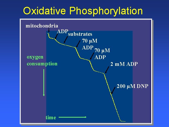
Oxidative Phosphorylation mitochondria ADPsubstrates 70 µM ADP 70 µM oxygen ADP consumption 2 m. M ADP 200 µM DNP time
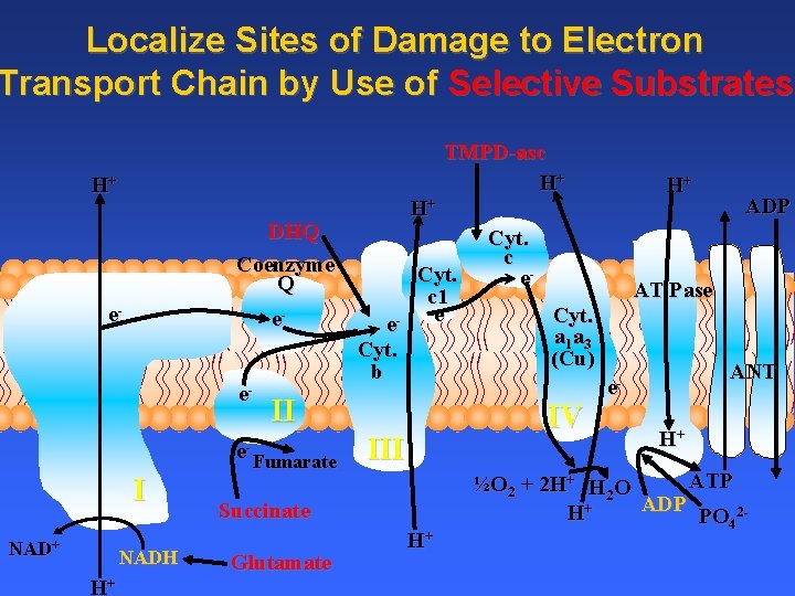
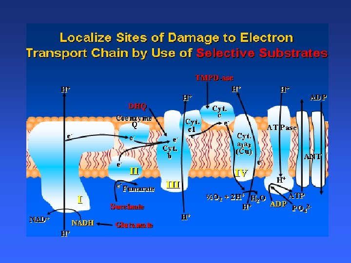
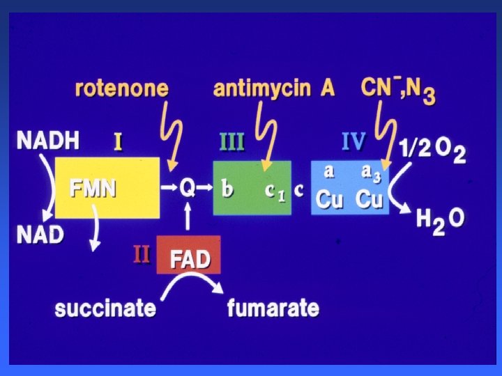
Localize Sites of Damage to Electron Transport Chain by Use of Selective Substrates TMPD-asc H+ H+ DHQ Coenzyme Q e- eee- I NAD+ NADH H+ H+ Cyt. c 1 e- e Cyt. b II Fumarate Cyt. c e- III H+ ADP AT Pase Cyt. a 1 a 3 (Cu) IV Succinate Glutamate H+ ANT e. H+ ATP ½O 2 + 2 H+ H 2 O ADP H+ PO 42 -
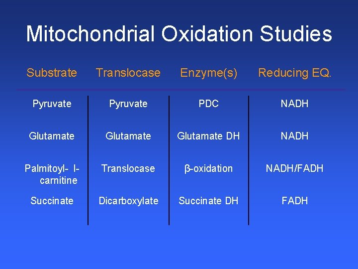
Mitochondrial Oxidation Studies Substrate Translocase Enzyme(s) Reducing EQ. Pyruvate PDC NADH Glutamate DH NADH Palmitoyl- lcarnitine Translocase β-oxidation NADH/FADH Succinate Dicarboxylate Succinate DH FADH
Clinical Phenotype of Mitochondrial Disease • Why study Mitochondria • Mitochondrial Oxidative Phosphorylation • Mitochondrial function in Patients • Respirasomes ? ? Supercomplexes!! • Respirasomes in Heart Failure • Summary and Conclusions 2008 Mc. Kim Conference, Duluth, MN September 17, 2008
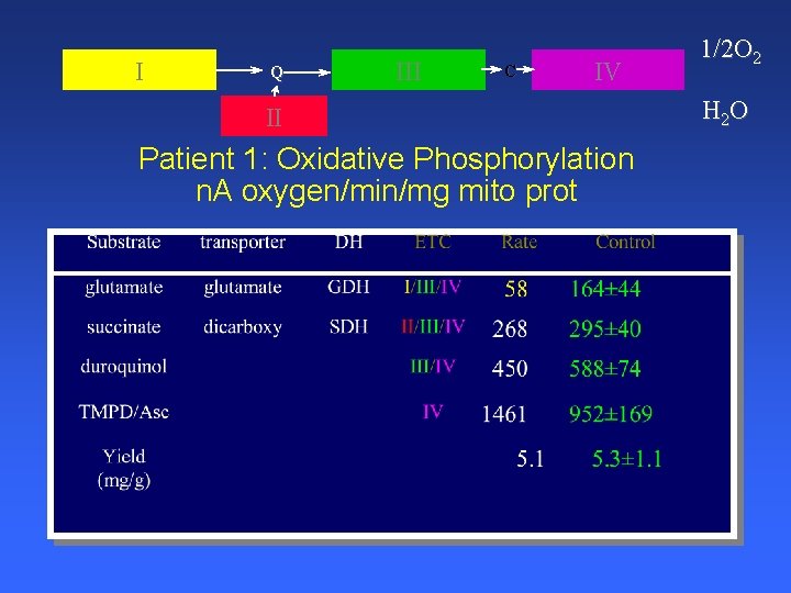
Patient 1 Ø 56 yo man CC: myalgia and fatigue Ø 2002 1 month after starting Lipitor (statin) Ø Generalized fatigue, myalgia and minimal weakness stopped Lipitor in early 2003 Ø Intermittently elevated CK 506 U/L (N=0 -232) Ø Continued symptoms off lipitor 5 years!! Ø Exercises regularly
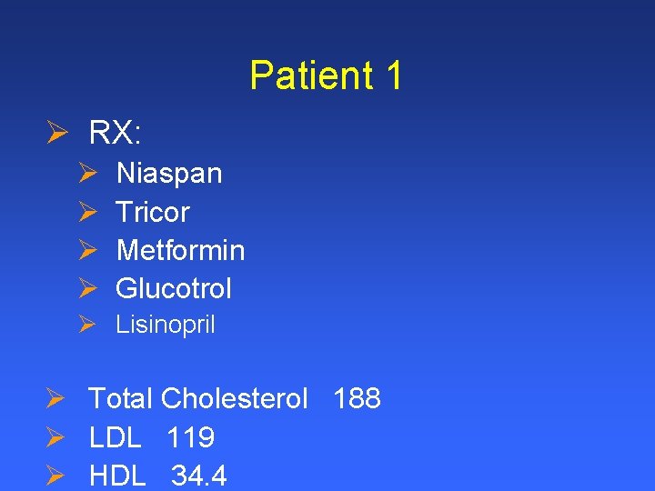
Patient 1 Ø RX: Ø Ø Niaspan Tricor Metformin Glucotrol Ø Lisinopril Ø Total Cholesterol 188 Ø LDL 119 Ø HDL 34. 4
I Q III C IV II Patient 1: Oxidative Phosphorylation n. A oxygen/min/mg mito prot 1/2 O 2 H 2 O
I Q III C IV II Patient 1: Skeletal Muscle Mitochondria ETC nmoles/min/mg mito prot
I Q III C IV II Patient 1: Skeletal Muscle ETC mol/min/g wet wt
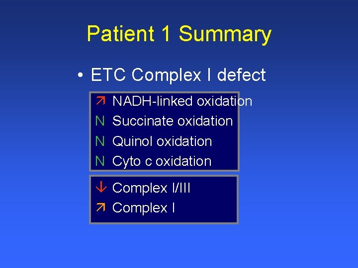
Patient 1 Summary • ETC Complex I defect ä N NADH-linked oxidation Succinate oxidation Quinol oxidation Cyto c oxidation â Complex I/III ä Complex I
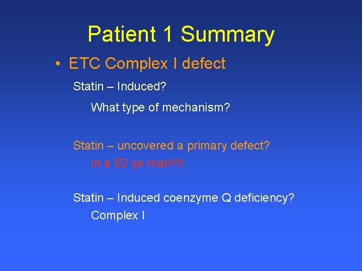
Patient 1 Summary • ETC Complex I defect Statin – Induced? What type of mechanism? Statin – uncovered a primary defect? In a 52 yo man!!!! Statin – Induced coenzyme Q deficiency? Complex I
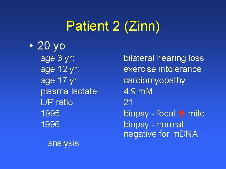
Patient 2 (Zinn) • 20 yo age 3 yr: age 12 yr: age 17 yr: plasma lactate L/P ratio 1995 1996 analysis bilateral hearing loss exercise intolerance cardiomyopathy 4. 9 m. M 21 biopsy - focal mito biopsy - normal negative for m. DNA
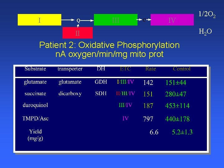
I Q III C IV II Patient 2: Oxidative Phosphorylation n. A oxygen/min/mg mito prot 1/2 O 2 H 2 O
I Q III C IV II Patient 2: Skeletal Muscle Mitochondria ETC nmoles/min/mg mito prot
I Q III C IV II Patient 2: Skeletal Muscle ETC mol/min/g wet wt
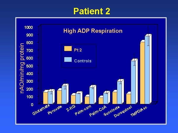
Patient 2 1000 High ADP Respiration n. AO/min/mg protein 900 800 Pt 2 700 600 Controls 500 400 300 200 100 0 Gl te te a a m v uta yru P G 2 -K rn A te ol sc o a n ca i n A C i / u c lm PD oq lm uc r a Pa M S u P T D
Patient 2 Summary • ETC Complex III defect N â ê N NADH-linked oxidation Succinate oxidation Quinol oxidation Cyto c oxidation â Complex I/III ê Complex III
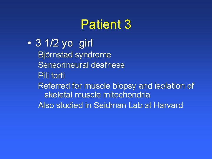
Patient 3 • 3 1/2 yo girl Björnstad syndrome Sensorineural deafness Pili torti Referred for muscle biopsy and isolation of skeletal muscle mitochondria Also studied in Seidman Lab at Harvard
I Q III C IV II Patient 3: Oxidative Phosphorylation n. A oxygen/min/mg mito prot 1/2 O 2 H 2 O
I Q III C IV II Patient 3: Skeletal Muscle Mitochondria ETC nmoles/min/mg mito prot
I Q III C IV II Patient 3: Skeletal Muscle ETC mol/min/g wet wt
Patient 3 Summary • ETC Complex III defect N N ê N NADH-linked oxidation Succinate oxidation Quinol oxidation Cyto c oxidation â Complex I/III and II/III ê Complex III
Original Article Missense Mutations in the BCS 1 L Gene as a Cause of the Björnstad Syndrome J. Travis Hinson, B. A. , Valeria R. Fantin, Ph. D. , Jost Schönberger, M. D. , Noralv Breivik, M. D. , Geir Siem, M. D. , Barbara Mc. Donough, R. N. , Pankaj Sharma, M. D. , Ph. D. , Ivan Keogh, M. D. , Ricardo Godinho, M. D. , Ph. D. , Felipe Santos, M. D. , Alfonso Esparza, M. D. , Yamileth Nicolau, M. D. , Edgar Selvaag, M. D. , Ph. D. , M. H. A. , Bruce H. Cohen, M. D. , Charles L. Hoppel, M. D. , Lisbeth Tranebjærg, M. D. , Ph. D. , Roland D. Eavey, M. D. , S. M. , J. G. Seidman, Ph. D. , and Christine E. Seidman, M. D. N Engl J Med Volume 356(8): 809 -819 February 22, 2007
Conclusion Ø BCS 1 L mutations cause disease phenotypes ranging from highly restricted pili torti and sensorineural hearing loss (the Björnstad syndrome) to profound multisystem organ failure (complex III deficiency and the GRACILE syndrome) Ø All BCS 1 L mutations disrupted the assembly of mitochondrial respirasomes (the basic unit for respiration in human mitochondria), but the clinical expression of the mutations was correlated with the production of reactive oxygen species Ø Mutations that cause the Björnstad syndrome illustrate the exquisite sensitivity of ear and hair tissues to mitochondrial function, particularly to the production of reactive oxygen species
Clinical Phenotype of Mitochondrial Disease • Why study Mitochondria • Mitochondrial Oxidative Phosphorylation • Mitochondrial function in Patients • Respirasomes ? ? Supercomplexes!! • Respirasomes in Heart Failure • Summary and Conclusions 2008 Mc. Kim Conference, Duluth, MN September 17, 2008
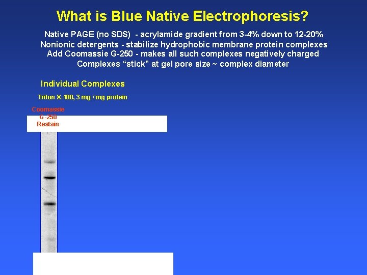
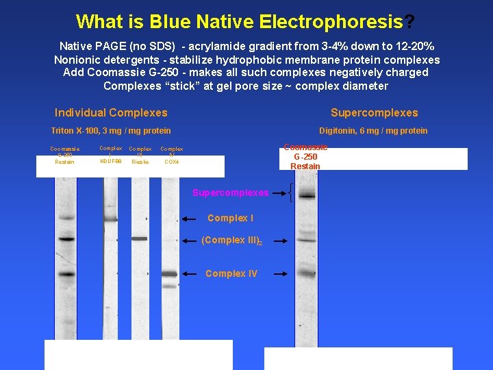
What is Blue Native Electrophoresis? Native PAGE (no SDS) - acrylamide gradient from 3 -4% down to 12 -20% Nonionic detergents - stabilize hydrophobic membrane protein complexes Add Coomassie G-250 - makes all such complexes negatively charged Complexes “stick” at gel pore size ~ complex diameter Individual Complexes Triton X-100, 3 mg / mg protein Coomassie G-250 Restain
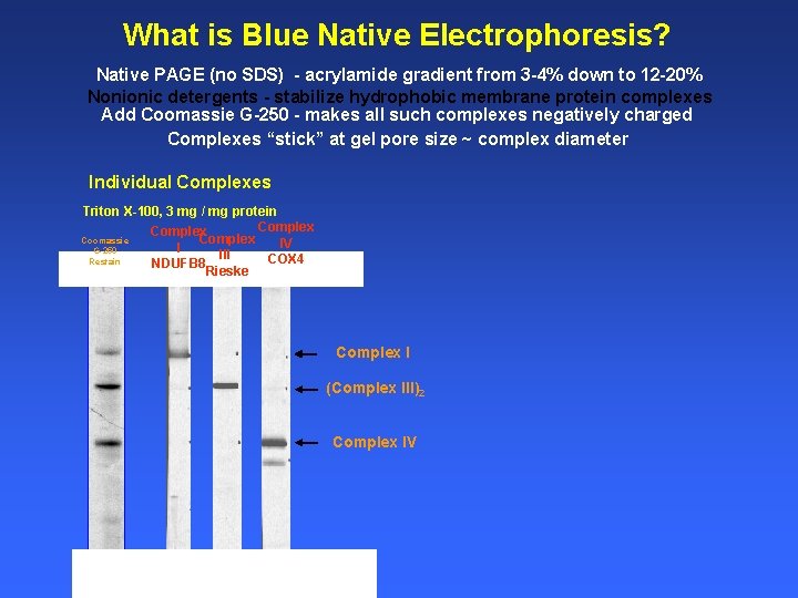
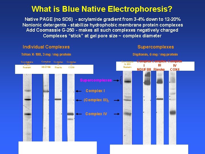
What is Blue Native Electrophoresis? Native PAGE (no SDS) - acrylamide gradient from 3 -4% down to 12 -20% Nonionic detergents - stabilize hydrophobic membrane protein complexes Add Coomassie G-250 - makes all such complexes negatively charged Complexes “stick” at gel pore size ~ complex diameter Individual Complexes Triton X-100, 3 mg / mg protein Complex Coomassie Complex IV I G-250 III COX 4 Restain NDUFB 8 Rieske Complex I (Complex III)2 Complex IV
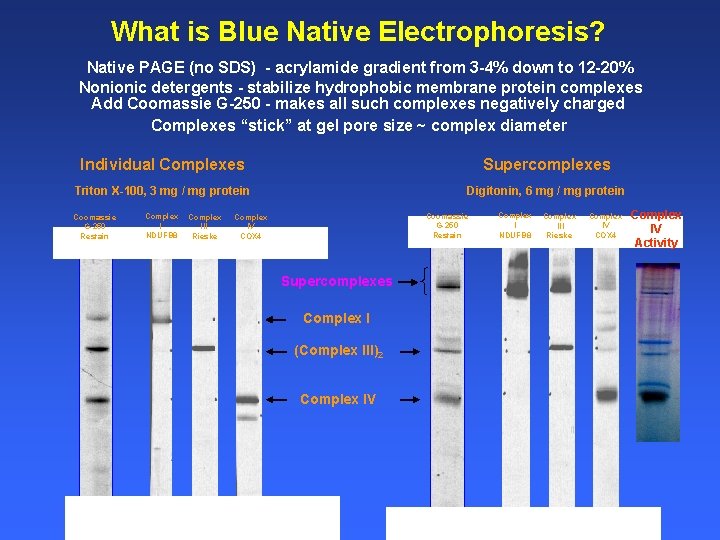
What is Blue Native Electrophoresis? Native PAGE (no SDS) - acrylamide gradient from 3 -4% down to 12 -20% Nonionic detergents - stabilize hydrophobic membrane protein complexes Add Coomassie G-250 - makes all such complexes negatively charged Complexes “stick” at gel pore size ~ complex diameter Individual Complexes Supercomplexes Triton X-100, 3 mg / mg protein Digitonin, 6 mg / mg protein Coomassie G-250 Restain Complex I NDUFB 8 Complex III Rieske Coomassie G-250 Restain Complex IV COX 4 Supercomplexes Complex I (Complex III)2 Complex IV
What is Blue Native Electrophoresis? Native PAGE (no SDS) - acrylamide gradient from 3 -4% down to 12 -20% Nonionic detergents - stabilize hydrophobic membrane protein complexes Add Coomassie G-250 - makes all such complexes negatively charged Complexes “stick” at gel pore size ~ complex diameter Individual Complexes Supercomplexes Triton X-100, 3 mg / mg protein Digitonin, 6 mg / mg protein Coomassie G-250 Restain Complex I NDUFB 8 Complex III Rieske Coomassie G-250 Restain Complex IV COX 4 Supercomplexes Complex I (Complex III)2 Complex IV Complex III I IV Rieske NDUFB 8 COX 4
What is Blue Native Electrophoresis? Native PAGE (no SDS) - acrylamide gradient from 3 -4% down to 12 -20% Nonionic detergents - stabilize hydrophobic membrane protein complexes Add Coomassie G-250 - makes all such complexes negatively charged Complexes “stick” at gel pore size ~ complex diameter Individual Complexes Supercomplexes Triton X-100, 3 mg / mg protein Digitonin, 6 mg / mg protein Coomassie G-250 Restain Complex I NDUFB 8 Complex III Rieske Coomassie G-250 Restain Complex IV COX 4 Supercomplexes Complex I (Complex III)2 Complex IV Complex I NDUFB 8 Complex III Rieske Complex IV COX 4 Complex IV Activity
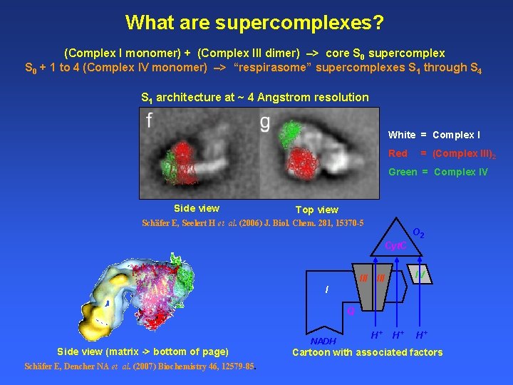
What are supercomplexes? (Complex I monomer) + (Complex III dimer) –> core S 0 supercomplex S 0 + 1 to 4 (Complex IV monomer) –> “respirasome” supercomplexes S 1 through S 4 S 1 architecture at ~ 4 Angstrom resolution White = Complex I Red = (Complex III)2 Green = Complex IV Side view Top view Schäfer E, Seelert H et al. (2006) J. Biol. Chem. 281, 15370 -5 O 2 Cyt. C III IV III I Q Side view (matrix -> bottom of page) Schäfer E, Dencher NA et al. (2007) Biochemistry 46, 12579 -85. NADH H+ H+ H+ Cartoon with associated factors
Clinical Phenotype of Mitochondrial Disease • Why study Mitochondria • Mitochondrial Oxidative Phosphorylation • Mitochondrial function in Patients • Respirasomes ? ? Supercomplexes!! • Respirasomes in Heart Failure • Summary and Conclusions 2008 Mc. Kim Conference, Duluth, MN September 17, 2008
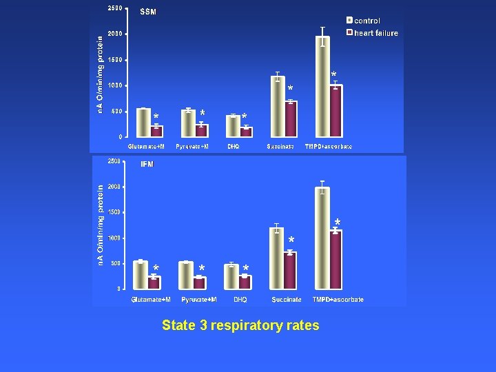
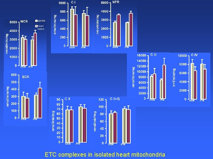
State 3 respiratory rates
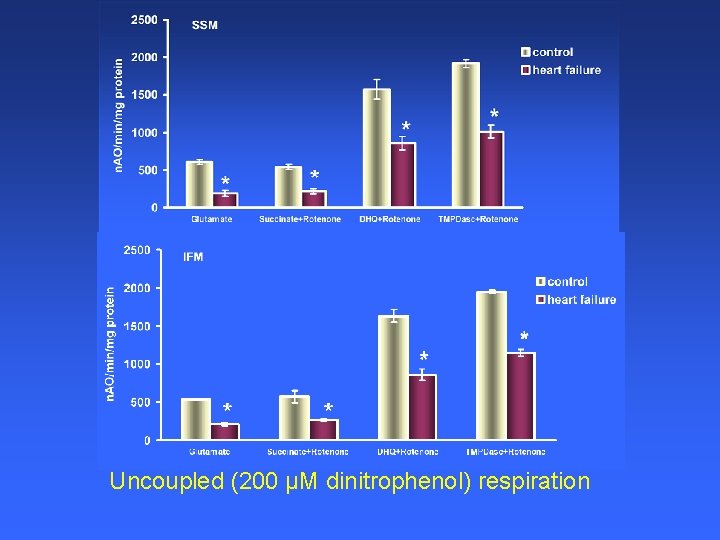
Uncoupled (200 µM dinitrophenol) respiration
ETC complexes in isolated heart mitochondria
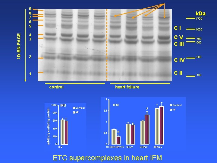
1 D-BN-PAGE 9 8 7 6 5 k. Da 1700 4 3 2 CI 1000 CV C III 750 500 C IV C II 1 control heart failure ETC supercomplexes in heart IFM 200 130
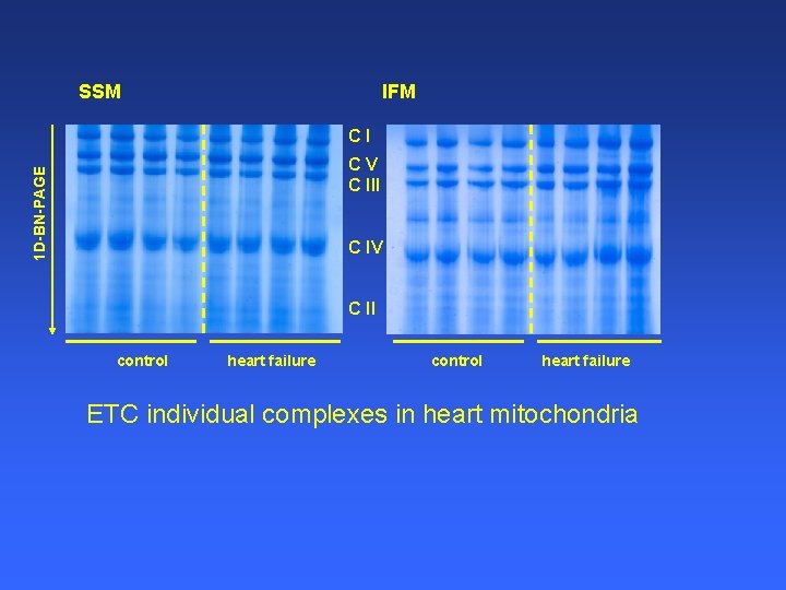
SSM IFM CI 1 D-BN-PAGE CV C III C IV C II control heart failure ETC individual complexes in heart mitochondria
CONCLUSION In HF the mitochondrial defect lies in the supermolecular assembly of the ETC in functional respirasomes. We propose that phosphorylation of specific complex IV subunits is involved in disruption of supercomplex assembly of the mitochondrial ETC in HF.
Acknowledgements PPG - Laboratory • Mariana Rosca, M. D. Collaborators • Hani Sabbah, Ph. D. Henry Ford Hospital Detroit, MI • Edwin Vazquez, B. S. • William Stanley, Ph. D. • Janos Kerner, Ph. D. University of Maryland Baltimore, MD • Margaret Chandler, Ph. D. Center for Inherited Disorders of Energy Metabolism • William Parland, Ph. D. Clinical • David Kehres, Ph. D. • Bruce Cohen, MD • Hiral Patel • Sumit Parikh, MD • Douglas Kerr, MD, Ph. D • Colin Naples • Shawn Mc. Candless Funding Support: PPG P 01 HL 074237 EPrint P 01 AG 015885 Cardiovascular Research R 01 HL 64848 August 18, 2008