
Clinical Pathological Conference Elizabeth Ross M D n
Clinical Pathological Conference Elizabeth Ross, M. D. n Chief Resident Department of Medicine n October 12 th, 2007 n n
Chief Complaint n A 46 year old Dominican woman presents with 3 months of increasing abdominal distention and one month of diffuse epigastric pain
History of Present Illness n n 2 -3 years prior to admission: patient first noticed easy bruisability, she was diagnosed with “anemia” and iron supplementation was started. 3 months pta: she noticed abdominal distention and was started on a “water pill”. 1 -2 months pta: Her abdominal distention progressed, she felt like she looked pregnant. 2 -3 weeks pta: unrelenting diffuse epigastric pain and discomfort.
HPI, continued n n Her pain persisted so she sought medical attention and was admitted to an outside hospital Imaging and lab studies revealed abnormal LFTs and portal and splenic vein thrombosis She was started on a heparin drip and transferred to Bellevue Repeat imaging confirmed IVC and hepatic vein thrombosis and also showed portal and splenic vein thrombosis
Additonal History Past Medical History: As above Past Surg History: Tuboligation 15 years ago Medications: iron, multivitamin On transfer: heparin drip Allergies: none Family History: Denies history of: clotting disorders, bleeding disorders, malignancy Social History: Born in Dominican Republic, has lived in the US for 10 years, no recent travel. Ten pack-year tobacco history, quit 9 years ago. No etoh, no illicit drug use. Lives with husband. Worked as HHA until four months ago.
Review of Symptoms n Monthly, regular menstruation since menarche, with heavy bleeding
Physical Exam n n n General: well-developed woman with apparent ascites, moaning in pain, appears stated age, mildly jaundice Vital signs: BP 127/82, HR 108 and regular, RR 18, Temp 97. 6, Sp. O 2 97% room air HEENT: oropharynx dry, mild scleral icterus Lymph: no cervical, axillary or inguinal lymphadenopathy Neck: supple, no jugular venous distension Pulmonary: clear to auscultation bilaterally
Physical Exam, continued n n n n Heart: tachycardic, regular rhythm, normal heart sounds, no murmurs Abdominal: Distended, diffusely tender, shifting dullness present, fluid wave present, no masses palpable Extremities: trace lower extremity edema bilaterally, 2+ peripheral pulses Skin: no rashes Rectal: guaiac negative Neuro: Alert and oriented to person, place and time Asterixis present
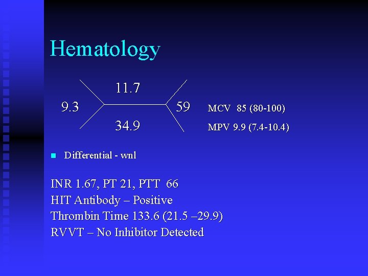
Hematology 11. 7 9. 3 59 34. 9 n MCV 85 (80 -100) MPV 9. 9 (7. 4 -10. 4) Differential - wnl INR 1. 67, PT 21, PTT 66 HIT Antibody – Positive Thrombin Time 133. 6 (21. 5 – 29. 9) RVVT – No Inhibitor Detected
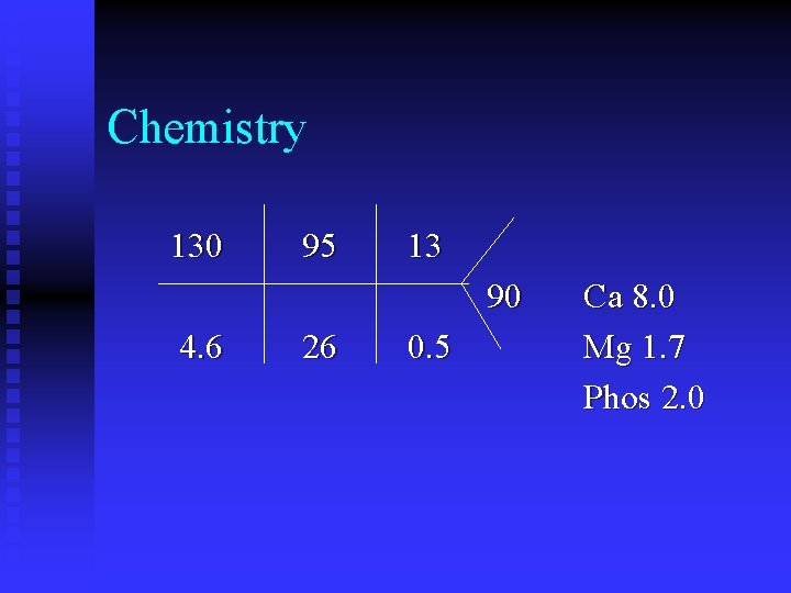
Chemistry 130 95 13 90 4. 6 26 0. 5 Ca 8. 0 Mg 1. 7 Phos 2. 0
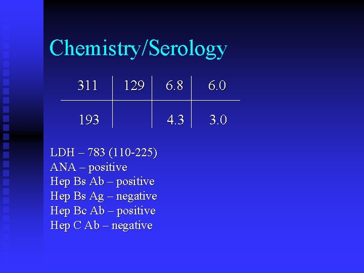
Chemistry/Serology 311 129 193 LDH – 783 (110 -225) ANA – positive Hep Bs Ab – positive Hep Bs Ag – negative Hep Bc Ab – positive Hep C Ab – negative 6. 8 6. 0 4. 3 3. 0
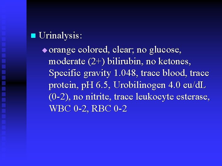
n Urinalysis: u orange colored, clear; no glucose, moderate (2+) bilirubin, no ketones, Specific gravity 1. 048, trace blood, trace protein, p. H 6. 5, Urobilinogen 4. 0 eu/d. L (0 -2), no nitrite, trace leukocyte esterase, WBC 0 -2, RBC 0 -2
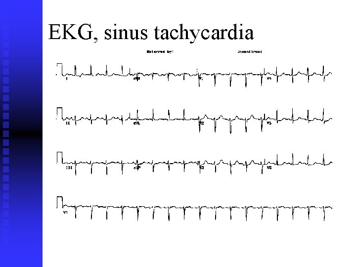
EKG, sinus tachycardia
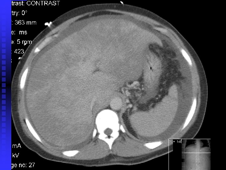
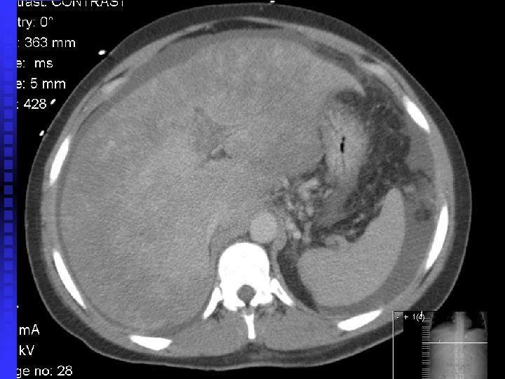
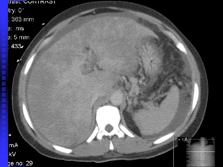
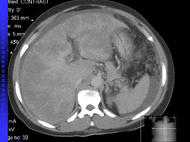
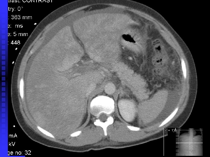
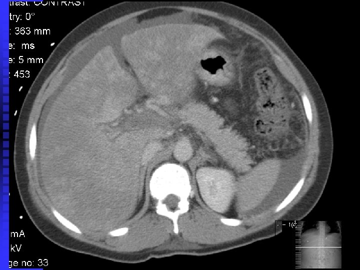
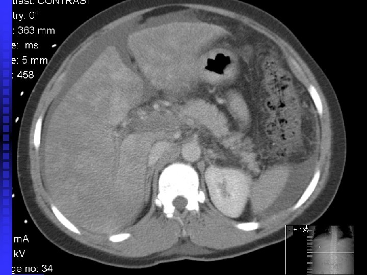
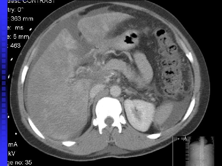
Abd/Pelvic CT with contrast n Hepatic vein, portal vein, splenic vein and IVC thromboses n … A diagnostic procedure was performed
Student Discussants Marty Wolf – Paroxysmal Nocturnal Hematuria n Leo Drozhinin – Anti-phospholipid Antibody Syndrome n Marci Cherit – Autoimmune Hepatitis n Cristobal Gao – Gastric Cancer n
Abdominal/pelvic CT with IV contrast Dr. Kay Cho
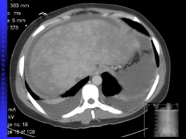
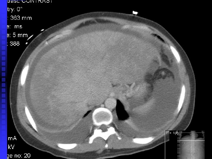
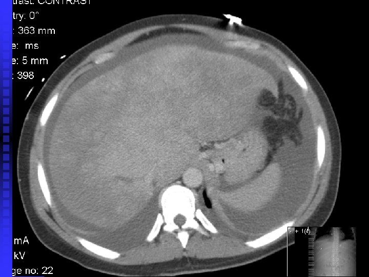
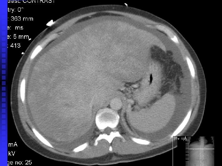
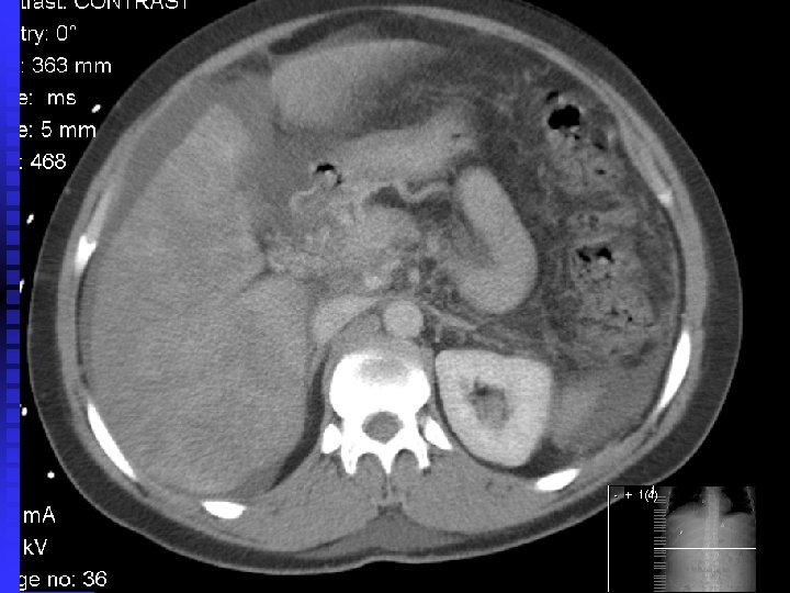
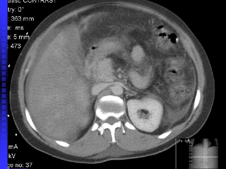

Faculty Discussant Dr. Mitchell Charap

A DIAGNOSTIC PROCEDURE WAS PERFORMED

Hematologist/Hematopathologist Dr. David Araten
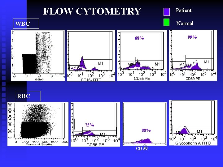
FLOW CYTOMETRY Patient Normal WBC 68% RBC 75% 88% CD 59 99%
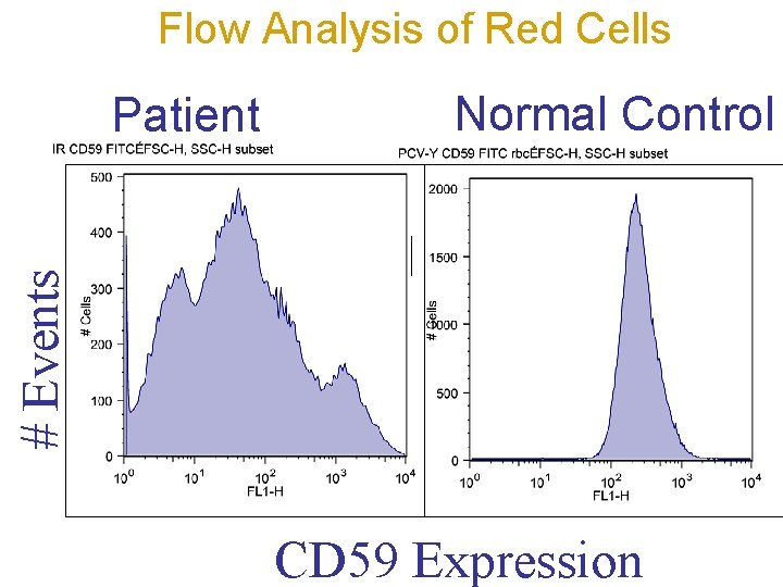
Flow Analysis of Red Cells Normal Control # Events Patient CD 59 Expression
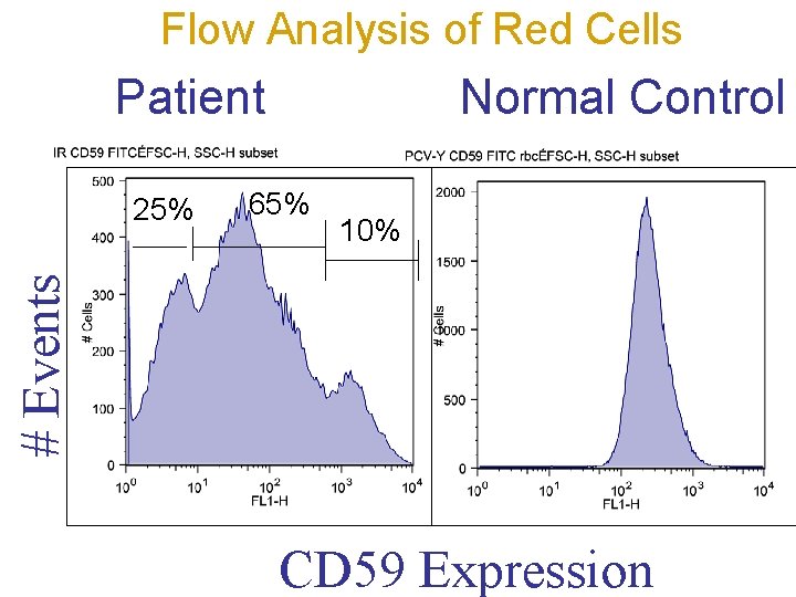
Flow Analysis of Red Cells Patient 65% 10% # Events 25% Normal Control CD 59 Expression
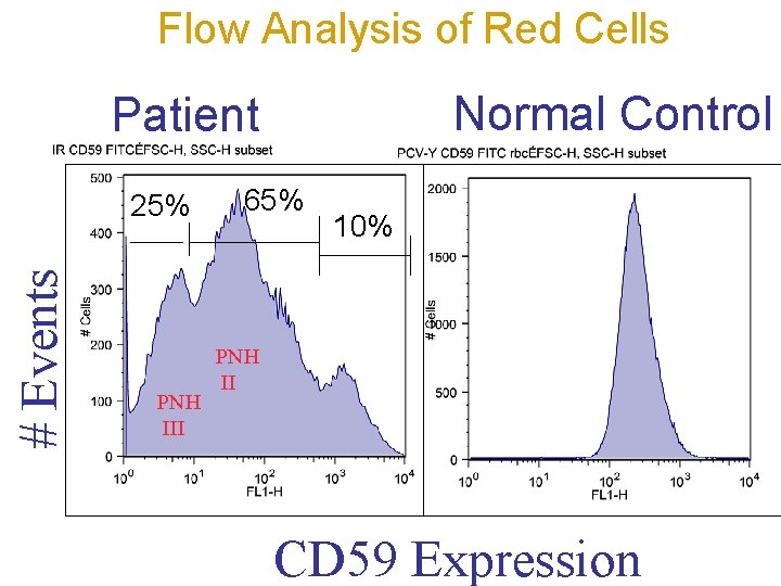
Flow Analysis of Red Cells Normal Control Patient # Events 25% PNH III 65% 10% PNH II CD 59 Expression
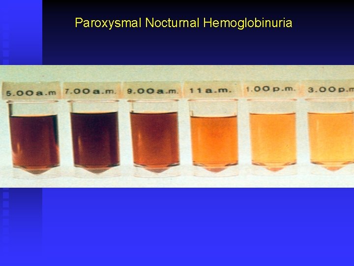
Paroxysmal Nocturnal Hemoglobinuria
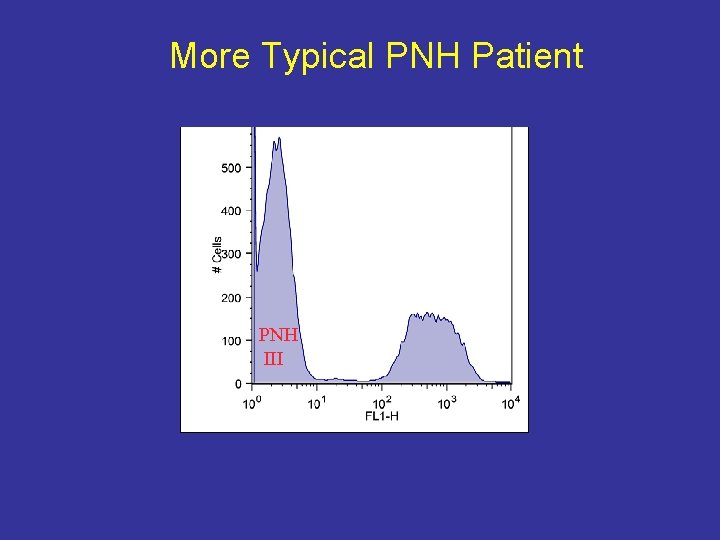
More Typical PNH Patient PNH III
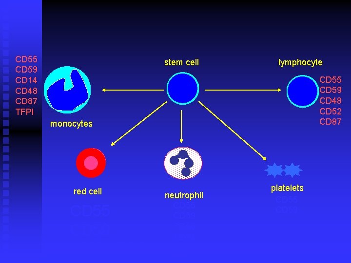
CD 55 CD 59 CD 14 CD 48 CD 87 TFPI stem cell lymphocyte CD 55 CD 59 CD 48 CD 52 CD 87 monocytes red cell CD 55 CD 59 acetylcholinesterase neutrophil CD 55 CD 59 CD 66 CD 24 CD 16 platelets CD 55 CD 59
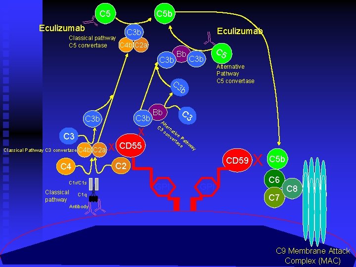
C 5 Eculizumab C 5 b Eculizumab C 3 b Classical pathway C 5 convertase C 4 b C 2 a C 3 b Bb C 3 b X C 3 Classical Pathway C 3 convertase C 4 b X CD 55 C 1 r/C 1 s C 1 q Alternative Pathway C 5 convertase C 3 Al C 3 tern co ativ nv e er Pa ta th se w a y CD 59 C 2 C 4 Classical pathway C 2 a Bb C 5 GPI X C 5 b C 6 C 7 C 8 Antibody C 9 Membrane Attack Complex (MAC)
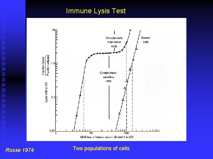
Immune Lysis Test Rosse 1974 Two populations of cells
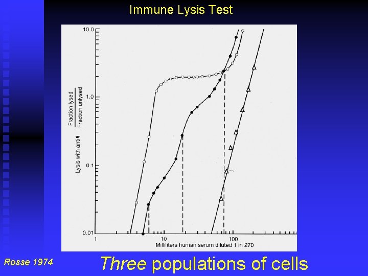
Immune Lysis Test Rosse 1974 Three populations of cells
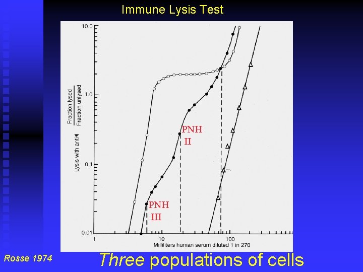
Immune Lysis Test PNH III Rosse 1974 Three populations of cells
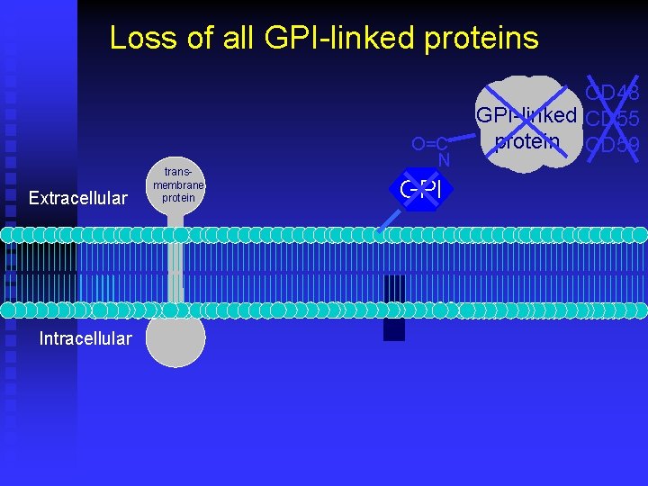
Loss of all GPI-linked proteins Extracellular Intracellular transmembrane protein O=C N GPI CD 48 GPI-linked CD 55 protein CD 59
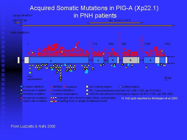
Acquired Somatic Mutations in PIG-A (Xp 22. 1) Large deletion in PNH patients del 735 bp del exons 3 -4 -5 . ** * * Ava I polymorphism in frame deletion missense mutation inherited mutation nonsense mutation splice site mutation * * ** 3 2 1 ** ** * 716 H 849 4 * * * 1 ** ** Null mutations * * 982 5 *1189 * * * 1452 6 50 bp deletion insertion non coding region insertion/deletion TM, Transmembrane domain (nt 1246 -1326, aa 415 -442) Glc. NAc transferase homologous region (nt 912 -1185, aa 304 -395) insertion/duplication changes very close to each other, presumably H: Hot spot reported by Mortazavi et al 2003 resulting from a single mutational event * From Luzzatto & Nafa 2000
Marked Geographical Disparity n High rates of thrombosis reported in the United States, Europe and India n Much lower rates in Mexico, Japan, China, and Thailand
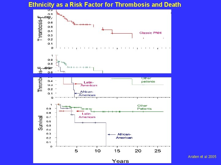
Ethnicity as a Risk Factor for Thrombosis and Death Araten et al 2005

Wrap-Up Dr. Elizabeth Ross
Final Diagnosis: Paroxysmal Nocturnal Hemoglobinuria -with granulocyte and erythrocyte clone size greater than 50%
PNH An acquired hemolytic anemia somatic mutation of the PIG-A gene in a multipotent hematopoietic stem cell n This results in the impaired synthesis of the GPI anchor so proteins (CD 55, 59) can’t link antigens to cell surface n Used to be diagnosed with Ham’s test, illustrating red cells’ susceptibility to lysis by complement, now flow cytometry is used n
PNH See intravascular hemolysis, cytopenias and venous thrombosis n Venous thrombosis occurs in ~50% of patients which is the most common cause of death n When granulocyte clone size is larger than 50%, patients are at increased risk of thrombosis (Hall et al, Blood, 2003) n PNH accounts for 15 -25% of patients with hepatic venous thrombosis n
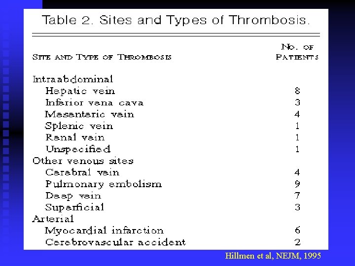
Hillmen et al, NEJM, 1995
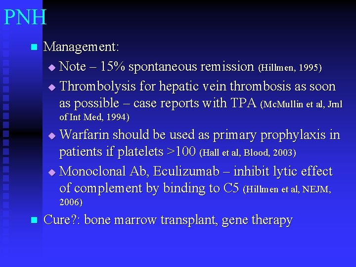
PNH n Management: u Note – 15% spontaneous remission (Hillmen, 1995) u Thrombolysis for hepatic vein thrombosis as soon as possible – case reports with TPA (Mc. Mullin et al, Jrnl of Int Med, 1994) Warfarin should be used as primary prophylaxis in patients if platelets >100 (Hall et al, Blood, 2003) u Monoclonal Ab, Eculizumab – inhibit lytic effect of complement by binding to C 5 (Hillmen et al, NEJM, u 2006) n Cure? : bone marrow transplant, gene therapy
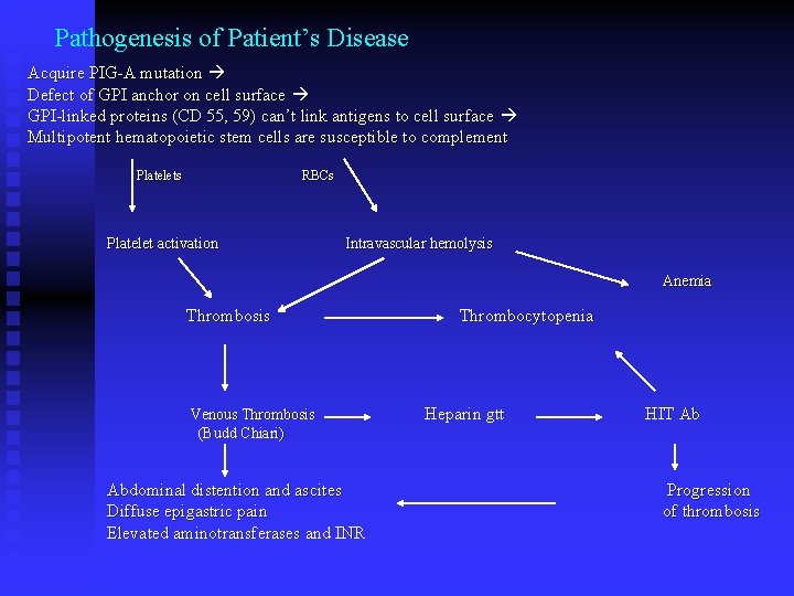
Pathogenesis of Patient’s Disease Acquire PIG-A mutation Defect of GPI anchor on cell surface GPI-linked proteins (CD 55, 59) can’t link antigens to cell surface Multipotent hematopoietic stem cells are susceptible to complement Platelets RBCs Platelet activation Intravascular hemolysis Anemia Thrombosis Venous Thrombosis (Budd Chiari) Abdominal distention and ascites Diffuse epigastric pain Elevated aminotransferases and INR Thrombocytopenia Heparin gtt HIT Ab Progression of thrombosis
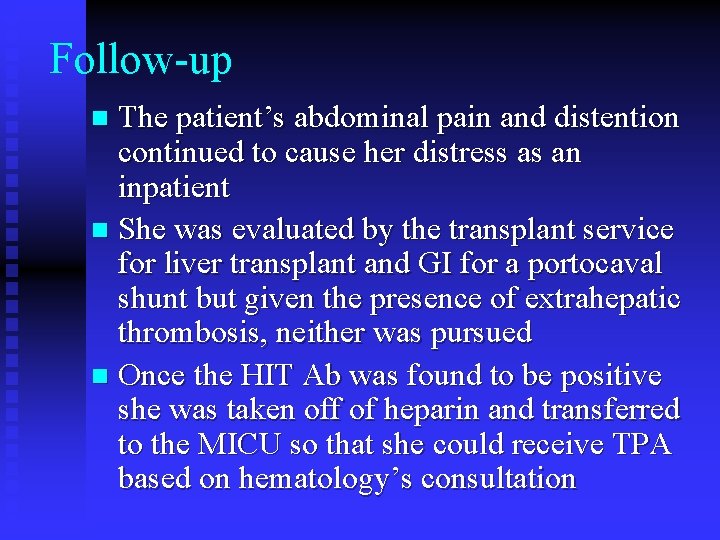
Follow-up The patient’s abdominal pain and distention continued to cause her distress as an inpatient n She was evaluated by the transplant service for liver transplant and GI for a portocaval shunt but given the presence of extrahepatic thrombosis, neither was pursued n Once the HIT Ab was found to be positive she was taken off of heparin and transferred to the MICU so that she could receive TPA based on hematology’s consultation n
Follow-up n She received 6 cycles of tissue plasminogen activator (Alteplase) – which was stopped secondary to a thigh hematoma and she was started on lepirudin (Refludan) and started on warfarin with a goal INR 2. 8 -3. 4 n Abd/Pel CT and doppler ultrasound 2 weeks after admission (approximately 1 week after the TPA) revealed some flow through the hepatic vein
Follow-up n n Once her ascites and epigastric pain improved, and her INR was at goal, she was discharged Approximately one year later she had an abd ultrasound with doppler which revealed: -patent main portal vein, collateral flow in the porta hepatis supplying the left portal vein, patent hepatic and splenic veins. No ascites. Abd/pel CT showed: portal venous hypertension Her platelets remain near 90, 000
Thank You Martin Blaser, MD Mitchell Charap, MD David Araten, MD Kay Cho, MD Josh Olstein, MD
- Slides: 59