Clinical microbiology By Dr Hussein Al Naji Clinical

Clinical microbiology By Dr. Hussein Al. Naji

Clinical microbiology It is a branch of medical science concerned with the prevention, diagnosis and treatment of infectious diseases. In addition, this field of science studies various clinical applications of microbes for the improvement of health. There are four kinds of microorganisms that cause infectiou disease: bacteria, fungi, parasites and viruses, and one type of infectious protein called prion. This lecture focuses on the Gram stain, bacterial morphology, and metabolic characteristics, all of which enable the clinician to rapidly determine the organism causing infection.
 Because bacteria are colorless")
Demonstration of an infectious agent A. Direct smear (GRAM STAIN) Because bacteria are colorless and usually invisible to light microscopy, colorful stains have been developed to visualize them. The most useful is the Gram stain, which separates organisms into 2 groups: gram-positive bugs and gram-negative bugs. This stain also allows the clinician to determine whether the organism is round or rod-shaped. For any stain you must first smear the substance to be stained (sputum, pus, etc. ) onto a slide and then heat it to fix the bacteria on the slide.
 Pour on crystal violet stain")
There are 4 steps to the Gram stain: 1) Pour on crystal violet stain (a blue dye) and wait 60 seconds. 2) Wash off with water and flood with iodine solution. Wait 60 seconds. 3) Wash off with water and then "decolorize" with 95% alcohol. 4) Finally, counter-stain with safranin (a red dye). Wait 30 seconds and wash off with water. When the slide is studied microscopically, cells that absorb the crystal violet and hold onto it will appear blue. These are called gram-positive organisms. However, if the crystal violet is washed off by the alcohol, these cells will absorb the safranin and appear red. These are called gram-negative organisms. The different stains are the result of differences in the cell walls of gram-positive and gram-negative bacteria.

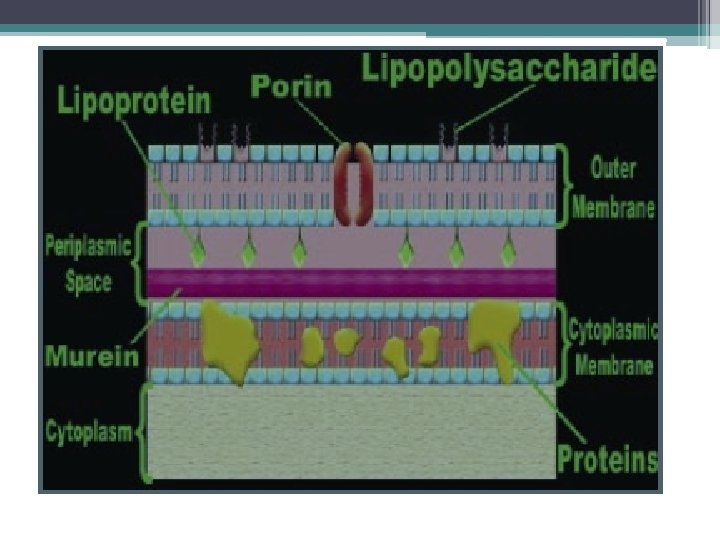
STRUCTURE OF GRAM-NEGATIVE BACTERIA. A. Cell Wall 1. outer membrane serves as the primary permeability barrier of the cell and helps to retain proteins in the periplasmic space. Porins are water-filled channels in the outer membrane that facilitate transport of nutrients and low molecular weight substances, including antimicrobial agents, into the cell. Bacteria vary in the number and types of porins they contain. 2. Lipopolysaccharides are found on the surface of the cell and are the major component of endotoxin. They contribute to the bacterium’s ability to cause disease and they give gram-negative bacteria their net negative charge.

4. Lipoproteins attach the outer membrane to the murein layer. 5. The peptidoglycan layer of gram-negative bacteria is a relatively thin polymer consisting of cross-linked N-acetylmuramic acid and N -acetylglucosamine. It is often referred to as the murein layer or cell wall and is responsible for maintaining the shape of the organism. It is located within the periplasmic space. 6. The periplasmic space lies between the outer membrane and the cytoplasmic membrane. Periplasmic proteins include binding proteins for specific substrates, hydrolytic enzymes and detoxifying enzymes.

c. Cytoplasmic Membrane The cytoplasmic membrane surrounds the cytoplasm of the cell and contains proteins and phospholipids. Many of the proteins contained in the cell membrane are enzymes responsible for cellular metabolism. The cytoplasmic membrane also serves as a permeability barrier and a permeability link for substances entering the cell. d. Cytoplasm and Other Internal Components The cell cytoplasm contains the chromosome, ribosomes and other internal structures. The vast majority of bacteria have a single chromosome but a few, such as Vibrio cholera, have two chromosomes.

STRUCTURE OF GRAM-POSITIVE BACTERIA Cell Wall Since the gram-positive cell wall contains only two major components it is much less complicated than the gram-negative cell wall. • Teichoic acids are polymers that are interwoven in the peptidoglycan layer and extend as hair-like projections beyond the surface of the gram-positive cell. They also are major surface antigens in those organisms that possess them. • The peptidoglycan layer, or murein layer, of gram-positive bacteria is much thicker than that of gram-negative bacteria. It is responsible for maintaining the shape of the organism and often is referred to as the cell wall.

The Cytoplasmic Membrane, Cytoplasm, and Other Internal Components These structures are very similar in both gram-positive and gram-negative bacteria.

Gram-Positive Cells 2 Layers: 1. Inner cytoplasmic membrane 2. Outer thick peptidoglycan layer (60 -100% peptidoglycan Gram- Negative cells 3 Layers: 1. Inner cytoplasmic membrane 2. Thin peptidoglycan layer (510% peptidoglycan) 3. Outer membrane with lipopolysaccharide (LPS) Low lipid content NO endotoxin (except Listeria High lipid content Endotoxin (LPS) - lipid A monocytogenes) NO periplasmic space Periplasmic space NO porin channel Vulnerable to lysozyme and porin channel Resistant to lysozyme and
 Cocci: spherical. 2) Bacilli: rods. Short bacilli are")
Bacteria have 4 major shapes: 1) Cocci: spherical. 2) Bacilli: rods. Short bacilli are called coccobacilli. 3) Spiral forms: comma-shaped, S-shaped, or spiral- shaped. 4) Pleomorphic: lacking a distinct shape (like jello). The different shaped creatures organize together into more complex patterns, such as pairs (diplococci), clusters, strips, and single bacteria with flagella.
- Slides: 12