Clinical disorders of acidbase balance part I Mohamed
 Mohamed Osama Ezwaie, MD Associate")
Clinical disorders of acid-base balance ( part I ) Mohamed Osama Ezwaie, MD Associate professor of nephrology ezwaie@yahoo. com 2019
 • Definitions • Human body acids •")
Learning objectives ( part I & II) • Definitions • Human body acids • Chemical and mathematical basis for acid base disorders • Physiology of acid base balance/ homeostasis • ECF ionic composition • Clinical disorders of acid base disorders • Step-wise diagnostic approach

Acid- base definitions • Acid - substance that generates H+ in solutions, When an acid releases a proton it is converted to conjugate base. • Base - substance that generate OH- in solutions, When a base binds a proton it is converted to conjugate acid. • An acid base disorder is a change in the normal value of extracellular p. H that may result when renal or respiratory function is abnormal or when an acid or base load overwhelms excretory capacity.

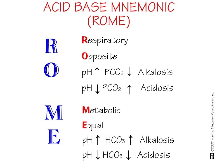
Acid- base definitions • Acidemia - Reduction in the blood p. H below normal range of 7. 35 -7. 45 • Alkalemia - Elevation in blood p. H above the normal range of 7. 35 – 7. 45 • Acidosis – process that increases [H+] by increasing PCO 2 or by reducing [HCO 3 • Alkalosis – process that reduces [H+] by reducing PCO 2 or by increasing [HCO 3 -] • Normal arterial acid base values Pa. CO 2 p. H ( mm. Hg) HCO 3 ( m. Eq/L) Range 7. 35 - 7. 45 36 -44 22 -26 Optimal value 7. 40 40 24

Acid- base definitions • Simple acid base disorders : Disorders that are either metabolic or respiratory. • Mixed acid base disoders: More than one acid base disturbance present. p. H may be normal or abnormal. • Though acidosis and alkalosis usually leads to acidemia and alkalemia respectively, the exception occurs when there is a mixed acid base disorder.

Acid- base disorders; definitions • Though acidosis and alkalosis usually leads to acidemia and alkalemia respectively, the exception occurs when there is a mixed acid base disorder. • In that situation, multiple acid base processes coexisting may lead to a normal p. H or a mixed picture. • Clinical disturbances of acid base metabolism classically are defined in terms of the HCO 3 - /CO 2 buffer system.

Human body acids • The human body generates acids continuously. A normal subject produces daily ; • 20 mmol of "volatile" acid (carbonic acid) and • 80 mmol of "nonvolatile" acids. • Most of the volatile acid is produced as CO 2 as result of cell respiration/ metabolism and is subject to the reaction: CO 2 + H 2 O ↔ H 2 CO 3 ↔ H+ + HCO 3 Occurs at pulmonary cap. level Occurs at tissue cap. level

Human body acids • Occurs at pulmonary cap. level Occurs at tissue cap. level

Human body acids • The non-volatile acids generated during metabolic processes dissociate and produce hydrogen ions: • Part of these ions is used in metabolism ( enzyme function) • Some fraction binds to oxygen in water to form hydronium ions ( hydrated protons H 3 O+, H 5 O 2+ ) • While rest remains free in plasma water, contributing to plasma p. H
![Human body acids • Because [H+] is so critical to enzyme function yet the](http://slidetodoc.com/presentation_image_h/25e3696e3d8417e1a7a2e76cc3261c32/image-10.jpg "Human body acids • Because [H+] is so critical to enzyme function yet the")
Human body acids • Because [H+] is so critical to enzyme function yet the absolute concentration is small • The hydrogen ion is kept in the body under rigorous control, its concentration is maintained within very narrow range: • [ H+]= 35 - 45 nanomols per liter of body fluid

Human body acids • The p. H of the body must be maintained within a narrow range • Most body systems function optimally at a p. H of near 7. 4 • As the p. H changes (either higher or lower); • Enzymes may cease to function • Nerve and muscle activity weakens • Finally all metabolic activity becomes deranged
![[Plasma ion] Vs. [hydrogen ion] Ion* nmoles/L H+ 40 K+ 4, 000 Ca++ 2,](http://slidetodoc.com/presentation_image_h/25e3696e3d8417e1a7a2e76cc3261c32/image-12.jpg "[Plasma ion] Vs. [hydrogen ion] Ion* nmoles/L H+ 40 K+ 4, 000 Ca++ 2,")
[Plasma ion] Vs. [hydrogen ion] Ion* nmoles/L H+ 40 K+ 4, 000 Ca++ 2, 500, 000 Mg++ 1, 000 Na+ 140, 000 *K+, Potassium ion; Ca++, calcium ion; Mg++, magnesium ion; Na+, sodium ion.

Concept of p. H; logarithmic scale • The p. H is a method of expressing extremely small concentrations (of acid in solution) in numbers, and has no unit • To understand logarithm, think of "power. " Thus 103 = 1000 and log (1000) = 3 • p. H is defined as the negative logarithm of the hydrogen ion (H+) concentration or activity p. H = - log [H+] • The scale is exponential — a change of one unit is actually a ten-fold change in[ H + ], a solution with p. H of 7 has ten times as much acid as a solution with p. H of 8.

Concept of p. H; logarithmic scale • The intracellular p. H is about 7. 0 at body temperature - fairly close to neutral and tightly maintained; it is where most of the body's chemistry occurs • The body maintains the blood at p. H 7. 4, which is about 0. 4 p. H units more alkaline than the intracellular p. H • This is equivalent to a 2. 5 -fold difference in concentration: extracellular 40 n. Mol/L; intracellular 100 n. Mol/L • Converting chemical formula to logarithmic formula ( scale/ equation), which is the standard equation used in acid-base clinical chemistry

Equations of acid –base chemistry • The conversion of chemical equation of carbonic acid system to mathematical equation CO 2 + H 2 O ↔ H 2 CO 3 ↔ H+ + HCO 3 - ( chemical equation) [H+] x [HCO 3 -] = K x [CO 2] x [H 2 O] (Henderson mathematical / equilibrium equation) “ K ” is equilibrium constant

Equations of acid –base chemistry • The next step is to write Henderson's equation in the right order with the water concentration omitted as a constant. [H+] = K x [CO 2] / [HCO 3 -] • Hasselbalch modified Henderson's elegant idea by regarding the water concentration as constant and taking logarithms of the remaining components (p. K is the negative logarithm of "K"). This resulted in the • Henderson-Hasselbalch Equation- primary form: p. H = p. K + log ( [HCO 3 -] / [CO 2] )

Henderson-Hasselbach logarithmic equation • The value of p. K in the bicarbonate buffer system (p. K = 6. 1) • The constant 0. 03 converts Pa. CO 2 from mm Hg to mmoles/L. may be substituted to come up with this final equation • Henderson-Hasselbalch Equation-final form: p. H = 6. 1 +log HCO 3 -/ (0. 03 x PCO 2)

Chemical to logarithmic equation CO 2 + H 2 O ↔ H 2 CO 3 ↔ H+ + HCO 3 - ( chemical equation) Henderson-Hasselbalch Equation: p. H = 6. 1 +log HCO 3 -/ (0. 03 x PCO 2)

Henderson-Hasselbach logarithmic equation • Henderson-Hasselbalch Equation:

Acid-Base clinical physiology • Body defenses against acid-base disturbances: • The chemical buffer systems ( ECF, and intracellular); acts immediately, first line of defense, but can not remove hydrogen ions from body • Renal system regulation; acts hours to days, eliminates excess acids or alkali from body • Bicarbonate reclammation; where 80 -90% reabsorbed by ( PCT ) • Net acid excretion ( NAE ); • Titrable acids ( H 2 Po 4 - ) by DCT • Non-titrable acids; ammniogensis ( NH 4+) by PCT • Respiratory system regulation; acts within minutes to eliminate CO 2 from lungs

Acid-Base clinical physiology • Body defenses against acid-base disturbances: • The chemical buffer systems ( ECF, and intracellular); acts immediately, can not remove hydrogen ions from body. • Renal system regulation; acts hours to days, eliminates excess acids or alkali from body • Respiratory system regulation; acts within minutes to eliminate CO 2 from lungs

Acid-Base clinical physiology • Body buffers: • First line of defense against acute changes in [ H ] • [ H ] ions are bufferred by both intracellular and extracellular buffers • The efficiency of a buffer system depends on; • Its buffering capacity ( molar concentration of both components ) • Direct connection to the respiratory system

Acid-Base clinical physiology • Body buffers: • The total buffering capacity of body fluids (15 mmol/kg of body weight), which is not enough to compensate the whole production of nonvolatile acids in the body ( 80 mmol/day) • Buffer systems in body fluids: Blood contains two basic buffer systems: Bicarbonate and nonbicarbonate. Each consists of a weak acid or and their conjugate base.

Acid-Base clinical physiology • Body buffers: Bicarbonate Nonbicarbonate Bicarbonate/ carbonic acid p. Ka = 6. 12 Diaphasic phosphate/ monophasic phosphate p. Ka= 6. 7 Proteinate/ protein p. Ka = 5. 4 - 9. 4 Hemoglobinate/ hemoglobin p. Ka = 5. 4 - 9. 4
: Bicarbonate Nonbicarbonate Bicarbonate/ carbonic acid p.")
Acid-Base clinical physiology • Body buffers ( ECF): Bicarbonate Nonbicarbonate Bicarbonate/ carbonic acid p. Ka = 6. 12 Diaphasic phosphate/ monophasic phosphate p. Ka= 6. 7 Proteinate/ protein p. Ka = 5. 4 - 9. 4 Hemoglobinate/ hemoglobin p. Ka = 5. 4 - 9. 4 ( intracellular )

Acid-Base clinical physiology • Carbonic acid-Bicarbonate buffering: • This buffering system is very effective because; • Most abundant buffer system in the body • Its the ability to convert carbonic acid to carbon dioxide (through the enzyme carbonic anhydrase) then remove CO 2 from the body through respiration • Since carbon dioxide is readily diffusible across all cell membranes, the results of buffering can be reflected quickly in intracellular compartments
 • Hemoglobin")
Intracellular buffers • Intracellular buffers include phosphates and cytosolic proteins ( hemoglobin) • Hemoglobin is one of the most important buffering agents. Its buffering capacity is due to the imidazole chain of the hemoglobin molecule, which contains histadines. • Imidazole can accept an extra proton or donate it back at the normal body p. H.

Intracellular buffers • Hemoglobin has an additional property that enables it to maintain p. H within the capillaries, known as the Bohr effect. When combined with oxygen in pulmonary capillaries, hemoglobin tends to release hydrogen ions that have attached to the imidazole chain (it becomes a stronger acid). At site of Co 2 washout

Intracellular buffers • When hemoglobin is exposed to acid and lower oxygen concentrations in the capillaries, it gives up the oxygen. It then becomes a weaker acid, taking up extra hydrogen ion. This change maintains the p. H in the capillaries essentially the same despite the higher CO 2 concentration. • An opposite change occurs when hemoglobin is exposed to the higher oxygen concentration in the lung. As it takes up oxygen, it becomes more acidic (more prone to release the hydrogen ion). • The hydrogen ions react with bicarbonate to form carbonic acid, which in turn is converted to carbon dioxide and released into the alveoli. Hemoglobin is therefore not only an oxygen-transporting molecule, but is also an acid-transporting system. •

Renal system regulation 1. Reabsorption of filtered HCO 3 by PCT ( HCO 3 reclamation): Reabsorption of filtered HCO 3 by PCT ( HCO 3 reclamation) • Huge amount of bicarbonate filtered by kidneys each day • 180 L/ day ( GFR) X 25 m. Eq/ L ( filtered conc. ) = 4500 m. Eq/ day of HCO 3 that must be reclaimed

Renal system regulation 1. Reabsorption of filtered HCO 3 by PCT ( HCO 3 reclamation): • This process simply keeps HCO 3 from being lost in the urine and therefore prevents metabolic acidosis • This process dose not add net HCO 3 to ECF, nor secrets net hydrogen ion into urine, dose not change hydrogen status of the body • This process has transport maximum (>26 m. Eq/L)

Renal system regulation 2. Renal excretion of hydrogen ions: Renal excretion of hydrogen ions • Elimination of enough hydrogen ions to equal the fixed acid produced each day, maintaining HCO 3 from being consumed and maintained as ECF buffer. • The removal of hydrogen ion from the body by kidney results in generation of new HCO 3 to replace the 50 -100 m. Eq/ day of HCO 3 that was used to buffer the daily production of fixed acids.

Renal system regulation 2. Renal excretion of hydrogen ions: this done by two Renal excretion of hydrogen ions mechanisms; • Active secretion hydrogen ion by an ATP-utilizing proton “ pump “ in the collecting tubule. One HCO 3 is produced for every hydrogen ion excreted, titratable acids. • Hydrolysis of glutamine in the proximal tubule generates NH 4 ( which excreted in urine) and HCO 3 ( which is returned to ECF), through process of ammoniogenesis. • The latter process is major way of generating HCO 3 than excretion of hydrogen ion in generating HCO 3

Respiratory system regulation • Respiratory / ventilatory compensation: • Changes in minute ventilation, through stimulation of respiratory center, by altered Pa. CO 2 ( >45 mm. Hg ) and Pa. O 2 ( <70 mm. Hg ) • Hypercapnic response is mediated by neurons located at floor of fourth ventricle of brain ( centrally) • Hypoxemic response mediated by chemoreceptors of the carotid bodies ( peripherally) • It requires 6 -12 hours for this response to be complete
 • Defined as the sum of the")
Total CO 2 ( venous blood ) • Defined as the sum of the carbonic acid and the bicarbonate, or TCO 2 = [H 2 CO 3] + [HCO 3 -] • As the normal ratio of bicarbonate to carbonic acid at physiologic p. H is around 20: 1, total CO 2 will therefore be about 5% higher than serum bicarbonate. • When you observe a difference between total CO 2 and bicarbonate that is larger than 5%, the patient is having acidosis
 Mohamed Osama Ezwaie, MD Associate")
Clinical disorders of acid-base balance ( part II ) Mohamed Osama Ezwaie, MD Associate professor of nephrology ezwaie@yahoo. com 2019

ECF Ionic composition • Anion composition • 80% bicarbonate + chloride = 'measured anions' • 20% protein (albumin), phosphate, sulphate, lactate, urate, organic anions etc = 'unmeasured anions‘ • Cation composition • 90% sodium and potassium • 10% calcium, magnesium , Ig = unmeasured cations
 • ECF is electro-neutral with regard to charges •")
Anion gap ( ECF ) • ECF is electro-neutral with regard to charges • Sum of positively charged ions = sum of negatively charged ions Na⁺+ K ⁺ +Unmeasured cations= Cl ⁻+HCO 3 ⁻ + unmeasured anions • AG defined as quantity of umeasured anions (20%) not balanced by unmeasured cations (10%)
 • AG defined as quantity of umeasured anions not")
Anion gap ( ECF ) • AG defined as quantity of umeasured anions not balanced by unmeasured cations AG = UA – UC = Na – Cl + HCO 3 ( normally 12 ± 4 m. Eq/L)
 [ Cations")
ECF anion gap Normal acid base status (AG 12± 4 m. Eq/l) [ Cations ] = [ Anions ] Unmeasured cations Unmeasured anions (Anion gap) K Na Hco 3 Cl
 • If the anion of the acid added to")
Anion gap ( ECF ) • If the anion of the acid added to plasma is Cl- , the anion gap will be normal (i. e. , the decrease in [HCO 3 -] is matched by an increase in [Cl-]). For example: HCl + Na. HCO 3 → Na. Cl + H 2 CO 3 → CO 2 + H 2 O • Clinically known as normal anion gap metabolic acidosis , NAGMA ( hyperchloremic acidosis)
 Normal anion gap acidosis Unmeasured cations")
Hypercholermic acidosis ( AG 12± 4 m. Eq/l) Normal anion gap acidosis Unmeasured cations Unmeasured anions (Anion gap) K Hco 3 Na Cl
 • In contrast, if the anion of the acid")
Anion gap ( ECF ) • In contrast, if the anion of the acid is not Cl- (e. g. lactate, βhydroxybutyrate), the anion gap will increase (i. e. the decrease in [HCO 3 -] is not matched by an increase in the [Cl-] but rather by an increase in the [unmeasured anion]: HA + Na. HCO 3 → Na. A + H 2 CO 3 → CO 2 + H 2 O ( A- is the unmeasured anion ) Results in high anion gap metabolic acidosis, AGMA

AG > 16 m. Eq/l High anion gap acidosis Unmeasured cations Unmeasured anions (Anion gap) K Hco 3 Na Cl

Clinical significance of Serum AG • High SAG; • D. D of metabolic acidosis; • AGMA • NAGMA • Low SAG; • Paraproteinemia, Immunoglobulins in multiple myeloma • Sever hyperkalemia • Sever hypercalcemia • Hypoalbuminemia (Mask underlying AGMA)

Clinical disorders of acid base balance Acid Base Disorder Initial Chemical Change Compensatory Response Respiratory Acidosis ↑ PCO 2 ↑HCO 3 - Respiratory Alkalosis ↓ PCO 2 ↓ HCO 3 - Metabolic Acidosis ↓ HCO 3 - ↓ PCO 2 Metabolic Alkalosis ↑ HCO 3 - ↑ PCO 2

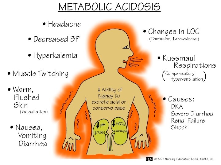
High anion gap metabolic acidosis • • Uremia Diabetic ketoacidosis Lactic acidosis salisylates Methanol Ethylene Glycol Paraldehyde

Non-Anion gap metabolic acidosis • Renal tubular acidosis • Type 1 ( failure of hydrogen ion excretion, DCT) • Type 2 ( loss of HCO 3, PCT) • • Diarrhea Uretrosigmoidostomy diversion Acetazolamide Spironolactone

Principles of correction of metabolic acidosis • Calculating dose of hypertonic Na. Hco 3 ( 892 m. Osm/L ); • We assume bicarbonate distributes in 50% of body wt. ( volume of distribution, vd ), and to raise p. H > 7. 2, and Hco 3 > 12 • One vial of 8. 4% hypertonic Na. Hco 3; 5 o ml contains 44. 6 m. Eq Hco 3 deficit ( m. Eq/L ) = 0. 5 X body wt. ( kg) X ( desired – measured ) • 50% of the required dose given at start slowly bolus infusion over 1 -2 hours, then monitor p. H, Hco 3 and Pco 2 to give the remaining amount • 70 kg patient whose arterial p. H 7. 0 and Hco 3 6 m. Eq/L, Pco 2 34, calculate the Hco 3 deficit ? ( 210 m. Eq/L, require 4. 7 vials of bicarbonate )

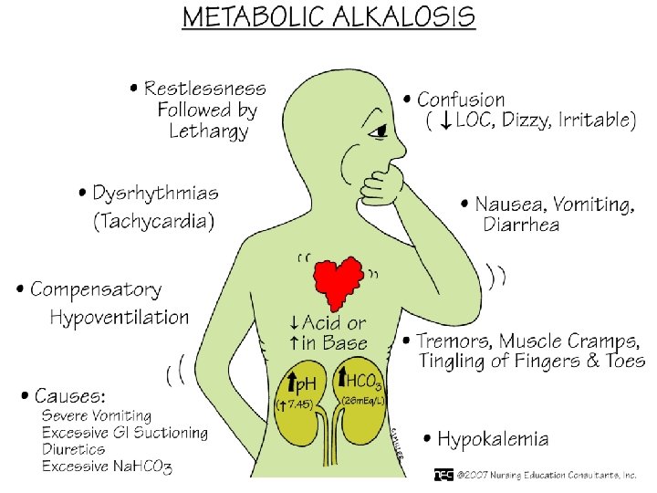
Metabolic alkalosis • Contraction alkalosis • Vomiting, NGT suction • Endocrine; conn’s syndrome, Bartter’s syndrome, cushing’s syndrome • Loop and thiazide diuretics • Excess alkali • Post-hypercapnia
: • • • CNS depression ( drugs/ CVA)")
Respiratory acidosis • Acute ( hypoventilation): • • • CNS depression ( drugs/ CVA) Airway obstruction Pneumonia Pulmonary edema Pneumothorax Myopathy • Chronic : • COPD • Restrictive lung disease

CO 2 retention

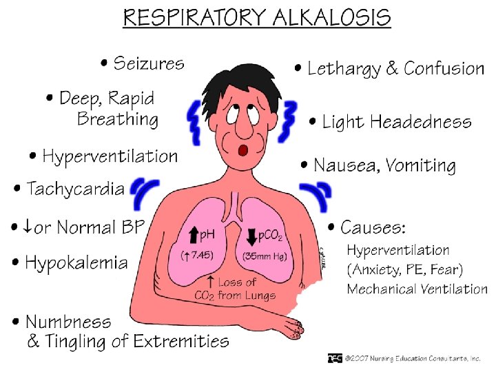
Respiratory alkalosis • Conditions associated with hyperventilation: • • • Hypoxia Anxiety CNS disease Mechanical ventilators Progesterone Salisylates/ sepsis

Compensatory responses - AG

Step-wise diagnostic approach • 1. History and physical examination ( clinical clues to dx. ) • 2. Look at the p. H • 3. Determine primary problem by comparing direction of change of p. CO 2 and p. H. Opposite directions (i. e. a falling p. H with a rising p. CO 2 ) indicates primary respiratory problem. Same direction indicates primary metabolic problem. • 4. Assess compensation by comparing direction of change of p. CO 2 and bicarbonate and applying a compensation rule. If no rule fits, then two problems are probably occurring simultaneously • 5. Anion gap • 6. AGMA; calculate delta ration ( ∆∕ ∆ ) • 7. NAGMA; calculate urine anion gap • 8. Additional investigation; to find a cause

Case # 1 • • p. H 7, 32 Pa. Co 2 28 mm. Hg HCO 3 14 m. Eq/ L Renal function and glucose normal • What is acid base status of this patient?

Case #2 • Patient his ABGs paper showed the following; p. H 7. 15, Pa. Co 2 26, Hco 3 12 mm. Hg, his blood chemistry; sodium 136 m. Eq/d. L, chloride 114 m. Eq/L, potassium 4. 5 m. Eq/d. L • Questions; • What ABB disorder patient is suffering from ? • What is d. d diagnosis of his ABB disorder ?

Case # 3 • 58 years lean man with diabetes mellitus, and chronic kidney disease, presents with abrupt dyspnea, and right sided chest pain for 2 hours. • p. O 2 sat. 84% • p. H 7, 48 • Pa. Co 2 20 mm. Hg • HCO 3 10 m. Eq/ L • Creatinine 2. 8 mg/ dl • Blood glucose 223 mg/ dl • What is acid base status of this patient?
- Slides: 62